A Practical Guide to CLIA Validation for IHC Assays: From Design to Compliance
This article provides a comprehensive, step-by-step framework for designing and executing a robust CLIA validation study for immunohistochemistry (IHC) assays.
A Practical Guide to CLIA Validation for IHC Assays: From Design to Compliance
Abstract
This article provides a comprehensive, step-by-step framework for designing and executing a robust CLIA validation study for immunohistochemistry (IHC) assays. Aimed at researchers and drug development professionals, it covers foundational principles of CLIA regulations and IHC validation, detailed methodological applications for assay design, strategies for troubleshooting and optimization, and a complete validation plan with comparative analysis. The guide synthesizes current regulatory expectations (CLIA, CAP, FDA) and best practices to ensure assays are reliable, reproducible, and clinically actionable for diagnostic use.
Understanding the Essentials: CLIA Regulations and IHC Validation Fundamentals
Purpose and Scope of CLIA The Clinical Laboratory Improvement Amendments (CLIA) of 1988 establish quality standards for all laboratory testing performed on human specimens in the United States to ensure the accuracy, reliability, and timeliness of patient test results. Its scope encompasses approximately 260,000 laboratory entities, regulating testing based on complexity—waived, moderate, and high—with increasing stringency of requirements.
Key Regulatory Bodies
- Centers for Medicare & Medicaid Services (CMS): The primary federal agency responsible for implementing and enforcing CLIA regulations. CMS issues certificates, conducts inspections, and has enforcement authority.
- College of American Pathologists (CAP): A premier non-profit laboratory accreditation organization approved by CMS to inspect laboratories under CLIA. Its standards often exceed baseline CLIA requirements, incorporating discipline-specific checklists.
Table 1: Key CLIA Regulatory Bodies and Roles
| Regulatory Body | Primary Role in CLIA Context | Key Function |
|---|---|---|
| Centers for Medicare & Medicaid Services (CMS) | Implementation & Enforcement | Issues CLIA certificates; conducts inspections; enforces federal regulations. |
| College of American Pathologists (CAP) | Accreditation & Oversight | Provides CMS-approved accreditation; inspects labs using CAP-specific checklists. |
Application Note: CLIA Framework for IHC Assay Validation Study Design
Within a thesis on Immunohistochemistry (IHC) assay validation, the CLIA framework provides the non-negotiable regulatory baseline. For a laboratory developing an IHC assay as a Laboratory Developed Test (LDT) for clinical use, the validation study design must explicitly address CLIA requirements for high-complexity testing. This includes establishing performance specifications for accuracy, precision, reportable range, reference range, and analytical sensitivity/specificity. The study must be documented to satisfy both CMS inspector review and the more rigorous CAP Laboratory General and Anatomic Pathology checklist requirements.
Protocol 1: Analytical Precision (Reproducibility) Testing for an IHC Assay Objective: To determine the intra-observer, inter-observer, inter-instrument, and inter-day precision of an IHC assay’s staining results as required by CLIA for high-complexity testing. Materials: See Scientist's Toolkit below. Methodology:
- Sample Selection: Select 20-30 formalin-fixed, paraffin-embedded (FFPE) tissue blocks representing the assay's target antigen expression spectrum (negative, weak, moderate, strong).
- Sectioning & Slide Preparation: Cut consecutive sections from each block. Assign slides to different testing batches across days, operators, and instruments.
- Staining Runs: Perform the IHC staining protocol according to the established procedure. Design the experiment to include:
- Intra-run: 3 repeats on the same instrument, same day, same operator.
- Inter-run: Staining over 5 separate days.
- Inter-operator: 3 trained technologists perform staining independently.
- Inter-instrument: Staining performed on 2 different, properly calibrated autostainers.
- Quantitative & Semi-Quantitative Assessment: All slides are evaluated by multiple pathologists/observers blinded to the run conditions. Use a validated scoring system (e.g., H-score, Allred score, percentage positivity).
- Statistical Analysis: Calculate Cohen’s kappa for observer agreement. For continuous scores, compute the coefficient of variation (CV%) across conditions. A CV of <20% for semi-quantitative scores is often targeted.
Table 2: Example Precision Study Results for a HER2 IHC Assay (Thesis Data Simulation)
| Precision Dimension | Condition Tested | Agreement Metric (Kappa) | Coefficient of Variation (CV%) |
|---|---|---|---|
| Inter-Observer | 3 Pathologists, Same Slide | 0.85 (Substantial Agreement) | N/A |
| Inter-Instrument | 2 Autostainers, 10 Specimens | N/A | 8.5% (H-Score) |
| Inter-Day | 5 Separate Runs, 5 Specimens | N/A | 12.1% (H-Score) |
The Scientist's Toolkit: Key Reagent Solutions for IHC Validation
| Item | Function in IHC Validation |
|---|---|
| FFPE Tissue Microarray (TMA) | Contains multiple tissue cores on one slide, enabling parallel staining of many specimens under identical conditions for precision and accuracy studies. |
| Validated Primary Antibodies | The key reagent for target detection. Must be clinically validated for specificity, sensitivity, and optimal dilution on FFPE tissue. |
| Reference Standard Materials | Well-characterized cell line controls, patient specimens with known status (via orthogonal method), or commercially available control slides essential for accuracy determination. |
| Detection System (Polymer-based) | Amplifies the primary antibody signal. Must be matched to the host species of the primary antibody and validated for minimal background. |
| Antigen Retrieval Solution | Critical for unmasking epitopes in FFPE tissue (e.g., citrate or EDTA buffer). pH and heating method must be optimized and controlled. |
| Automated Staining Platform | Provides consistent reagent application, incubation times, and temperatures, required for reproducible high-complexity testing under CLIA. |
Visualizations
CLIA Regulatory Structure Diagram
IHC Validation within CLIA Framework Workflow
Defining CLIA Validation vs. Research-Use-Only (RUO) Assay Development
Within the critical path of drug development, immunohistochemistry (IHC) assays serve as pivotal tools for patient stratification, pharmacodynamic assessment, and companion diagnostic development. The transition from a Research-Use-Only (RUO) assay to a Clinical Laboratory Improvement Amendments (CLIA)-validated test is a fundamental, regulated process that ensures analytical validity and reliability for clinical decision-making. This application note delineates the conceptual and practical distinctions between RUO assay development and CLIA validation, providing detailed protocols framed within a thesis on CLIA validation study design for IHC assays.
Core Definitions and Regulatory Landscape
Research-Use-Only (RUO) Assays
RUO assays are in vitro diagnostic products labeled, promoted, and sold for use in laboratory research. They are not intended for use in clinical diagnosis, patient management, or any other clinical purpose. Their development is governed by scientific rigor but not by specific regulatory performance standards.
CLIA-Validated Laboratory Developed Tests (LDTs)
A CLIA-validated test is a Laboratory Developed Test (LDT) for which the laboratory has established, through a defined validation study, the analytical performance specifications (e.g., accuracy, precision, reportable range) as required under the CLIA regulations (42 CFR Part 493). This validation is mandatory for any non-waived test used to report patient results.
Table 1: Primary Distinctions: RUO vs. CLIA Validation
| Aspect | Research-Use-Only (RUO) Assay | CLIA-Validated Assay (LDT) |
|---|---|---|
| Intended Use | Basic research, target discovery, preliminary assay feasibility. | Clinical diagnosis, patient management, clinical trial enrollment. |
| Regulatory Oversight | General labeling requirements (21 CFR 809.10(c)). | CLIA regulations (42 CFR Part 493); potential FDA oversight for high-risk LDTs. |
| Performance Standards | Scientific best practices; no mandated performance thresholds. | Mandatory validation of analytical performance characteristics. |
| Required Documentation | Experimental protocols, reagent data sheets. | Extensive validation plan, validation report, Standard Operating Procedures (SOPs), quality control (QC) records. |
| Quality Systems | Ad hoc, based on laboratory practice. | Formal Quality Management System (QMS) per CLIA. |
| Result Reporting | For research analysis only. | Authorized for patient reports influencing medical care. |
The Validation Pathway: From RUO to CLIA
The transition requires a formal, documented process to establish analytical validity.
Diagram Title: Pathway from RUO Assay to CLIA Validation
Core Components of CLIA Validation for IHC Assays
A comprehensive CLIA validation for an IHC assay must address key analytical performance characteristics.
Table 2: Essential Analytical Performance Characteristics for IHC CLIA Validation
| Characteristic | Definition | Typical IHC Study Design & Acceptance Criteria (Example) |
|---|---|---|
| Accuracy | Agreement with a reference method or material. | Compare results to a clinically validated assay or well-characterized cell line microarray. Target: ≥95% overall agreement. |
| Precision | Repeatability (within-run) and reproducibility (between-run, operator, day, instrument). | Test ≥3 positive and ≥3 negative cases across ≥3 runs, ≥2 operators, ≥3 days. Target: ≥90% intra- and inter-assay concordance. |
| Analytical Sensitivity | Lowest detectable level of analyte (e.g., low-expressing cell lines). | Titrate antibody on cell lines with known expression levels. Establish minimum detectable concentration. |
| Analytical Specificity | Assay's ability to measure only the intended analyte. Includes cross-reactivity and interference. | Test on tissues/cells with known homologous proteins or common interfering substances (e.g., melanin, hemoglobin). |
| Reportable Range | The range of analyte expression (e.g., 0-3+ staining intensity) that can be reliably quantified. | Establish through staining intensity scoring of a full range of positive and negative controls. |
| Reference Range | The range of results expected in a target population (e.g., "positive" vs. "negative"). | Determine by testing a relevant patient population cohort (e.g., n=50-100). |
Detailed Experimental Protocols
Protocol: Precision (Reproducibility) Study for IHC Assay
Objective: To determine inter-operator, inter-day, and inter-instrument reproducibility of IHC staining and scoring.
Materials: See "The Scientist's Toolkit" below. Procedure:
- Sample Selection: Select a minimum of 10 formalin-fixed, paraffin-embedded (FFPE) tissue cases spanning the expected result range (negative, weak positive, moderate positive, strong positive).
- Sectioning: Cut serial sections from each block and mount on charged slides. Label slides anonymously with a study code.
- Study Design: Create a staining schedule where each case is stained over three separate runs (days), by two different certified technologists, potentially using two identical but distinct autostainers.
- IHC Staining: Perform IHC according to the locked-down SOP. Include all controls (positive tissue, negative tissue, reagent negative) in each run.
- Blinded Review: All slides are scored by at least two qualified pathologists/readers who are blinded to the run conditions and previous scores.
- Data Analysis: Calculate percent agreement (positive/negative) and intraclass correlation coefficient (ICC) for semi-quantitative scores (e.g., H-score). Concordance should meet pre-defined criteria (e.g., ≥90% agreement, ICC >0.85).
Protocol: Analytical Specificity (Cross-Reactivity) Assessment
Objective: To evaluate potential cross-reactivity of the primary antibody with homologous proteins.
Procedure:
- Bioinformatic Analysis: Perform a protein BLAST search to identify human proteins with high sequence homology to the target epitope.
- Cell Line Selection: Procure or engineer cell lines expressing the primary target and each identified homologous protein.
- FFPE Block Preparation: Culture cells, fix in formalin, pellet, and embed in paraffin to create a multi-cell line microarray block.
- IHC Staining: Stain the microarray with the validated IHC assay under standard conditions.
- Analysis: Evaluate staining in the target-expressing cell line (positive control) and each homologous protein-expressing cell line. Significant staining in a homologous line indicates potential cross-reactivity requiring further investigation (e.g., antibody blocking with specific peptides).
Diagram Title: Decision Flow: Assay Purpose Dictates Development Path
The Scientist's Toolkit: Key Reagent Solutions for IHC Validation
Table 3: Essential Materials for IHC Assay Development & Validation
| Item | Function in Validation | Example/Notes |
|---|---|---|
| Well-Characterized FFPE Tissue Microarrays (TMAs) | Provide controlled, multi-tissue samples for precision, accuracy, and reportable range studies. | Commercial or internally constructed TMAs with known biomarker status. |
| Cell Line Xenograft FFPE Blocks | Source of reproducible, homogeneous material for sensitivity and specificity studies. | Cell lines with known target expression levels, grown as mouse xenografts. |
| Isotype/Relevance-Matched Control Antibodies | Critical for establishing assay specificity and background during optimization and validation. | Same host species, isotype, and conjugation as primary antibody, targeting an irrelevant antigen. |
| Validated Positive & Negative Tissue Controls | Required for daily run quality control and validation accuracy studies. | Tissues with known high expression and confirmed null expression of the target. |
| Antigen Retrieval Reagents (pH 6, pH 9 buffers) | Standardize the epitope recovery step, a key variable in IHC. | Citrate-based (pH 6.0) or EDTA/TRIS-based (pH 9.0) buffers. |
| Signal Detection System | Chromogenic or fluorescent detection kit. Must be locked down during validation. | Polymer-based HRP or AP systems (e.g., DAB, Permanent Red). |
| Automated Staining Platform | Ensures consistency and reproducibility essential for CLIA validation. | Platforms from Ventana, Leica, Agilent, etc. Protocol must be device-specific. |
| Whole Slide Imaging & Analysis System | Enables quantitative or semi-quantitative scoring, essential for objective precision studies. | Slide scanners coupled with image analysis software (e.g., HALO, Visiopharm). |
Within a CLIA validation study design for IHC assays, establishing robust performance characteristics is fundamental for ensuring reliable diagnostic and research outcomes. This application note details the core validation parameters—Accuracy, Precision, Sensitivity, Specificity, and Reportable Range—providing protocols and frameworks essential for assay qualification in a regulated environment.
Accuracy: Agreement with a Reference Standard
Accuracy assesses the degree of agreement between the IHC assay result and an accepted reference standard (e.g., another validated assay, molecular confirmation, or expert pathology consensus).
Protocol: Method Comparison for Accuracy
- Sample Selection: Obtain 30-50 formalin-fixed, paraffin-embedded (FFPE) tissue samples encompassing the entire spectrum of expected antigen expression (negative, weak, moderate, strong).
- Reference Method: Analyze all samples using the established reference method (e.g., FISH for HER2, PCR for mutation status, or IHC validated against MSI).
- Test Method: Perform IHC staining on serial sections from the same blocks using the assay under validation. Ensure blinding of the evaluator to reference results.
- Evaluation: Have at least two board-certified pathologists score the IHC results independently using the validated scoring criteria.
- Analysis: Calculate percent agreement (overall, positive, negative). For quantitative data, use correlation coefficients (e.g., Pearson’s) and Bland-Altman analysis.
Table 1: Example Accuracy Data for a Novel ER IHC Assay (N=45)
| Reference Standard Positive | Reference Standard Negative | Total | |
|---|---|---|---|
| Test Positive | 22 (True Positive) | 1 (False Positive) | 23 |
| Test Negative | 2 (False Negative) | 20 (True Negative) | 22 |
| Total | 24 | 21 | 45 |
| Metric | Value | Calculation | |
| Overall Agreement | 93.3% | (22+20)/45 | |
| Positive Percent Agreement (Sensitivity) | 91.7% | 22/24 | |
| Negative Percent Agreement (Specificity) | 95.2% | 20/21 |
Precision: Reproducibility of Results
Precision evaluates the closeness of agreement between independent results under stipulated conditions. It includes repeatability (intra-assay) and reproducibility (inter-assay, inter-operator, inter-instrument, inter-day).
Protocol: Precision (Reproducibility) Study
- Panel Design: Select 5-8 FFPE samples covering low positive, moderate positive, high positive, and negative expression levels.
- Experimental Runs: Perform the IHC assay across multiple runs, days, operators, and instruments as required. A typical design includes:
- 3 separate runs
- 2 different operators
- 2 identical instruments (if applicable)
- 3 replicates per sample per run
- Staining & Analysis: All slides are stained and scored independently by each operator. Use continuous scores (e.g., H-score) or categorical scores (0, 1+, 2+, 3+).
- Statistical Analysis: Calculate the coefficient of variation (%CV) for continuous data. For categorical data, calculate percent agreement and Cohen’s/Fleiss’ Kappa for inter-observer concordance.
Table 2: Precision Study Results (H-Score, %CV)
| Sample | Mean H-Score | Intra-Run %CV | Inter-Run %CV | Inter-Operator %CV |
|---|---|---|---|---|
| Negative | 5 | 8.2 | 12.1 | 15.3 |
| Low Positive | 55 | 6.5 | 9.8 | 11.7 |
| High Positive | 210 | 4.1 | 7.2 | 8.9 |
Sensitivity: Detection of Low Antigen Levels
Analytical sensitivity is the lowest amount of analyte that can be reliably distinguished from background. For IHC, this is often the minimum antigen concentration detectable.
Protocol: Limit of Detection (LOD) Determination
- Cell Line or Tissue Microarray (TMA): Utilize a TMA constructed from cell lines with known, titrated antigen expression levels or patient tissues with graded expression.
- Antigen Dilution: If using xenografts or cell pellets, create a dilution series of antigen-positive cells in antigen-negative cells (e.g., from 1:1 to 1:1000).
- Staining: Stain serial sections of the LOD TMA or blocks with the optimized IHC protocol.
- Evaluation: Pathologists score each spot/dilution as "Positive" or "Negative" against a defined staining threshold.
- Analysis: The LOD is the lowest concentration where ≥95% of replicates are consistently scored as positive.
Specificity: Binding to the Target Antigen
Specificity confirms that the observed signal originates from the antibody binding to its intended target epitope and not from non-specific interactions.
Protocol: Specificity Verification
- Blocking with Recombinant Protein: Pre-incubate the primary antibody with a 10-fold molar excess of the target peptide/protein for 30 minutes. Use this mixture for IHC staining alongside a standard control.
- Genetic Confirmation: Compare IHC results with genetic status (e.g., ALK rearrangements confirmed by FISH).
- Knockout/Knockdown Controls: Use isogenic cell lines or tissues with genetic knockout/knockdown of the target gene.
- Alternative Antibodies: Compare staining patterns with antibodies targeting different epitopes on the same protein.
- Expected Outcome: Specific signal should be abolished or markedly reduced in the blocked condition and correlate with genetic status.
Reportable Range: Dynamic Range of Quantification
The Reportable Range defines the span of results that can be reliably quantified, from the Lower Limit of Quantification (LLOQ) to the Upper Limit of Quantification (ULOQ).
Protocol: Establishing the Reportable Range
- Sample Set: Assemble a set of 20-30 samples that uniformly represent the entire dynamic range of expression (H-score from 0 to 300).
- Testing: Stain all samples in duplicate across two separate runs.
- Linearity & Quantification: Plot results against a reference method or a consensus score. The LLOQ is the lowest concentration where the %CV is <20% and bias is <±20%. The ULOQ is the highest concentration where signal saturation does not occur and quantitative scoring remains linear.
- Verification: Ensure the assay's clinical or research cutoff values fall within the verified Reportable Range.
Table 3: Reportable Range Verification for a Quantitative IHC Assay
| Parameter | Value | Acceptance Criterion |
|---|---|---|
| Lower Limit of Quantification (LLOQ) | H-score = 15 | %CV <20%, Bias <±20% |
| Upper Limit of Quantification (ULOQ) | H-score = 280 | No signal saturation, linearity maintained |
| Clinical Cutoff (Example) | H-score = 50 | Well within Reportable Range |
The Scientist's Toolkit: Key Research Reagent Solutions
Table 4: Essential Materials for IHC Validation Studies
| Item | Function & Importance in Validation |
|---|---|
| FFPE Tissue Microarray (TMA) | Contains multiple tissue cores on one slide, enabling high-throughput, simultaneous analysis of multiple samples under identical staining conditions. Critical for precision and sensitivity studies. |
| Isogenic Cell Line Pairs (WT/KO) | Genetically engineered control cell lines provide definitive negative controls for antibody specificity verification. |
| Recombinant Target Protein/Peptide | Used for competitive blocking experiments to confirm antibody-epitope binding specificity. |
| Validated Reference Antibody | An antibody with well-characterized performance serves as a comparator for accuracy determination. |
| Automated IHC Stainer | Ensures consistent reagent application, incubation times, and temperatures, reducing variability in precision studies. |
| Digital Image Analysis Software | Enables objective, quantitative scoring of IHC staining (e.g., H-score, percentage positivity), essential for continuous data in precision and reportable range studies. |
| Control Slides (Multitissue) | Slides containing known positive and negative tissues for the target antigen. Required for daily run validation and monitoring assay drift. |
Visualizations
Within the framework of a comprehensive thesis on CLIA (Clinical Laboratory Improvement Amendments) validation study design for Immunohistochemistry (IHC) assays, the initial and most critical step is the precise definition of the assay's intended use and the associated clinical claim. This foundational element dictates every subsequent decision in the validation plan, from sample cohort selection to statistical endpoints. For a test developed to guide therapy decisions in non-small cell lung cancer (NSCLC), for instance, a claim of "detection of PD-L1 expression to identify patients for pembrolizumab therapy" establishes a completely different validation pathway compared to a purely prognostic claim.
Defining Key Terms and Their Impact on Study Design
The intended use describes the purpose of the in vitro diagnostic device, including the type of specimen, the analyte, and the clinical setting. The clinical claim is a specific statement about the association between the test result and a clinical condition, diagnosis, prognosis, or prediction of response to therapy.
Table 1: Impact of Clinical Claim Type on Validation Study Design Parameters
| Clinical Claim Type | Primary Statistical Endpoint | Required Comparator | Sample Cohort Characteristics | Key Challenge |
|---|---|---|---|---|
| Diagnostic (Detects presence of disease) | Sensitivity & Specificity | Gold-standard diagnostic method (e.g., histopathology) | Known disease status (positive/negative) | Imperfect reference standard |
| Prognostic (Predicts disease outcome independent of therapy) | Hazard Ratio (e.g., Overall Survival) | Clinical outcome data | Cohort with uniform treatment (or no treatment) | Long follow-up times required |
| Predictive (Predicts response to a specific therapy) | Objective Response Rate (ORR) or Progression-Free Survival (PFS) | Treatment response data | Cohort treated with the specific drug of interest | Requires linked treatment and outcome data |
| Companion Diagnostic (Essential for safe and effective use of a drug) | Positive/Negative Predictive Value, Co-positivity/Co-negativity with reference assay | Clinical outcome + Reference method (if available) | Pre-treatment samples from pivotal drug trial | Alignment with drug trial parameters |
Experimental Protocol: Establishing Analytical Performance for a Predictive IHC Assay
This protocol outlines the foundational analytical validation steps required to support a predictive clinical claim for an IHC assay targeting a tumor marker.
Protocol: Analytical Validation of a Predictive IHC Assay Objective: To establish analytical sensitivity (limit of detection), precision (repeatability and reproducibility), and specificity for an IHC assay prior to clinical validation.
Materials & Reagents:
- Formalin-fixed, paraffin-embedded (FFPE) cell line pellets with known antigen expression levels (negative, low, medium, high).
- Patient-derived FFPE tumor tissue sections (positive and negative).
- Primary antibody specific to the target antigen, with optimized dilution.
- Validated IHC detection system (e.g., polymer-based HRP).
- Automated IHC staining platform or materials for manual staining.
- Antigen retrieval solution (e.g., citrate-based, pH 6.0).
- Counterstain (e.g., hematoxylin), dehydration reagents, and mounting medium.
- Brightfield microscope with digital imaging capability.
Procedure:
- Limit of Detection (LoD) Determination:
- Prepare serial dilutions of the primary antibody.
- Stain replicate sections of the low-expressing cell line pellet with each dilution.
- Have at least two qualified pathologists score the stains using the intended clinical scoring method.
- The LoD is defined as the lowest antibody concentration that yields a positive score in ≥95% of replicates.
Precision Testing:
- Repeatability (Intra-assay): A single operator stains the same set of samples (spanning negative, low, high) in triplicate on the same day using the same reagents and equipment.
- Reproducibility (Inter-assay, Inter-operator, Inter-site): Multiple operators at multiple sites stain the same sample set over multiple days (minimum 3 days, 3 operators, 2 sites).
- Scores are analyzed using statistical measures like percent agreement and Cohen's kappa for categorical scores, or coefficient of variation for continuous scores.
Specificity Testing:
- Analytical Specificity: Perform cross-reactivity studies using tissue microarrays containing known related isotypes or tissues with known homologous proteins.
- Interfering Substances: Stain samples treated with common interferents (e.g., hemoglobin, bilirubin, fixative variations) and compare to controls.
The Scientist's Toolkit: Research Reagent Solutions for IHC Validation
Table 2: Essential Materials for IHC Assay Development and Validation
| Item | Function in Validation |
|---|---|
| FFPE Cell Line Xenografts/ Pellets | Provide consistent, biologically relevant controls with defined antigen expression levels for precision and LoD studies. |
| Tissue Microarray (TMA) | Enables high-throughput analysis of assay performance across dozens to hundreds of unique tissue specimens on a single slide. |
| Isotype Control Antibody | A negative control antibody matching the host species and immunoglobulin class of the primary antibody, critical for assessing non-specific binding. |
| Automated IHC Stainer | Standardizes the staining process (timing, temperatures, reagent application) to minimize variability, essential for reproducibility studies. |
| Whole Slide Scanner & Image Analysis Software | Facilitates digital pathology review, enables quantitative analysis, and archives images for audit trails and re-review. |
| Commercial Positive Control Slides | Provide a stable, vendor-validated control tissue to monitor assay performance across multiple staining runs over time. |
Visualizing the Path from Intended Use to Validation Design
Design Logic for CLIA IHC Validation
Predictive IHC in Immuno-oncology
Table 3: Common Acceptance Criteria for Key IHC Validation Parameters
| Validation Parameter | Typical Minimum Acceptance Criterion (Predictive Claim) | Common Statistical Method |
|---|---|---|
| Analytical Sensitivity (LoD) | ≥95% positive calls at the established LoD | Binomial proportion confidence interval |
| Intra-assay Precision (Repeatability) | ≥90% Positive/Percent Agreement or Kappa ≥0.85 | Percent agreement, Cohen's Kappa |
| Inter-assay Precision (Reproducibility) | ≥85% Positive/Percent Agreement or Kappa ≥0.80 | Percent agreement, Fleiss' Kappa |
| Clinical Sensitivity | Lower bound of 95% CI >80% (varies by claim) | 95% Confidence Interval |
| Clinical Specificity | Lower bound of 95% CI >80% (varies by claim) | 95% Confidence Interval |
| Positive Predictive Value (PPV) | Point estimate aligned with drug's response rate | 95% Confidence Interval |
| Negative Predictive Value (NPV) | Point estimate supports clinical utility | 95% Confidence Interval |
A meticulously defined intended use and clinical claim serves as the blueprint for a defensible CLIA validation. It aligns analytical and clinical study designs with the real-world application of the test, ensuring the generated data robustly supports the safe and effective use of the IHC assay in patient care.
1. Introduction
Within the framework of a Clinical Laboratory Improvement Amendments (CLIA) validation study for immunohistochemistry (IHC) assays, the pre-analytical phase is paramount. This phase establishes the foundational reliability of the assay before formal analytical validation begins. Three interdependent pillars form this critical groundwork: rigorous antibody characterization, systematic protocol optimization, and comprehensive reagent qualification. Failure in any of these steps compromises the assay's specificity, sensitivity, and reproducibility, rendering subsequent validation data unreliable for clinical or drug development decisions. These Application Notes detail the protocols and considerations essential for robust pre-validation.
2. Antibody Characterization
Characterization defines the antibody's performance profile. Key parameters include specificity, sensitivity, and optimal dilution.
2.1 Specificity Assessment: Knockout/Knockdown Validation
- Objective: To confirm the antibody binds only to the target antigen.
- Protocol:
- Acquire isogenic cell line pairs (wild-type and CRISPR/Cas9-generated knockout) or utilize siRNA-mediated knockdown controls.
- Culture cells on chamber slides or prepare formalin-fixed, paraffin-embedded (FFPE) cell pellets from both lines.
- Process slides in parallel using a standardized IHC protocol.
- Compare staining intensity. Specific antibodies show strong signal in wild-type and absent or significantly reduced signal in knockout/knockdown samples.
- Complementary techniques like Western blot (WB) on cell lysates from the same lines are mandatory.
2.2 Sensitivity & Optimal Dilution Titration
- Objective: To determine the antibody concentration that provides maximal specific signal with minimal background.
- Protocol:
- Select a biologically relevant FFPE tissue control known to express the target at variable levels.
- Prepare a serial dilution series of the primary antibody (e.g., 1:50, 1:100, 1:200, 1:500, 1:1000).
- Run IHC on serial sections using identical conditions.
- Score slides for signal intensity, background staining, and signal-to-noise ratio. The optimal dilution is the highest dilution that yields strong, specific staining with clean background.
Table 1: Example Data from Anti-ERα Antibody Characterization
| Parameter | Test Method | Control | Result | Acceptance Criterion |
|---|---|---|---|---|
| Specificity | IHC (KO Validation) | MCF-7 WT vs. ERα KO cell pellet | No staining in KO pellet; strong nuclear staining in WT | ≥95% reduction in H-score in KO vs. WT |
| Specificity | Western Blot | MCF-7 WT vs. ERα KO lysate | Single band at ~66 kDa in WT; absent in KO | Single band at expected molecular weight |
| Optimal Dilution | IHC Titration | ER+ Breast Cancer FFPE | Strong specific signal at 1:100-1:200; high background at 1:50; weak signal at 1:500 | Maximum signal-to-noise ratio at chosen dilution |
| Sensitivity (LOD) | IHC on TMA | Tissue Microarray (TMA) with known ER expression spectrum | Detectable staining in samples with ≥1% tumor cells expressing ER (as per reference lab) | Correlation coefficient (r) ≥ 0.9 with reference standard |
3. Protocol Optimization
Optimization refines the assay conditions to maximize performance with the characterized antibody.
3.1 Antigen Retrieval Optimization
- Objective: To identify the optimal method (heat-induced epitope retrieval (HIER) vs. enzymatic) and conditions (pH, time) for unmasking the target epitope.
- Protocol:
- Test multiple retrieval buffers (e.g., citrate pH 6.0, Tris-EDTA pH 9.0, low-pH solution) on the same control tissue.
- Vary retrieval time (e.g., 10, 20, 30 minutes) in a pressure cooker or water bath.
- Process slides with the optimal antibody dilution and standard detection.
- Select the condition yielding the strongest specific signal with lowest non-specific background.
3.2 Detection System Optimization
- Objective: To balance amplification of signal with introduction of background.
- Protocol:
- Compare different commercially available detection kits (e.g., polymer-based, avidin-biotin).
- Optimize incubation times for secondary antibody/polymer and chromogen (e.g., DAB).
- Include appropriate controls for endogenous enzymes (peroxidase, phosphatase).
Table 2: Protocol Optimization Matrix Example
| Variable | Options Tested | Evaluation Metric | Optimal Condition Selected |
|---|---|---|---|
| Antigen Retrieval | Citrate pH 6.0 (20 min), Tris-EDTA pH 9.0 (20 min), Enzymatic (Protease, 5 min) | Signal Intensity, Background, Cellular Morphology | Tris-EDTA pH 9.0 (20 min) |
| Primary Ab Incubation | 30 min @ RT, 60 min @ RT, Overnight @ 4°C | Signal Intensity, Uniformity | 60 min @ Room Temperature |
| Detection System | Polymer System A, Polymer System B, ABC Kit | Signal-to-Noise Ratio, Non-Specific Background | Polymer System B |
| DAB Incubation Time | 30 sec, 1 min, 2 min, 5 min | Intensity Saturation, Background Development | 2 minutes |
4. Reagent Qualification
Qualification ensures all reagents perform consistently lot-to-lot before being locked down for validation.
4.1 Primary Antibody Lot-to-Lot Testing
- Protocol: Test new antibody lots in parallel with the qualified lot using the optimized protocol on a standard control tissue. Compare staining intensity, pattern, and background. Use quantitative image analysis or semi-quantitative scoring (H-score) to ensure differences are within pre-defined limits (e.g., ≤10% variance in H-score).
4.2 Critical Reagent Qualification
- Protocol: Qualify key detection reagents (polymer, chromogen, retrieval buffers) by running a complete assay with new lots alongside current qualified lots. Include positive, negative, and no-primary antibody controls. Establish acceptance criteria based on control slide performance.
The Scientist's Toolkit: Key Research Reagent Solutions
- Isogenic CRISPR/Cas9 Knockout Cell Lines: Essential for antibody specificity confirmation by providing genetically defined negative controls.
- Multiplex IHC/IF-Validated Antibodies: Antibodies specifically validated for use in multiplex assays, ensuring minimal cross-reactivity.
- Tissue Microarrays (TMAs): Contain multiple tissue cores on one slide, enabling high-throughput optimization and qualification across diverse samples.
- Validated Positive Control FFPE Tissues: Tissues with known, stable expression levels of the target, critical for run-to-run monitoring.
- Automated Staining Platform Reagents: Detection kits and buffers optimized for specific automated stainers, ensuring reproducibility and consistency.
- Chromogen Alternatives to DAB: Such as permanent red or vector blue, for multiplexing or compatibility with specific counterstains/scanners.
- Digital Image Analysis Software: Enables quantitative, objective assessment of staining intensity and percentage for qualification and optimization.
5. Experimental Protocols
Protocol 5.1: Comprehensive Antibody Characterization via IHC and WB
- Sample Preparation: Generate FFPE blocks from wild-type and knockout cell line pellets. Fix cells in 10% NBF for 24 hours, process, and embed.
- IHC Staining: Section pellets at 4-5 µm. Deparaffinize and rehydrate. Perform antigen retrieval as per initial guidelines. Apply endogenous peroxidase block. Apply primary antibody at starting dilution (e.g., manufacturer's recommendation) alongside a no-primary control. Apply labeled polymer-HRP secondary. Develop with DAB for a standardized time (e.g., 5 min). Counterstain, dehydrate, and mount.
- Western Blot: Lyse parallel cell pellets in RIPA buffer. Separate 20 µg protein by SDS-PAGE. Transfer to PVDF membrane. Block and incubate with the same primary antibody used for IHC (diluted in blocking buffer). Incubate with HRP-conjugated secondary. Develop with chemiluminescent substrate and image.
- Analysis: Compare IHC staining intensity and pattern between WT and KO. For WB, confirm a single band at the expected molecular weight in WT lane only.
Protocol 5.2: Checkerboard Titration for Protocol Optimization
- Design Matrix: Create a grid where the X-axis represents a series of primary antibody dilutions (e.g., 1:50, 1:100, 1:200, 1:500). The Y-axis represents different antigen retrieval conditions (e.g., Buffer A, Buffer B; 10 min, 20 min).
- Staining: Apply the matrix to serial sections of a well-characterized positive control tissue using an automated stainer or manual protocol where all other variables are constant.
- Scoring: Blind-score each condition for signal intensity (0-3+), percentage of positive cells, background staining (0-3+), and overall signal-to-noise ratio.
- Selection: Choose the condition at the intersection of the highest antibody dilution and most efficient retrieval that yields a maximum intensity score with a minimum background score.
6. Visualizations
Pre-Validation in CLIA IHC Study Workflow
Antibody Characterization Key Pathways
Iterative Protocol Optimization Workflow
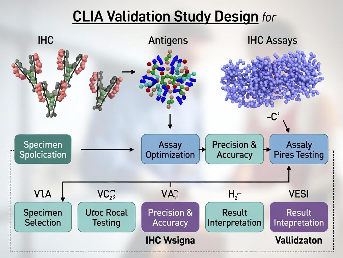
Building Your Blueprint: A Step-by-Step CLIA IHC Validation Study Design
Within the framework of a comprehensive thesis on CLIA validation study design for immunohistochemistry (IHC) assays, the establishment of robust acceptance criteria is the cornerstone of analytical validation. These criteria, derived directly from clinical requirements and regulatory guidance, define the success metrics for assay performance, ensuring reliability and reproducibility in patient diagnosis and therapy selection.
Application Notes on Critical Acceptance Criteria
Acceptance criteria must be prospectively defined and justified. For IHC assays, they are anchored in the assay’s intended use (IU) and its clinical performance requirements. Key parameters include analytical sensitivity, analytical specificity, precision, and accuracy.
Table 1: Core Acceptance Criteria for IHC Assay Validation
| Performance Characteristic | Typical Metric | Example Acceptance Criterion (e.g., HER2 IHC) | Primary Regulatory Guidance Reference |
|---|---|---|---|
| Analytical Sensitivity (Detection Limit) | Minimum detectable antigen concentration or cell line reactivity. | 100% detection of appropriate cell lines with ≥1+ staining intensity at the defined antibody dilution. | CLIA '88; CAP Laboratory Standards. |
| Analytical Specificity | Cross-reactivity/interference; staining in negative tissues/cells. | ≤5% background staining in known negative tissue sections; no cross-reactivity with related antigens per in silico/biotinylation analysis. | FDA IHC Assay Development Guidance. |
| Precision (Repeatability & Reproducibility) | Intra-run, inter-run, inter-operator, inter-instrument agreement. | ≥95% inter-operator concordance (Cohen’s kappa ≥0.90) for scoring categories (0, 1+, 2+, 3+). | CLSI Guideline EP12-A2, EP05-A3. |
| Accuracy (Comparator Method) | Concordance with a validated reference method (e.g., FISH, another IHC assay). | Overall Percent Agreement (OPA) ≥90% and Positive Percent Agreement (PPA) ≥95% versus FISH for binary clinical call (Positive vs. Negative). | FDA Guidance on Companion Diagnostics. |
| Robustness | Tolerance to deliberate variations in pre-analytical/analytical conditions. | Acceptable staining (meeting accuracy/precision criteria) across specified ranges of antigen retrieval time (±3 min), primary antibody incubation time (±10%), and room temperature (±2°C). | ICH Guideline Q2(R1). |
Detailed Experimental Protocols
Protocol 1: Determining Analytical Specificity (Cross-Reactivity)
- Objective: To assess potential cross-reactivity of the primary antibody with unrelated or similar epitopes.
- Materials: See "The Scientist's Toolkit" below.
- Method:
- In Silico Analysis: Perform BLAST alignment of the antibody's target epitope sequence against human proteome databases.
- Peptide Blocking: Pre-incubate the primary antibody with a 10-fold molar excess of the target peptide versus a control peptide for 1 hour at RT.
- Parallel Staining: Apply the pre-absorbed antibody mixtures to a multi-tissue block containing tissues known to express the target and homologous proteins.
- Evaluation: Loss of staining only in the target peptide-blocked sample confirms specificity. Persistent staining with both indicates cross-reactivity.
Protocol 2: Assessing Inter-Operator Reproducibility for Scoring
- Objective: To quantify concordance between multiple trained pathologists/scorers.
- Materials: A validated IHC assay, a set of 50 pre-stained patient tissue samples spanning all score categories, standardized scoring guidelines.
- Method:
- Blinded Review: Three independent, qualified operators score each sample according to the clinical algorithm (e.g., 0, 1+, 2+, 3+) in a blinded fashion.
- Data Collection: Record individual scores for each sample-operator pair.
- Statistical Analysis: Calculate pairwise percent agreement and Cohen’s kappa statistic for all operator combinations. Use Fleiss' kappa for overall agreement.
- Criterion Assessment: Compare the lower bound of the 95% confidence interval for kappa to the pre-defined acceptance criterion (e.g., ≥0.90).
Visualizations
Deriving Acceptance Criteria Flowchart
IHC Validation Protocol Workflow
The Scientist's Toolkit: Key Research Reagent Solutions
| Item | Function in IHC Validation |
|---|---|
| Formalin-Fixed, Paraffin-Embedded (FFPE) Cell Lines | Provide consistent, quantifiable antigen-positive and negative controls for sensitivity/specificity runs. |
| Multi-Tissue Microarrays (MTAs) | Enable high-throughput assessment of staining specificity across dozens of tissue types on a single slide. |
| Recombinant Target Protein / Peptide | Used for antibody blocking experiments to confirm epitope specificity. |
| Validated Primary Antibody Clone | The critical reagent; specificity, lot consistency, and optimal dilution must be rigorously defined. |
| Polymer-Based Detection System | Amplifies signal while reducing background; choice impacts sensitivity and specificity. |
| Automated Staining Platform | Essential for standardizing the analytical phase and minimizing variability in precision studies. |
| Digital Pathology & Image Analysis Software | Enables quantitative, objective scoring for critical parameters like H-Score, reducing operator subjectivity. |
| Reference Standard Slides (e.g., HER2 CTR) | Commercially available controls with known score values, used for daily run validation and proficiency testing. |
Application Notes
Within a Clinical Laboratory Improvement Amendments (CLIA) validation study for immunohistochemistry (IHC) assays, rigorous sample cohort selection is foundational. The cohort must reflect the intended clinical use population to ensure the assay's analytical sensitivity, specificity, precision, and reportable range are valid. Key considerations include the cohort size (statistical power), representation of relevant tissue types and disease states, and adherence to standardized biospecimen quality requirements. Failure in any domain introduces pre-analytical variables that can invalidate subsequent validation data.
Cohort Size Justification
Cohort size is determined by statistical requirements for precision (e.g., 95% confidence intervals) around key performance metrics. For rare biomarkers, enrichment strategies may be required.
Table 1: Recommended Minimum Cohort Sizes for CLIA IHC Assay Validation
| Validation Parameter | Typical Minimum Sample Number | Statistical Rationale | Regulatory Guidance Reference |
|---|---|---|---|
| Analytical Sensitivity (Detection Limit) | 5-10 positive, 5-10 negative | Estimate lower limit of detection | CAP Checklist ANP.22900 |
| Analytical Specificity (Interference) | 10-20 with known interfering conditions (e.g., necrosis, edge artifact) | Assess potential false positives/negatives | CLIA '88; CAP ANP.12200 |
| Within-Run & Between-Run Precision | 20-30 samples, spanning expression range (negative, weak, moderate, strong) | Calculate CV; ensure reproducibility | CLSI EP05-A3 |
| Reportable Range (Staining Intensity) | 30-50 samples, covering all expected scores (0, 1+, 2+, 3+) | Establish dynamic range and linearity (if quantitative) | CAP ANP.22850 |
| Comparison to a Reference Method | 50-100+ samples, with ~50% prevalence of marker | For 95% CI width of ~±10% for sensitivity/specificity | CLSI EP12-A2 |
Tissue Types and Phenotype Requirements
The cohort must encompass the full spectrum of tissue types and morphologies expected in clinical practice. This ensures the assay's robustness across matrix effects.
Table 2: Essential Tissue Cohort Composition for a Broad-Spectrum IHC Assay Validation
| Tissue Category | Sub-types & Examples | Purpose in Validation | Minimum Recommended Cases |
|---|---|---|---|
| Target-Positive Tissues | Tissues with known expression of the target antigen (e.g., tumor types, normal tissues). | Establish assay sensitivity and expected staining patterns. | 20-30 |
| Target-Negative Tissues | Tissues with known absence of the target antigen. | Establish assay specificity and background levels. | 10-15 |
| Tissue Mimics & Challenging Morphologies | Necrotic tissue, crush artifact, inflamed stroma, fatty tissue, bone decalcified sections. | Test for staining artifacts and interference. | 5-10 of each relevant type |
| Normal/Counterstain Controls | Relevant normal adjacent tissues (e.g., skin, colon, lymph node). | Assess specificity of staining and internal positive/negative controls. | 5-10 |
| Previous Lot/Platform Comparison | Cases previously tested on a legacy assay or platform. | Demonstrate consistency and comparability. | 20-30 |
Biospecimen Requirements
Pre-analytical variables are a major source of error. Standardizing biospecimen criteria is non-negotiable.
Table 3: Critical Biospecimen Pre-Analytical Parameters for IHC Validation
| Parameter | Acceptance Criteria for Validation Cohort | Impact on IHC Results |
|---|---|---|
| Fixation Type & Time | Neutral buffered formalin (10%), fixation time 6-72 hours. | Under-fixation: poor morphology, antigen loss; Over-fixation: antigen masking. |
| Tissue Ischemia Time | Cold ischemia time documented, ideally <1 hour. | Hypoxia can degrade antigens and induce false expression patterns. |
| Processing & Embedding | Standardized paraffin embedding protocol; no excessive heat. | Incomplete processing affects sectioning and antibody penetration. |
| Section Thickness | 4-5 micron sections, cut with clean, sharp microtome blades. | Thick sections cause uneven staining; torn sections damage morphology. |
| Slide Storage | Sections used within 6 weeks of cutting, stored desiccated at 4°C. | Antigenicity can degrade over time on glass slides. |
| Tissue Age (FFPE Block) | Preferably <5 years old, with known storage conditions (cool, dark). | Long-term storage can lead to oxidation and antigen degradation. |
Experimental Protocols
Protocol 1: Tissue Microarray (TMA) Construction for Validation Cohort
Purpose: To efficiently array the selected cohort samples onto a single slide for simultaneous staining under identical conditions, enabling high-throughput, controlled comparison.
Materials: Recipient paraffin block, tissue cores (0.6-2.0mm), hollow needle, TMA construction instrument or manual arrayer, heated plate, histology slides.
Methodology:
- Cohort Map Design: Create a digital map assigning each core a unique ID linked to case metadata. Include replicates of key samples.
- Donor Block Review: H&E-stained sections from each donor FFPE block are marked by a pathologist to define representative regions for coring.
- Recipient Block Preparation: Pour a standard paraffin block in a mold; allow to solidify completely.
- Coring and Arraying: a. Using the hollow needle, extract a core from the recipient block to create an empty hole. b. Immediately extract a core from the designated region of the donor block. c. Transfer the donor core into the pre-formed hole in the recipient block. d. Repeat in a grid pattern according to the design map.
- Block Fusion: Place the completed TMA block on a 37-42°C heated plate for 10-15 minutes to slightly melt paraffin and fuse cores. Apply gentle pressure with a slide. Allow to cool.
- Sectioning: Cut 4-5 micron sections using a standard microtome. Float sections on a water bath and collect on charged slides.
- Validation: Stain one TMA section with H&E to verify tissue presence and morphology.
Protocol 2: Staining Optimization and Validation Run on Cohort
Purpose: To perform the IHC assay on the entire validation cohort under optimized, locked-down conditions to generate data for performance characterization.
Materials: Optimized primary antibody, detection system (e.g., polymer-HRP), antigen retrieval solution (e.g., citrate buffer, pH 6.0), blocking serum, DAB chromogen, hematoxylin counterstain, automated IHC stainer or manual setup.
Methodology:
- Slide Baking and Deparaffinization: Bake slides at 60°C for 1 hour. Deparaffinize in xylene and rehydrate through graded alcohols to water.
- Antigen Retrieval: Perform heat-induced epitope retrieval in pre-heated retrieval buffer using a pressure cooker, steamer, or water bath (e.g., 95-100°C for 20-40 minutes). Cool slides.
- Endogenous Peroxidase Block: Apply 3% hydrogen peroxide solution for 10 minutes to block endogenous peroxidase activity. Rinse.
- Protein Block: Apply normal serum or protein block from the detection system for 10 minutes to reduce non-specific binding.
- Primary Antibody Incubation: Apply optimized primary antibody at predetermined dilution and incubate for the specified time (30-60 minutes at room temp or overnight at 4°C). Rinse.
- Detection System: Apply labeled polymer (e.g., HRP polymer) for 30 minutes. Rinse.
- Chromogen Development: Apply DAB substrate solution for 5-10 minutes, monitoring under a microscope. Stop development in water.
- Counterstaining and Mounting: Counterstain with hematoxylin, bluing step, dehydrate, clear, and mount with a permanent medium.
- Batch Staining: Process the entire validation cohort (full sections or TMAs) in a single batch with appropriate positive and negative controls on each slide/run.
Protocol 3: Digital Image Analysis and Scoring of Validation Cohort
Purpose: To objectively quantify or semi-quantify IHC staining results across the cohort for statistical analysis.
Materials: Whole slide scanner, digital image analysis software (e.g., HALO, QuPath, Aperio ImageScope), scoring rubric.
Methodology:
- Slide Digitization: Scan all stained cohort slides at 20x or 40x magnification.
- Algorithm Training: For quantitative assays, train a software algorithm on a subset of images. a. Annotate representative regions for positive staining, negative staining, and background. b. Define parameters: color deconvolution (separate DAB from hematoxylin), intensity thresholds, and region of interest (tumor vs. stroma). c. Validate algorithm performance against pathologist manual scores on a training set.
- Batch Analysis: Apply the validated algorithm to all cohort images.
- Data Extraction: Export quantitative metrics (e.g., H-score, % positive nuclei, staining intensity mean) for each case into a spreadsheet.
- Statistical Analysis: Calculate performance metrics (sensitivity, specificity, precision) using the quantitative data against the reference standard truth.
Diagrams
Title: CLIA IHC Validation Cohort Selection Workflow
Title: Key Factors in IHC Assay Validation
The Scientist's Toolkit
Table 4: Essential Research Reagent Solutions for IHC Validation Studies
| Item | Function | Key Considerations |
|---|---|---|
| FFPE Tissue Biospecimens | The core test material containing the target antigen in its native, fixed state. | Must have documented pre-analytical history (fixation, ischemia). Sourced from accredited biorepositories. |
| Validated Primary Antibody | Binds specifically to the target antigen of interest. | Clone specificity, host species, recommended dilution for IHC on FFPE tissue. Requires prior analytical validation. |
| Polymer-Based Detection System | Amplifies the primary antibody signal for visualization. | High sensitivity, low background. Common formats: HRP-polymer or AP-polymer with chromogen (DAB/Vector Red). |
| Antigen Retrieval Buffer | Reverses formaldehyde-induced cross-links to expose epitopes. | Choice of pH (e.g., citrate pH6.0, EDTA/TRIS pH9.0) is antigen-specific and must be optimized. |
| Automated IHC Stainer | Provides standardized, reproducible staining conditions for a cohort. | Essential for high-precision, batch processing in validation. Reduces inter-run variability. |
| Whole Slide Scanner | Digitizes entire tissue sections for archiving and analysis. | Enables digital pathology workflows, remote review, and quantitative image analysis. |
| Digital Image Analysis Software | Quantifies staining intensity and percentage of positive cells. | Reduces scorer subjectivity, provides continuous data for statistical analysis, essential for biomarker quantification. |
| Multitissue Control Slides | Slides containing multiple known positive/negative tissues. | Run with every batch to monitor staining consistency, assay sensitivity, and specificity. |
Application Notes and Protocols
Thesis Context: Within the framework of a CLIA (Clinical Laboratory Improvement Amendments) validation study for immunohistochemistry (IHC) assays, the formal assessment of precision—encompassing repeatability (intra-assay, intra-run, intra-observer) and reproducibility (inter-assay, inter-run, inter-site, inter-observer)—is a cornerstone requirement. This document details the experimental design and protocols to generate robust precision data, ensuring the IHC assay's reliability for clinical use in drug development companion diagnostics.
1. Foundational Definitions & Key Metrics
Precision is quantified through statistical analysis of agreement. The core metrics are summarized below:
Table 1: Key Precision Metrics for IHC CLIA Validation
| Metric | Definition | Typical Target for CLIA | Calculation Method |
|---|---|---|---|
| Percent Agreement | Proportion of identical scores between repeated measurements. | ≥90% for critical positive/negative calls. | (Number of Agreements / Total Comparisons) x 100. |
| Cohen's Kappa (κ) | Measures inter-observer agreement for categorical scores, correcting for chance. | κ ≥ 0.6 (Substantial); κ ≥ 0.8 (Almost Perfect). | Statistical software (e.g., R, MedCalc). |
| Intraclass Correlation Coefficient (ICC) | Measures consistency for continuous data (e.g., H-score, % positivity). | ICC ≥ 0.9 (Excellent consistency). | Two-way random-effects or mixed-effects models. |
| Coefficient of Variation (CV%) | Ratio of standard deviation to mean for continuous data across replicates. | ≤20% (Run-to-run; lower for within-run). | (Standard Deviation / Mean) x 100. |
2. Core Experimental Design Protocol
Protocol 2.1: Hierarchical Precision Study for IHC Biomarker Quantification
Objective: To estimate variance components attributable to different factors (repeatability and reproducibility) in the IHC staining and scoring process.
Materials & Reagents: See "The Scientist's Toolkit" below.
Methodology:
- Sample Selection: Select 20-30 formalin-fixed, paraffin-embedded (FFPE) tissue samples spanning the assay's dynamic range (negative, low, medium, high expression). Ensure sample adequacy for consecutive sectioning.
- Experimental Matrix:
- Factor 1 - Operator: 2-3 distinct, trained technologists.
- Factor 2 - Run/Day: Each operator performs the assay on 3 separate, non-consecutive days.
- Factor 3 - Replicate: Within each run, each sample is stained in duplicate (adjacent tissue sections).
- Total Slides: 20 samples x 3 operators x 3 days x 2 replicates = 360 slides.
- Randomization: Pre-determine a randomized slide run order for each operator and day to avoid batch effects.
- Blinded Analysis: After staining, slides are coded. Each operator scores their own slides and a shuffled set scored by all operators (for inter-observer reproducibility).
- Data Capture: Record quantitative data (e.g., H-score) and/or categorical data (0, 1+, 2+, 3+) per predefined scoring guidelines.
- Statistical Analysis:
- Calculate Percent Agreement and Cohen's Kappa for categorical scores.
- Perform ANOVA or linear mixed-effects modeling to partition total variance into components: between-sample, between-operator, between-run, and residual (repeatability) variance.
- Calculate ICC and CV% as per Table 1.
Protocol 2.2: Inter-Site Reproducibility Protocol
Objective: To validate assay performance across multiple laboratory sites, as required for multicenter trials.
Methodology:
- Master Kit & Sample Distribution: A central site prepares and distributes identical lots of pre-qualified reagents, pre-titered antibody, control slides, and the same set of 10-15 FFPE tissue blocks/sections to 3 participating laboratories.
- Standardized Protocol: All sites follow the identical, validated IHC protocol (retrieval conditions, antibody dilution, incubation times, detection system, instrument platform).
- Execution: Each site conducts the assay in 2 independent runs, with duplicate samples per run, following a pre-specified layout.
- Data Centralization & Analysis: All stained slides are digitized. Scoring is performed centrally by 2-3 pathologists and also by local site readers. Analyze using ICC and Kappa statistics comparing sites and central vs. local reads.
The Scientist's Toolkit: Key Research Reagent Solutions
Table 2: Essential Materials for IHC Precision Studies
| Item | Function & Importance for Precision |
|---|---|
| Certified Reference FFPE Tissue Microarrays (TMAs) | Contain multiple tissue types with known biomarker expression levels. Provide a consistent sample substrate across runs for controlling staining variability. |
| Pre-Diluted, Ready-to-Use Antibody Cocktails | Eliminates operator-induced variability in antibody dilution, a major source of reproducibility error. Essential for inter-site studies. |
| Automated Staining Platforms | (e.g., Ventana Benchmark, Leica BOND, Agilent/Dako Omnis). Provides superior repeatability over manual staining by precisely controlling reagent incubation times and temperatures. |
| Whole Slide Scanners & Image Analysis Software | Enables quantitative, objective scoring (nuclear, membrane, H-score analysis) minimizing subjective inter-observer variability. Allows for re-analysis and audit trails. |
| CLIA-Grade, Lot-Controlled Detection Kits | Chromogenic detection systems (e.g., DAB) with consistent lot-to-lot performance are critical. Includes enzyme conjugates, polymer detection, and chromogen substrates. |
| Commercial Antigen Retrieval Buffers | Standardized, pH-balanced buffers (e.g., EDTA, citrate) ensure consistent epitope retrieval, a key pre-analytical variable. |
3. Visualization of Experimental Workflow and Statistical Relationship
Diagram Title: IHC Precision Study Workflow & Variance Partitioning
Diagram Title: IHC Process Map with Critical Control Points for Precision
Introduction Within the broader thesis on CLIA validation for IHC assays, the design of accuracy studies is paramount. Accuracy, defined as the closeness of agreement between a test result and the accepted reference value, is established through comparison to a reference standard or a well-characterized comparator assay. This application note details the framework and protocols for designing these critical studies, ensuring robust analytical validation for drug development and clinical research.
1. Defining the Reference Framework Accuracy can be assessed via comparison to a reference standard (gold standard) or a validated comparator method. The choice dictates study design.
- Reference Standard: A method that provides the definitive measure of the analyte (e.g., mass spectrometry for protein quantification, sequencing for genetic alterations). Its error is considered negligible.
- Comparator Assay: An existing, fully validated method (e.g., a previously approved IVD assay or a research assay with established performance) used when a true reference standard is unavailable. Discrepancies require resolution.
Table 1: Comparison of Reference Approaches
| Feature | Reference Standard | Comparator Assay |
|---|---|---|
| Definition | Definitive, highest order method | Well-characterized, validated method |
| Basis of Truth | Incontrovertible | Pragmatic, based on prior validation |
| Study Goal | Establish absolute accuracy | Establish concordance/equivalence |
| Discrepancy Analysis | Not applicable; test result is inaccurate | Required; may involve adjudication with a third method |
| Common Use in IHC | Less common; possible with digital pathology/quantitative imaging vs. reference counts | Common (e.g., vs. another clinical IHC assay, vs. ISH/FISH results) |
2. Key Components of Study Design
- Sample Cohort: Must be representative of the intended-use population, covering the full spectrum of antigen expression (negative, low, moderate, high) and relevant tissue types/topographies.
- Sample Size: Justified statistically to achieve desired confidence intervals for metrics like Positive/Negative Percent Agreement. A minimum of 60 positive and 60 negative samples is often a starting point for precision and accuracy studies.
- Blinding: Testing with the candidate and reference/comparator assays must be performed independently and blinded to the other method's result to avoid bias.
- Pre-defined Acceptance Criteria: Criteria for accuracy (e.g., lower bound of 95% CI for Overall Percent Agreement > 85%) must be established a priori.
3. Experimental Protocols
Protocol 1: Accuracy Assessment vs. a Reference Standard Objective: Determine the quantitative accuracy of a new IHC assay (e.g., H-score via digital image analysis) against a quantitative reference method (e.g., mass spectrometry-based quantification). Materials: See "The Scientist's Toolkit" below. Procedure:
- Sample Selection & Processing: Select FFPE tissue blocks (n=100-150) spanning the expression range. From each block, prepare:
- One section (4-5 µm) for IHC staining.
- Ten consecutive sections (10 µm each) for macro-dissection and mass spectrometry.
- Reference Testing (Mass Spectrometry):
- Mark tumor-rich areas on H&E-stained guide slides.
- Macro-dissect corresponding areas from unstained curls.
- Extract proteins, digest with trypsin.
- Perform targeted LC-MS/MS using stable isotope-labeled peptide standards for absolute quantification of the target protein (results in ng/mg tissue).
- Candidate IHC Testing:
- Stain the 4-5 µm section using the fully optimized IHC protocol.
- Digitize slides at 20x magnification.
- Annotate the same tumor regions analyzed by MS.
- Use image analysis software to calculate the H-score (0-300) within annotations.
- Data Analysis & Correlation:
- Pair MS result (ng/mg) with H-score for each sample region.
- Perform Deming regression analysis to account for error in both measurements.
- Calculate the correlation coefficient (e.g., Pearson's r) and the 95% CI.
Protocol 2: Concordance Study with a Comparator Assay Objective: Establish the diagnostic concordance of a new IHC assay with an existing clinical assay for a binary readout (Positive/Negative). Materials: See "The Scientist's Toolkit" below. Procedure:
- Sample Selection: Select archival FFPE samples (n=120) with known comparator assay result (60 positive, 60 negative).
- Blinded Testing: Re-cut all blocks. Test all samples with the new IHC assay in a single batch by personnel blinded to the comparator results.
- Result Adjudication: A pathologist, blinded to both results, reviews any stained sample where the new IHC result is ambiguous.
- Discrepancy Analysis: For samples with discordant results (New IHC+ / Comparator -, or vice versa), subject them to an adjudication method (e.g., a different antibody clone, RNA-seq, PCR). This establishes a "referee" result.
- Statistical Analysis:
- Create a 2x2 concordance table vs. the comparator.
- Calculate Positive Percent Agreement (PPA), Negative Percent Agreement (NPA), and Overall Percent Agreement (OPA) with 95% CIs.
- If adjudicated, recalculate agreement statistics versus the final adjudicated truth.
Table 2: Example Concordance Results (vs. Comparator Assay)
| Metric | Formula | Result (95% CI) | Acceptance Met? |
|---|---|---|---|
| Positive Percent Agreement (Sensitivity) | [True Pos / (True Pos + False Neg)] x 100 | 96.7% (88.7-99.6%) | Yes (>90%) |
| Negative Percent Agreement (Specificity) | [True Neg / (True Neg + False Pos)] x 100 | 93.3% (84.1-97.4%) | Yes (>90%) |
| Overall Percent Agreement | [(True Pos + True Neg) / Total] x 100 | 95.0% (89.6-97.7%) | Yes (>85%) |
4. Visualizing Study Workflows and Relationships
Title: Accuracy Study Design Decision Flow
Title: Protocol 1: Quantitative Accuracy Workflow
Title: Protocol 2: Concordance & Adjudication Workflow
5. The Scientist's Toolkit: Essential Research Reagent Solutions
| Item | Function in Accuracy Studies |
|---|---|
| FFPE Tissue Microarray (TMA) | Contains multiple tissue cores on one slide, enabling high-throughput, simultaneous staining of diverse samples under identical conditions, reducing run-to-run variability. |
| CRISPR/Cas9-engineered Cell Lines | Provide isogenic controls with defined expression levels (knock-out, low, high) of the target antigen. Essential for creating standardized controls and calibration curves. |
| Recombinant Antigen Protein Spikes | Used to spike negative tissue lysates or cell pellets for recovery experiments in quantitative assays, helping establish linearity and limit of detection. |
| Validated, High-Specificity Antibodies (Primary & Secondary) | The core reagent for IHC. Specificity validation (KO/KD validation) is critical to ensure the accuracy of the signal. Conjugated secondaries enable multiplexing. |
| Chromogenic & Fluorescent Detection Kits | Amplify the primary antibody signal for visualization. Selection depends on platform (brightfield vs. multiplex fluorescence). Must be optimized for sensitivity and low background. |
| Digital Pathology & Image Analysis Software | Enables objective, quantitative assessment of IHC staining (e.g., H-score, % positive cells, staining intensity). Crucial for quantitative correlation studies. |
| Stable Isotope-Labeled Peptide Standards (AQUA) | Used in mass spectrometry-based reference methods for the absolute, targeted quantification of specific proteins in complex tissue digests. |
Establishing Analytic Sensitivity (Limit of Detection) and Specificity (Interference Testing)
Within a comprehensive CLIA validation thesis for IHC assays, establishing robust analytical sensitivity (Limit of Detection, LoD) and specificity (through interference testing) is paramount. These parameters are critical for ensuring the assay reliably detects the target analyte at low concentrations and does not cross-react with interfering substances. This application note provides detailed protocols and methodologies for these cornerstone validation studies, aligning with CLIA, CAP, and FDA guidelines for assay development in drug and diagnostic research.
Establishing Analytic Sensitivity (Limit of Detection)
The LoD is the lowest concentration of an analyte that can be consistently detected by the assay. For IHC, this is often expressed as the minimum antigen level detectable above a negative control with a defined confidence level (e.g., 95%).
Key Experimental Protocol: Determining LoD Using a Serial Dilution Approach
Objective: To empirically determine the lowest detectable concentration of target antigen in a controlled matrix.
Materials & Reagents:
- Cell Line or Tissue Microarray (TMA): Containing cells/tissues with a known, quantified expression gradient of the target antigen.
- Reference Standard: Recombinant protein or purified antigen for spiking studies in negative matrix.
- Negative Control Matrix: Cell line or tissue known to be null for the target antigen.
- Validated Primary Antibody: Against the target epitope.
- Detection System: Chromogenic or fluorescent IHC detection kit (e.g., HRP-polymer/DAB).
- Image Analysis System: Quantitative pathology tool for measuring stain intensity (e.g., H-score, % positive cells).
Methodology:
- Sample Preparation:
- Prepare a serial dilution of the antigen-positive sample into the confirmed negative matrix. This can be achieved via:
- Cell Pellet Mixtures: Mixing known ratios of positive and negative cells.
- Tissue Lysate Spiking: Spiking negative tissue lysate with recombinant antigen.
- Pre-characterized TMA: Utilizing a TMA with cores representing a known concentration gradient.
- Prepare a serial dilution of the antigen-positive sample into the confirmed negative matrix. This can be achieved via:
- Assay Run: Subject all dilution levels (including replicates, typically n=5-10) and negative controls to the full, standardized IHC protocol.
- Quantitative Assessment: Using digital pathology software, assign a quantitative score (e.g., H-score = Σ (pi * i), where pi is % of cells with intensity i) to each replicate.
- Statistical Analysis:
- Calculate the mean and standard deviation (SD) of the negative control replicates.
- The provisional LoD is often set as the mean of the negative control + 3 SDs.
- Identify the lowest dilution level where ≥95% of replicates (e.g., 19/20) give a signal above the provisional LoD. This is the empirical LoD.
Data Presentation: LoD Determination for PD-L1 IHC Assay
Table 1: LoD Determination Using Serial Dilutions of a PD-L1+ Cell Line in a Null Matrix
| Dilution Factor (% Positive Cells) | Replicate H-Scores (n=5) | Mean H-Score | SD | Detection Rate (% Replicates > Cutoff) |
|---|---|---|---|---|
| 100% | 285, 290, 278, 295, 282 | 286.0 | 6.8 | 100% |
| 50% | 145, 138, 152, 142, 148 | 145.0 | 5.3 | 100% |
| 25% | 72, 68, 75, 70, 65 | 70.0 | 3.9 | 100% |
| 12.5% | 38, 35, 40, 32, 36 | 36.2 | 3.0 | 100% |
| 6.25% | 20, 18, 22, 16, 19 | 19.0 | 2.2 | 100% |
| 3.125% | 12, 10, 11, 8, 9 | 10.0 | 1.6 | 80% |
| 1.562% | 7, 5, 6, 4, 5 | 5.4 | 1.1 | 20% |
| Negative Control (0%) | 4, 3, 5, 2, 3 | 3.4 | 1.2 | -- |
| Cutoff (Mean NC + 3SD) | 7.0 |
Conclusion: The LoD for this assay is determined to be the 6.25% dilution, as it is the lowest concentration with a 100% detection rate above the cutoff of 7.0 H-score.
Establishing Analytic Specificity (Interference Testing)
Specificity for IHC includes both analytical specificity (ability to distinguish the target from similar epitopes) and assay robustness against common interfering substances.
Key Experimental Protocol: Interference Testing with Endogenous and Exogenous Substances
Objective: To evaluate the impact of potential interferents on assay signal.
Materials & Reagents:
- Test Samples: Positive tissues with known, moderate expression of the target.
- Interferents:
- Endogenous: Hemoglobin (hemolyzed blood), bilirubin, lipids, melanin.
- Exogenous: Tissue fixatives (e.g., residual formalin, alternative fixatives), decalcifying agents, common therapeutic drugs, dyes.
- Controls: Positive and negative tissue controls processed without interferents.
Methodology:
- Sample Preparation (Pre-treatment Spiking):
- Incubate tissue sections with clinically relevant concentrations of interferents prior to staining. Alternatively, use tissues naturally containing the interferent (e.g., hemorrhagic, icteric).
- For fixation interference, process identical tissue samples in different fixatives (e.g., 10% NBF, 95% ethanol, Bouin's) for varying durations.
- Assay Run: Stain all test and control samples in a single run to minimize variability.
- Quantitative & Qualitative Assessment:
- Compare the H-score (or equivalent) and staining pattern of the interferent-treated samples to the matched control.
- A significant change (e.g., >20% deviation in H-score or a notable alteration in localization) indicates interference.
Data Presentation: Interference Testing for an ER IHC Assay
Table 2: Effect of Common Pre-Analytical Variables on ER IHC Staining Intensity (H-score)
| Potential Interferent | Test Condition | Mean H-Score (n=3) | % Change vs. Control | Interpretation |
|---|---|---|---|---|
| Control (10% NBF, 24h) | Standard fixation | 180 | -- | Baseline |
| Fixation Time | 10% NBF, 48h | 175 | -2.8% | No interference |
| 10% NBF, 6h | 110 | -38.9% | Significant Interference | |
| Fixative Type | 95% Ethanol, 24h | 185 | +2.8% | No interference |
| Bouin's, 24h | 50 | -72.2% | Significant Interference | |
| Decalcification | EDTA, 14 days | 178 | -1.1% | No interference |
| 5% Nitric Acid, 48h | 95 | -47.2% | Significant Interference | |
| Hemoglobin (2 mg/mL) | Superimposed on section | 177 | -1.7% | No interference |
| Bilirubin (20 mg/dL) | Superimposed on section | 182 | +1.1% | No interference |
Visualizations
Diagram 1: CLIA IHC Validation Workflow for Sensitivity & Specificity
Diagram 2: Protocol for Limit of Detection Determination in IHC
The Scientist's Toolkit: Key Research Reagent Solutions
Table 3: Essential Materials for Sensitivity & Specificity Studies in IHC Validation
| Item | Function in Validation | Example/Note |
|---|---|---|
| CRMs & Cell Lines | Provide a source of antigen with known, traceable concentration for LoD and calibration. | NCI-60 cell lines, commercial CRM for phosphorylated proteins. |
| Tissue Microarray (TMA) | Enables high-throughput analysis of multiple tissue types, dilutions, or interferents on a single slide. | Custom TMA with cores of positive, negative, and gradient tissues. |
| Isotype/Concentration-Matched Control Antibodies | Critical for assessing non-specific binding and confirming primary antibody specificity. | Mouse IgG1κ for a mouse monoclonal IgG1κ primary. |
| Antigen Retrieval Buffers (pH 6, pH 9) | Optimizing retrieval is key to exposing the target epitope; pH can affect sensitivity. | Tris-EDTA (pH 9), Citrate (pH 6) buffers. |
| Detection System Amplification Kits | Polymer-based HRP/AP systems increase sensitivity and are essential for detecting low-abundance targets. | ImmPRESS polymer, EnVision+ systems. |
| Chromogens (DAB, AEC) | The precipitating substrate for visualization. DAB is most common; choice affects contrast and stability. | Liquid DAB+ for consistency and low background. |
| Digital Pathology & Image Analysis Software | Allows objective, quantitative assessment of staining intensity and percentage for LoD calculations. | HALO, QuPath, Visiopharm. |
| Automated Staining Platforms | Ensures protocol reproducibility, critical for generating consistent data across validation studies. | Leica Bond, Ventana Benchmark, Agilent Dako Omnis. |
| Interferent Stocks | Prepared solutions of known concentration (hemoglobin, bilirubin, drugs) for spiking studies. | Prepare in PBS and filter sterilize. |
Within the framework of CLIA validation study design for Immunohistochemistry (IHC) assays, rigorous documentation is paramount. The Validation Plan and Procedure (VPP) serves as the master protocol, defining objectives, acceptance criteria, and methodologies. Master Data Logs provide the raw, traceable record of all experimental execution. This documentation strategy ensures data integrity, regulatory compliance (CLIA, CAP, FDA), and reproducibility essential for drug development and clinical research.
Core Components of the VPP for IHC CLIA Validation
A comprehensive VPP for an IHC assay must address the following key analytical performance characteristics as per current regulatory and accreditation guidelines.
Table 1: Mandatory Validation Parameters for a Qualitative IHC Assay
| Performance Characteristic | Experimental Goal | Typical Acceptance Criterion (Example) | CLIA Requirement Reference |
|---|---|---|---|
| Accuracy (Concordance) | Agreement with a reference method or expected result. | ≥ 95% Positive Percent Agreement (PPA) and Negative Percent Agreement (NPA). | CLIA §493.1253(b)(2) |
| Precision | |||
| - Repeatability | Agreement under identical conditions (same run, operator, instrument). | ≥ 90% Intra-run concordance. | CLIA §493.1253(b)(3) |
| - Reproducibility | Agreement under varying conditions (different days, operators, instruments). | ≥ 85% Inter-run concordance. | |
| Analytical Sensitivity (Limit of Detection) | Lowest analyte concentration reliably detected. | Consistent positive staining in ≥ 95% of replicates using a low-expressing control. | CLIA §493.1253(b)(1) |
| Analytical Specificity | |||
| - Interference | Assessment of factors (e.g., necrosis, edge artifact) that may affect staining. | No significant qualitative change in staining pattern. | CLIA §493.1253(b)(4) |
| - Cross-reactivity | Staining assessment in tissues with known off-target protein expression. | No unacceptable off-target staining in relevant tissues. | |
| Robustness/Ruggedness | Ability to withstand deliberate, small variations in pre-analytical and analytical conditions (e.g., pH, incubation time, temperature). | Method remains within predefined acceptance criteria. | Implied by CLIA quality systems |
| Reportable Range | All specimen types and conditions for which the test is validated. | Clear definition of validated specimen types (e.g., FFPE, resection, biopsy). | CLIA §493.1253(b)(5) |
Detailed Experimental Protocols
Protocol 1: Accuracy (Concordance) Study
Objective: To demonstrate agreement of the IHC assay with a validated comparator method (e.g., another IHC assay, ISH, PCR). Materials: See "The Scientist's Toolkit" (Section 6). Procedure:
- Select a cohort of 60-100 patient specimens spanning the expected range of expression (negative, weak, moderate, strong).
- Test all specimens using the novel IHC assay under validation and the predefined comparator method.
- Ensure testing is performed in a blinded manner by independent operators.
- Compare results and calculate Positive Percent Agreement (PPA) and Negative Percent Agreement (NPA). Data Analysis: Construct a 2x2 contingency table. Calculate PPA = [a/(a+c)]100 and NPA = [d/(b+d)]100, where a=True Positives, b=False Positives, c=False Negatives, d=True Negatives.
Protocol 2: Precision (Repeatability & Reproducibility) Study
Objective: To assess the assay's consistency across runs, days, operators, and instruments. Materials: A minimum of 3 control specimens (negative, low-positive, high-positive). Procedure:
- Repeatability: A single operator runs the 3 controls in triplicate in a single assay run.
- Reproducibility: At least two operators run the 3 controls in duplicate across three separate days (total of 18 data points per control).
- All staining is scored by at least two independent, blinded pathologists using the validated scoring criteria. Data Analysis: Calculate percent agreement (exact and within one score) for both intra-run and inter-run comparisons. Use Kappa statistics for categorical scores to assess inter-observer agreement.
Protocol 3: Analytical Sensitivity (Limit of Detection - LOD) Study
Objective: To establish the lowest level of analyte detectable by the assay. Materials: A cell line microarray or tissue cohort with a characterized gradient of target expression. Procedure:
- Assay a serial dilution of a known positive cell line pellet or a tissue cohort with progressively lower expression levels.
- Include a known negative control.
- Perform testing in replicates (n≥5) across different runs. Data Analysis: The LOD is the lowest concentration where ≥95% of replicates yield a positive result (score > 0).
Master Data Logs: Structure and Function
Master Data Logs are controlled documents that capture the primary record of validation activities. They must be contemporaneous, attributable, legible, contemporaneous, original, accurate, and enduring (ALCOA+ principles).
Table 2: Essential Master Data Logs for IHC Validation
| Log Name | Purpose | Key Data Fields |
|---|---|---|
| Specimen Management Log | Tracks all validation specimens from receipt to disposal. | Accession ID, Tissue Type, Fixation Time, Block Date, QC Status, Location. |
| Reagent & Lot Log | Documents all reagents, lots, and preparation dates used in validation. | Reagent Name, Catalog #, Lot #, Expiry Date, Preparation Date/Time, Preparer Initials. |
| Instrument Maintenance & Calibration Log | Records use, maintenance, and calibration of all equipment. | Instrument ID, Date, Procedure, Performer, Results/Notes, Next Due Date. |
| Assay Run Log | The primary record for each individual validation experiment. | Run ID, Date, Specimen List, Reagent Lot #s, Instrument IDs, Protocol Version, Deviations, Operator. |
| Raw Data & Scoring Log | Captures primary staining results and scores from each reader. | Run ID, Specimen ID, Slide ID, Reader ID, Score(s), Scan Image File Path, Reading Date. |
| Deviation & Non-Conformance Log | Documents any event that deviates from the VPP. | Deviation ID, Date, Description, Impact Assessment, Corrective Action, Sign-off. |
Visualized Workflows and Relationships
Diagram 1: VPP and Master Data Logs Relationship
Diagram 2: CLIA IHC Validation Documentation Workflow
The Scientist's Toolkit: Key Research Reagent Solutions
Table 3: Essential Materials for IHC Validation Studies
| Item | Function in Validation | Key Considerations |
|---|---|---|
| Certified Reference Materials (CRMs) | Provide biologically defined controls for accuracy and precision studies. | Use well-characterized cell line pellets or commercial tissue microarrays (TMAs) with known expression. |
| Multiplex Fluorescence IHC Kits | Enable simultaneous detection of target and co-markers for specificity studies. | Validate antibody pairs for lack of cross-reactivity; ensure spectral unmixing is robust. |
| Automated Stainers | Standardize the analytical phase, critical for reproducibility. | Must be validated per CLIA; maintenance logs are part of Master Data. |
| Digital Pathology Scanner & Image Analysis Software | Provides objective, quantifiable data for scoring; essential for precision studies. | FDA-cleared or validated algorithms preferred; scanner calibration must be documented. |
| Antibody Validation Packs | Includes positive/negative control slides, data sheets, and recommended protocols. | Ensure vendor provides detailed specificity data (e.g., knockout cell line validation). |
| Pre-analytical Control Kits | Monitor and standardize tissue fixation, processing, and antigen retrieval. | Includes controls for fixation time, cold ischemia time, and processing quality. |
Navigating Challenges: Common Pitfalls and Optimization Strategies in IHC Validation
Within a CLIA validation study design for IHC assays, the pre-analytical phase is the most significant source of variability, directly impacting assay precision, accuracy, and reproducibility. This document provides application notes and protocols to identify, troubleshoot, and control key pre-analytical variables: fixation, tissue processing, and antigen retrieval. Rigorous standardization of these steps is foundational to generating robust, reliable IHC data suitable for clinical and drug development decision-making.
Section 1: Fixation Variables & Protocols
Fixation preserves tissue morphology and antigenicity. Inconsistent fixation is a primary cause of failed IHC validation.
Table 1: Impact of Formalin Fixation Time on Antigen Signal Intensity
| Antigen Target | Fixation Time (Hours) | Mean Signal Intensity (Score 0-3) | Coefficient of Variation (%) | Interpretive Result |
|---|---|---|---|---|
| ER (Nuclear) | <6 | 2.8 | 15% | Strong, Reliable |
| ER (Nuclear) | 6-24 (Optimal) | 3.0 | 8% | Optimal |
| ER (Nuclear) | 48 | 1.2 | 35% | Weak, Variable |
| HER2 (Membrane) | <6 | 2.5 | 25% | Variable |
| HER2 (Membrane) | 6-24 (Optimal) | 2.9 | 10% | Optimal |
| HER2 (Membrane) | 72 | 0.5 | 50% | Very Weak, False Negative |
| Ki-67 (Nuclear) | 12-24 | 2.9 | 7% | Optimal |
| Cytokeratin | 18-24 | 3.0 | 5% | Optimal |
Protocol 1.1: Standardized Neutral Buffered Formalin (NBF) Fixation for Validation Studies Objective: Ensure consistent, uniform fixation for all tissue specimens in a validation cohort. Materials: 10% Neutral Buffered Formalin (pH 7.2-7.4), sterile specimen containers, calibrated timer. Procedure:
- Grossing: Trim tissue specimen to a maximum thickness of 5mm.
- Immersion: Immediately immerse tissue in a volume of NBF at least 10x the tissue volume.
- Fixation Time: Start fixation timer upon immersion. For most tissues, fix for 18-24 hours at room temperature (20-25°C).
- Validation Control: Include a control tissue of known reactivity (e.g., tonsil) fixed alongside test samples for the identical duration.
- Post-Fixation: After fixation, transfer tissue to 70% ethanol for storage or proceed to processing. Do not leave in formalin beyond 72 hours.
Protocol 1.2: Assessing Fixation Adequacy Objective: Quantify the impact of under- or over-fixation. Experiment: Fix replicate slices of a uniform tissue (e.g., mouse xenograft) for times ranging from 1 to 72 hours. Process identically and stain for a labile antigen (e.g., ER), a stable antigen (e.g., Cytokeratin), and a nuclear antigen (Ki-67). Score intensity and homogeneity.
Section 2: Tissue Processing & Embedding
Processing dehydrates and infiltrates tissue with paraffin. Incomplete infiltration causes sectioning artifacts.
Table 2: Effects of Processing Cycle Variations on Section Quality
| Processing Variable | Standard Protocol | Sub-Optimal Protocol | Observed Effect on IHC |
|---|---|---|---|
| Ethanol Dehydration | 70%, 80%, 95%, 100% (1 hr each) | 70%, 95%, 100% (30 min each) | Poor infiltration, tissue crumbling, non-specific background. |
| Xylene Clearing | Two changes, 1 hr each | One change, 1 hr | Hazy sections, paraffin-ethanol incompatibility. |
| Paraffin Infiltration | Two changes, 1 hr each at 60°C | One change, 1 hr at 60°C | Soft blocks, sections compress, difficulty floating on water bath. |
| Embedding Orientation | Consistent, planar face down | Inconsistent | Variable antigen presentation across slides, invalid comparison. |
Protocol 2.1: Optimized Manual Tissue Processing Objective: Provide a reliable protocol for small-batch processing. Schedule:
| Step | Reagent | Time | Temperature |
|---|---|---|---|
| 1 | 70% Ethanol | 1 hour | RT |
| 2 | 80% Ethanol | 1 hour | RT |
| 3 | 95% Ethanol | 1 hour | RT |
| 4 | 100% Ethanol | 1 hour | RT |
| 5 | 100% Ethanol | 1 hour | RT |
| 6 | Xylene | 1 hour | RT |
| 7 | Xylene | 1 hour | RT |
| 8 | Paraffin Wax | 1 hour | 60°C |
| 9 | Paraffin Wax | 1 hour | 60°C |
Note: For dense or fatty tissues, increase time in 100% ethanol and xylene by 30-50%.
Section 3: Antigen Retrieval Optimization
Antigen retrieval (AR) reverses formaldehyde-induced crosslinks. It is the most critical tunable variable in IHC.
Table 3: Antigen Retrieval Method Comparison for Common Targets
| Target | Recommended AR Method | Buffer (pH) | Time/Temp | Key Consideration for Validation |
|---|---|---|---|---|
| Nuclear (ER, PR, p53) | Heat-Induced Epitope Retrieval (HIER) | Citrate (6.0) | 20 min/97°C | pH critical; high pH may destroy epitope. |
| Membrane (HER2, CD20) | HIER | Tris-EDTA (9.0) | 20 min/97°C | High pH often needed for robust signal. |
| Cytoplasmic (Cytokeratin) | HIER | Citrate (6.0) or Tris-EDTA (9.0) | 20 min/97°C | Validate both buffers. |
| Phospho-Proteins (pAKT) | Protease-Induced Epitope Retrieval (PIER) | Protease XXIV | 5-10 min/37°C | HIER may denature phospho-epitope; gentle protease required. |
| Labile/Sequential Stains | HIER (mild) | Citrate (6.0) | 10 min/95°C | Over-retrieval can damage tissue architecture for subsequent stains. |
Protocol 3.1: Titration of Antigen Retrieval Conditions Objective: Empirically determine optimal AR for a new antibody during assay development. Materials: Serial sections of well-fixed control tissue, citrate buffer (pH 6.0), Tris-EDTA buffer (pH 9.0), decloaking chamber or pressure cooker, slide staining setup. Procedure:
- Buffer & pH Matrix: Create two sets of slides. Set A: Retrieve in Citrate (pH 6.0). Set B: Retrieve in Tris-EDTA (pH 9.0).
- Time/Temp Gradient: For each buffer set, retrieve slides for 5, 10, 20, and 30 minutes at the standard retrieval temperature (97-100°C).
- Staining: Process all slides in the same IHC run with the primary antibody of interest.
- Analysis: Evaluate for (a) maximal specific signal, (b) minimal background, and (c) best preservation of morphology. The condition achieving all three is optimal.
Protocol 3.2: Standardized HIER Protocol for Validation Studies Objective: Ensure reproducible retrieval once optimal conditions are defined. Procedure:
- Deparaffinize and Hydrate slides.
- Place slides in a pre-filled, room temperature AR buffer container. Use 1-2L of buffer.
- Heat retrieval vessel in a decloaking chamber or pressure cooker to 97-100°C.
- Submerge slide rack and start timer for the pre-determined optimal time (e.g., 20 minutes).
- Cool the container at room temperature for 20-30 minutes.
- Rinse slides in distilled water, then proceed to IHC staining.
Diagrams
Pre-Analytical Variables Impact on IHC Outcome
Troubleshooting Pre-Analytical IHC Problems
The Scientist's Toolkit: Key Research Reagent Solutions
| Item | Function in Pre-Analytical Phase | Key Consideration for CLIA Validation |
|---|---|---|
| 10% NBF, pH 7.2-7.4 | Standard fixative for morphology and antigen preservation. | Must be fresh (<1 year old); monitor pH monthly. |
| Validated Control Tissue Microarray (TMA) | Contains cores with known positive/negative reactivity for multiple targets. | Essential for monitoring inter-run precision of the entire pre-analytical chain. |
| Certified Antigen Retrieval Buffers (Citrate pH 6.0, Tris-EDTA pH 9.0) | Standardized solutions for HIER. | Use commercially prepared, lot-controlled buffers for run-to-run consistency. |
| Automated Tissue Processor | Provides consistent dehydration, clearing, and infiltration. | Must be validated; ensure reagent change schedules are strictly followed. |
| Calibrated Timer/Tracker | Tracks fixation and retrieval times precisely. | Critical documentation for audit trails. |
| Low-Binding Microtome Blades | Produce thin, non-compressed tissue sections. | Reduces variability in antigen presentation across slides. |
| Adhesive positively-charged Slides | Prevents tissue section loss during AR and staining. | Lot qualification required to ensure consistent adhesion. |
| Protease Enzyme (e.g., Pepsin, Trypsin) | For Protease-Induced Epitope Retrieval (PIER). | Concentration and time must be tightly optimized and controlled. |
| Decloaking Chamber/Pressure Cooker | Provides standardized, high-temperature HIER. | More reproducible than microwave methods; temperature must be verified. |
Within the framework of a CLIA (Clinical Laboratory Improvement Amendments) validation study design for immunohistochemistry (IHC) assays, managing analytical variability is paramount. This variability, stemming from staining inconsistency, batch effects, and instrument calibration drift, directly impacts assay precision, reproducibility, and ultimately, the validity of clinical and research data. This document provides detailed application notes and protocols to identify, quantify, and mitigate these sources of error, ensuring robust IHC assay performance suitable for regulated environments.
Table 1: Common Sources of IHC Analytical Variability and Their Typical Impact
| Variability Source | Measured Parameter | Typical Coefficient of Variation (CV) Range (Pre-Mitigation) | Target CV Post-Protocol Implementation | Key Contributing Factors |
|---|---|---|---|---|
| Staining Inconsistency | H-Score / DAB Intensity | 15-35% | <10% | Antigen retrieval time/temp, reagent incubation time, antibody lot, slide washing. |
| Reagent Batch Effects | Positive Control Signal | 10-25% (between lots) | <5% | Primary antibody affinity/concentration, polymer detection system composition, DAB chromogen formulation. |
| Instrument Calibration | Scan Intensity (AU) | 8-20% (day-to-day) | <3% | Light source intensity, filter alignment, scanner focus, digital camera settings. |
| Overall Assay Run-to-Run | Quantified Biomarker Expression | 20-40% | <15% | Combined effect of all above, plus operator technique and environmental conditions. |
Table 2: Key Performance Indicators for CLIA-Validation Ready IHC Assays
| Performance Indicator | Acceptability Criterion | Method of Assessment | Frequency |
|---|---|---|---|
| Positive Control Linearity | R² > 0.95 across dilution series | Serial dilution of cell line or tissue control | Each new reagent lot |
| Inter-Run Precision | CV ≤ 15% for positive control | Consecutive runs over 20 days | During full validation study |
| Inter-Observer Concordance | Intraclass Correlation Coefficient (ICC) > 0.90 | Scoring by multiple trained personnel | Annually and after re-training |
| Limit of Detection (LOD) | Consistent weak-positive signal vs. negative | Titration of primary antibody to extinction | During assay development/optimization |
Detailed Experimental Protocols
Protocol 3.1: Longitudinal Monitoring for Staining Inconsistency & Batch Effects
Objective: To systematically quantify staining variability using a controlled multibatch slide set. Materials: See "The Scientist's Toolkit" (Section 6). Procedure:
- Control Slide Preparation: From a large, homogeneous block of well-characterized positive tissue (e.g., cell line microarray), cut 150 serial sections at 4µm.
- Batch Design: Divide slides into 5 "batch" groups (n=30). Designate one group as the "Master Lot" group (Batch A). The remaining groups (B-E) will be used over subsequent months.
- Staining with Intentional Variability:
- Batch A: Stain all 30 slides in a single, optimized run using Master Lot reagents.
- Batches B-E: Stain each group in separate weekly runs over four months. Intentionally introduce one pre-planned, controlled variable per batch (e.g., Batch B: new primary antibody lot; Batch C: new detection kit lot; Batch D: different antigen retrieval time ±10%; Batch E: different slide washer).
- Digital Image Acquisition & Analysis: Scan all slides on a calibrated scanner using identical settings. Using image analysis software, measure the average optical density (OD) or H-Score within a fixed, annotated region of interest (ROI) for every slide.
- Statistical Analysis: Calculate the mean, standard deviation (SD), and CV for Batch A (establishing baseline). Compare the means of Batches B-E to Batch A using ANOVA. Plot a Levey-Jennings chart with the mean OD of 2 control slides from each run to visualize drift.
Protocol 3.2: Comprehensive Instrument Calibration Verification
Objective: To ensure digital imaging instruments produce quantitatively consistent data over time. Materials: Calibrated photometric filter set, NIST-traceable density slide, uniform light source checkerboard slide. Procedure:
- Daily (Pre-Run) Checks:
- Power on scanner and allow 15-minute warm-up.
- Perform automated flat-field calibration if instrument function.
- Scan the uniform light source slide. Analyze for vignetting or dust artifacts (CV of intensity across image must be <2%).
- Weekly Linearity & Dynamic Range Verification:
- Scan the NIST-traceable density slide containing certified optical density patches (e.g., 0.0 to 3.0 OD).
- In analysis software, measure the mean pixel intensity (8-bit or 16-bit) for each patch.
- Plot measured Intensity vs. Certified OD. The curve should be monotonic. Calculate the R² value for the linear portion of the curve (typically 0.0 to 2.0 OD). Accept if R² > 0.98.
- Monthly Color Calibration (Brightfield Scanners):
- Scan a color calibration slide (e.g., stained tissue microarray with RGB controls).
- Use vendor software to verify color fidelity and white balance. Record Delta-E values against baseline; accept if ΔE < 5.
Mitigation Strategies for CLIA Validation
For Staining Inconsistency: Implement a robotic autostainer with fixed, validated protocols. Use pre-diluted, aliquoted reagents where possible. Include a full set of controls (positive, negative, isotype) on every slide run, not just once per batch.
For Batch Effects: Establish a "bridge study" protocol. When a new lot of any critical reagent (primary antibody, detection kit) is required, stain a minimum of 10 pre-cut control slides from the Master Block alongside 10 slides stained with the expiring lot. The mean signal difference must be ≤10% and variance must be statistically equivalent (F-test, p > 0.05).
For Instrument Calibration: Adopt a preventive maintenance schedule with documented logs. Create Standard Operating Procedures (SOPs) for all calibration steps. Define acceptance criteria and escalation paths for out-of-spec results.
Diagrams
Diagram 1: CLIA IHC Validation Workflow
Diagram 2: Batch Effect Bridge Study Protocol
The Scientist's Toolkit: Key Research Reagent Solutions
Table 3: Essential Materials for Variability Control in IHC
| Item | Function & Rationale |
|---|---|
| Certified Multi-Tissue Control Microarray (Positive/Negative) | Provides consistent internal controls across all runs for monitoring staining performance and batch effects. |
| Cell Line Pellet Block with Known Antigen Expression Gradient | Offers a homogeneous, renewable source for linearity and limit of detection studies, superior to patient tissue for quantification. |
| NIST-Traceable Optical Density Calibration Slide | Provides an absolute standard for verifying the linearity and dynamic range of digital scanners and image analysis systems. |
| Pre-Diluted, Ready-to-Use Primary Antibody Clones | Minimizes operator-induced variability in dilution. Aliquoting prevents freeze-thaw cycles. Critical for high-complexity CLIA tests. |
| Automated Staining Platform with Environmental Controls | Robotic liquid handling ensures precise reagent application times and volumes. Heated plate compartments stabilize incubation temperatures. |
| Whole Slide Scanner with Daily Calibration Module | Integrated calibration ensures field uniformity and color fidelity, essential for quantitative digital pathology. |
| Image Analysis Software with Batch Processing & ROI Tools | Enables high-throughput, objective quantification of biomarker expression while minimizing observer bias. |
Application Notes
In the context of CLIA validation for IHC assays, observer variability remains a significant threat to analytical accuracy and clinical utility. Robust training, structured proficiency testing, and bias mitigation strategies are non-negotiable components of a rigorous validation study design. These processes ensure that the assay's analytical performance, as established in the validation, is consistently maintained in routine clinical or research application, thereby supporting reliable patient stratification in drug development.
Key principles include:
- Standardization: The creation of objective, granular scoring criteria with definitive, image-based examples for each score.
- Calibration: Formal initial training sessions to align all scorers (observers) with the reference standard.
- Competency Assessment: Ongoing, blinded proficiency testing with quantitative monitoring of inter- and intra-observer concordance.
- Bias Mitigation: Implementation of scoring workflows that minimize the influence of known clinical or sample metadata.
Quantitative Data on Observer Variability and Training Efficacy
Table 1: Impact of Structured Training on Scoring Concordance
| Metric | Pre-Training (Mean ± SD) | Post-Training (Mean ± SD) | Improvement |
|---|---|---|---|
| Inter-Observer Concordance (Kappa) | 0.52 ± 0.15 | 0.85 ± 0.07 | 63.5% |
| Intra-Observer Concordance (Kappa) | 0.78 ± 0.10 | 0.94 ± 0.04 | 20.5% |
| Scoring Time per Sample (seconds) | 45 ± 12 | 32 ± 8 | -28.9% |
Table 2: Proficiency Testing Benchmarks for IHC Assay Validation
| Performance Tier | Minimum Acceptable Inter-Observer Kappa | Minimum Intra-Observer Concordance | Required Score Accuracy vs. Reference Standard |
|---|---|---|---|
| High-Stakes (Clinical Trial) | ≥ 0.80 | ≥ 0.90 | ≥ 95% |
| Research-Use (Biomarker Discovery) | ≥ 0.70 | ≥ 0.85 | ≥ 90% |
| Developmental/Exploratory | ≥ 0.60 | ≥ 0.80 | ≥ 85% |
Detailed Protocols
Protocol 1: Initial Observer Training and Calibration
Objective: To align all observers with the established, reference scoring criteria.
- Pre-Training Assessment: Distribute a set of 20 pre-characterized IHC slides (stained for the target antigen, e.g., PD-L1) to each observer. Instruct them to score each slide using the draft criteria without guidance. Calculate baseline inter-observer agreement.
- Didactic Session: Conduct a 2-hour session reviewing the scoring manual. Focus on antigen localization (membranous, cytoplasmic, nuclear), scoring thresholds (e.g., Tumor Proportion Score for PD-L1), and artifact identification.
- Image Library Review: Systematically review a validated digital image library containing definitive examples for each score (0, 1+, 2+, 3+). Discuss borderline cases.
- Practical Calibration: Using a multi-head microscope or digital pathology platform, score 30 training slides in real-time as a group, discussing discrepancies until consensus is reached.
- Post-Training Assessment: Administer a new set of 20 slides. Performance must meet the predefined benchmark (e.g., Kappa ≥0.80 vs. reference scores) to proceed.
Protocol 2: Ongoing Proficiency Testing & Bias Reduction
Objective: To ensure sustained scoring accuracy and minimize observer drift and bias.
- Proficiency Panel Creation: Quarterly, assemble a panel of 10-15 challenging cases. The panel should include known positives, negatives, borderline scores, and common pitfalls. Reference scores are established by a panel of 3 expert pathologists.
- Blinded Scoring Workflow: a. Slides or digital images are assigned random, non-sequential IDs. b. All patient/sample identifiers and associated clinical data (e.g., treatment arm, outcome) are obscured. c. Observers score the panel within a defined timeframe.
- Data Analysis & Feedback: a. Calculate each observer's percent agreement and Cohen's Kappa versus the reference standard and versus the group mean. b. Perform intra-observer variability analysis by including 1-2 duplicate slides (with different IDs) within the panel. c. Generate individual and aggregate performance reports. Hold a feedback session to review discrepancies.
Visualizations
Title: Proficiency Testing & Training Cycle for IHC Observers
Title: Common Observer Biases and Corresponding Mitigation Strategies
The Scientist's Toolkit: Key Research Reagent & Material Solutions
Table 3: Essential Materials for IHC Scoring Validation Studies
| Item | Function in Validation Context |
|---|---|
| Validated IHC Reference Slides | Pre-characterized tissue microarrays (TMAs) or whole slides with consensus scores from an expert panel. Serve as the gold standard for training and proficiency testing. |
| Digital Pathology & Image Analysis Software | Enables whole-slide imaging, centralized blinded review, annotation, and can provide initial algorithmic scores to compare against human observer scores. |
| Scoring Manual & Digital Image Atlas | The definitive document and visual guide defining scoring categories (0, 1+, 2+, 3+), localization, and artifact exclusion. Critical for standardization. |
| Statistical Software (e.g., R, MedCalc) | For calculating inter-observer agreement (Cohen's/Fleiss' Kappa, ICC), intra-observer concordance, and generating statistical process control charts for longitudinal monitoring. |
| Blinding Tools (Slide Masking Tape, Digital Blinding Module) | Physical or digital tools to obscure patient IDs and block metadata during scoring sessions to reduce information bias. |
| Calibrated Multi-Head Microscope | Allows simultaneous viewing by trainer and trainees during calibration sessions, ensuring discussion is centered on the exact same field of view. |
Within the framework of a CLIA (Clinical Laboratory Improvement Amendments) validation study for immunohistochemistry (IHC) assays, the analytical sensitivity and specificity are paramount. The broader research thesis emphasizes that robust validation design must explicitly address the inherent risks of diagnostic misclassification. False positives (FP) and false negatives (FN) can directly impact patient management, clinical trial outcomes, and drug development decisions. This application note details protocols and controls essential for identifying, managing, and mitigating these risks, with a focus on threshold determination and borderline case adjudication.
Key Controls for Risk Mitigation
Effective mitigation requires a comprehensive panel of controls integrated into every run and validation phase.
- Positive Tissue Control (PTC): A tissue section with known, heterogeneous expression of the target antigen at expected levels. It verifies assay sensitivity and staining pattern.
- Negative Tissue Control (NTC): A tissue section known to be negative for the target antigen. It assesses assay specificity and background/non-specific staining.
- Internal Controls: Normal adjacent tissue or cells with known expression status (e.g., stromal cells, non-neoplastic epithelium) present on the patient sample slide. They control for pre-analytical variables and reagent application.
- Reagent Controls:
- Primary Antibody Omission/Isotype Control: Replaces specific primary antibody with buffer or isotype-matched immunoglobulin. Identifies non-specific binding from detection system.
- Expression Control: An antibody against a ubiquitously expressed protein (e.g., Beta-actin) to assess sample integrity.
- System Suitability Control (SSC): A standardized cell line or tissue microarray containing pre-defined expression levels, used to monitor inter-run precision and plate-to-plate variability.
Table 1: Estimated Reduction in Error Rates Through Implemented Controls
| Control Type | Primary Function | Potential Impact on False Positives | Potential Impact on False Negatives |
|---|---|---|---|
| Negative Tissue Control (NTC) | Assess specificity & background | Reduce by 60-80% (by identifying nonspecific staining) | Minimal direct impact |
| Positive Tissue Control (PTC) | Assess sensitivity & protocol | Minimal direct impact | Reduce by 70-85% (by identifying protocol failure) |
| Primary Antibody Omission | Identify detection system noise | Reduce by 40-60% | None |
| Internal Control (On-Slide) | Control for sample integrity | Indirect reduction | Reduce by 50-70% (by identifying degraded samples) |
| System Suitability Control | Monitor inter-run precision | Reduce by 30-50% (run-to-run) | Reduce by 30-50% (run-to-run) |
Threshold Determination Protocol: The H-Score Method
A critical step in validation is establishing a reproducible, quantitative scoring threshold to dichotomize positive vs. negative results.
Protocol: H-Score Calculation and Threshold Optimization
- Objective: To establish an objective, continuous scoring metric and determine its optimal cut-off for clinical relevance.
- Materials: Validated IHC assay, scanner and image analysis software (or microscope), cohort of samples with known outcome (e.g., response data).
- Methodology:
- Staining & Digitization: Perform IHC on a training cohort (n≥60) representing the disease spectrum. Digitize slides at 20x magnification.
- Pathologist Annotation: A pathologist annotates representative tumor regions, excluding necrosis and artifacts.
- Image Analysis: Software quantifies staining intensity (categorized as 0, 1+, 2+, 3+) and the percentage of positive cells for each intensity.
- H-Score Calculation: For each case, compute the H-Score:
H-Score = (% cells 1+ * 1) + (% cells 2+ * 2) + (% cells 3+ * 3). Range is 0 to 300. - Threshold Determination: Using the linked clinical outcome data (e.g., progression-free survival), perform Receiver Operating Characteristic (ROC) analysis. The optimal cut-off is the H-Score value that maximizes the sum of sensitivity and specificity (Youden's J statistic).
- Validation: Lock the threshold and test its performance on a separate, blinded validation cohort.
Borderline Case Adjudication Protocol
Cases with results near the defined threshold (e.g., H-Score ± 10% of cut-off) require a standardized adjudication process.
Protocol: Borderline Case Review and Consensus
- Objective: To ensure consistent and accurate classification of cases with ambiguous staining results.
- Materials: Borderline case slides, multi-head microscope or digital pathology platform, standardized scoring form.
- Methodology:
- Identification: During scoring, flag all cases within the pre-defined "borderline zone" around the clinical threshold.
- Independent Review: Two board-certified pathologists, blinded to the initial score and each other's assessment, re-score the borderline cases using the validated protocol.
- Consensus Meeting: If the two scores are concordant (same positive/negative call), that result is final. If discordant, a third expert pathologist reviews the case.
- Tie-Breaker & Final Call: The third pathologist's review breaks the tie. Alternatively, all three meet to discuss the case and reach a consensus diagnosis.
- Documentation: All individual scores and the final adjudicated result are documented in the study record. The frequency of borderline cases is reported as a metric of assay clarity.
Visualization: Experimental Workflow & Decision Pathway
Diagram Title: IHC Result Decision Workflow with Borderline Adjudication
Diagram Title: Risk & Control Mapping Across IHC Workflow Phases
The Scientist's Toolkit: Key Research Reagent Solutions
Table 2: Essential Materials for IHC Validation Studies
| Item | Function & Rationale |
|---|---|
| Validated Primary Antibody (RUO/IVD) | Core detection reagent. Must have documented specificity (e.g., siRNA knockdown, MS validation) and optimal dilution determined for IHC. |
| Multitissue Control Microarray (TMA) | Contains cores of positive, negative, and gradient expression tissues. Enables simultaneous screening of controls and threshold calibration. |
| Isotype Control (Matched Host/Clonality) | Critical for distinguishing specific signal from background caused by Fc receptor binding or non-specific protein interactions. |
| Polymer-based Detection System | Amplifies signal while minimizing background. Selection (anti-mouse/rabbit, HRP/AP) must be compatible with primary antibody host species. |
| Chromogen (DAB, AEC) | Produces the visible precipitate. DAB is permanent and common; choice impacts contrast and compatibility with automated scanners. |
| Automated Staining Platform | Ensures standardized, reproducible reagent application, incubation times, and temperatures, reducing operator-dependent variability. |
| Digital Pathology Scanner & IA Software | Enables quantitative, continuous scoring (H-score, % positivity), reduces reader subjectivity, and facilitates remote adjudication. |
| Cell Line Blocks (Overexpressing/Knockout) | Engineered controls providing unequivocal positive and negative biological material for system suitability and extreme value assessment. |
Strategies for Revalidating and Managing Assay Changes (Post-Implementation)
1. Introduction: Context within CLIA Validation Study Design for IHC Assays Within the framework of Clinical Laboratory Improvement Amendments (CLIA) compliance, validation of immunohistochemistry (IHC) assays is not a static event but a lifecycle process. The broader thesis on CLIA validation study design must account for post-implementation changes, which are inevitable due to reagent lot changes, equipment upgrades, protocol optimization, or corrective actions. This document outlines structured strategies for revalidating and managing such changes to ensure continuous assay reliability, accuracy, and clinical utility.
2. Framework for Categorizing Assay Changes A risk-based approach is fundamental for determining the extent of revalidation required. Changes are categorized based on their potential impact on assay performance.
Table 1: Categorization of Assay Changes and Required Revalidation Actions
| Change Category | Description & Examples | Recommended Revalidation Action |
|---|---|---|
| Major/Substantial | Change likely to alter assay performance characteristics. E.g., new primary antibody clone, new detection system, change in antigen retrieval method. | Full or partial validation per initial CLIA validation study design. Must include all relevant performance characteristics (accuracy, precision, reportable range, reference range). |
| Moderate | Change with potential moderate impact. E.g., new lot of critical reagent (primary antibody), change in tissue fixation time, new slide stainer of same model. | Comparative (Bridging) Study: Direct and statistically powered comparison of old vs. new condition using pre-characterized samples covering entire assay range. |
| Minor | Change with minimal expected impact. E.g., new lot of non-critical reagent (buffer, mounting medium), routine preventative maintenance, software patch. | Limited Verification: Testing of a small set of known positive and negative samples to confirm performance is unchanged. |
3. Experimental Protocols for Key Revalidation Studies
Protocol 3.1: Comparative (Bridging) Study for a New Antibody Lot Objective: To demonstrate equivalence between the current (old) and new lot of a primary antibody. Materials: See "Scientist's Toolkit" (Section 6). Procedure:
- Sample Selection: Select a minimum of 20 pre-characterized patient samples that span the expected expression range (negative, weak, moderate, strong positive). Include challenging cases (e.g., heterogeneous staining, edge artifacts).
- Staining Run: Stain all selected samples in a single run using both the old (control) and new (test) antibody lots. Employ a batch design that randomizes samples across stainers/slides to avoid bias.
- Blinded Evaluation: Have at least two qualified pathologists/analysts score the slides in a blinded manner. Use the established scoring system (e.g., H-score, percentage positivity).
- Data Analysis: Calculate inter-observer concordance. Use statistical methods (e.g., Passing-Bablok regression, Bland-Altman analysis, Cohen's kappa for categorical scores) to compare scores from old vs. new lots. Pre-defined acceptance criteria must be met (e.g., >90% concordance, kappa >0.8, no significant bias).
Protocol 3.2: Limited Verification for a Stainer Software Update Objective: To verify assay performance after a minor software update on an automated stainer. Procedure:
- Sample Selection: Select 5 known samples: 1 negative, 1 weak positive, 2 moderate positive, 1 strong positive.
- Staining Run: Stain the sample set in triplicate using the updated software.
- Assessment: Evaluate for correct staining pattern, intensity, and absence of artifacts. Compare to historical data or a concurrently stained control slide using the old software (if possible).
- Acceptance Criteria: All replicates must yield the expected result within pre-defined variance limits for intensity scores.
4. Data Presentation & Management A structured change management log is critical for audit trails and process control.
Table 2: Example Post-Implementation Change Management Log
| Date | Change Description (Category) | Rationale | Revalidation Protocol Executed | Data Summary & Analysis | Conclusion (Met Criteria?) | Approved By |
|---|---|---|---|---|---|---|
| MM/DD/YYYY | New lot #XYZ of primary antibody CD3 (Moderate) | Depletion of previous lot | Comparative study (n=25 cases) | Passing-Bablok slope: 1.02 (CI: 0.98-1.06). Mean ΔH-score: +2.1. | Yes, equivalence demonstrated. | J. Smith, Lab Director |
| MM/DD/YYYY | Automated stainer OS update v2.1.5 (Minor) | Security patch | Limited verification (n=5 cases, triplicate) | All scores within ±5% of historical mean. No artifacts observed. | Yes, performance verified. | A. Johnson, QA Officer |
5. Visualization of Key Processes
Title: Post-Implementation Change Management Workflow
Title: IHC Revalidation Study Design Matrix
6. The Scientist's Toolkit: Research Reagent Solutions
Table 3: Essential Materials for IHC Revalidation Studies
| Item | Function & Role in Revalidation |
|---|---|
| Formalin-Fixed, Paraffin-Embedded (FFPE) Tissue Microarray (TMA) | Contains multiple pre-characterized tissue cores on one slide. Enables high-throughput, simultaneous staining of diverse controls and samples under identical conditions, essential for comparative studies. |
| Cell Line Xenografts or Controls | Provides standardized, reproducible material with known antigen expression levels. Critical for precision studies (inter-run variability) and establishing continuous reportable ranges. |
| Isotype & Negative Control Reagents | Verify assay specificity. Must be included in every revalidation run to confirm lack of non-specific binding with new reagent lots or conditions. |
| Reference Standard Slides | Archival slides with well-documented staining patterns and scores. Serve as the "gold standard" for comparison in bridging studies and for training/scoring calibration. |
| Digital Pathology & Image Analysis Software | Enables quantitative, objective assessment of staining intensity (e.g., H-score, percentage positivity). Reduces observer bias and allows for robust statistical comparison (e.g., Bland-Altman plots). |
| Automated Stainer with Audit Trail | Ensures consistent, reproducible protocol execution. The audit trail function is mandatory for CLIA compliance to document all process parameters during the revalidation runs. |
Demonstrating Compliance: Executing the Validation and Comparative Performance Analysis
Application Notes
Within a CLIA validation study for IHC assays, the Validation Report is the definitive document that synthesizes experimental data, statistical analysis, and conformance to pre-defined acceptance criteria. It serves as the evidence-based conclusion of the assay's fitness for its intended clinical purpose. This report is not a mere summary but a rigorous argument for analytical validity, structured to withstand regulatory scrutiny. Its core components are the systematic presentation of performance data (Accuracy, Precision, Analytical Sensitivity, Specificity, Reportable Range, and Reference Interval), the statistical methods applied, and a clear pass/fail determination against the validation plan's acceptance criteria. The report must explicitly address any deviations and provide a final statement on the assay's validated status.
Protocols
Protocol 1: Statistical Analysis of Accuracy (Concordance) Data
Objective: To calculate the percentage agreement between the test IHC assay and a comparator method (e.g., another validated IHC assay, ISH, or PCR) for a set of characterized clinical specimens.
Materials:
- Test IHC Assay reagents and platform.
- Comparator method reagents and platform.
- Research Reagent Solutions: Formalin-fixed, paraffin-embedded (FFPE) tissue microarray (TMA) containing
nunique cases with known status for the target antigen, covering the full range of expected expression (negative, weak, moderate, strong).
Procedure:
- Stain the TMA using the test IHC assay according to the optimized Standard Operating Procedure (SOP).
- Scores from the test and comparator method are recorded in a 2x2 (for binary results) or larger contingency table.
- Calculate Overall Percent Agreement (OPA), Positive Percent Agreement (PPA), and Negative Percent Agreement (NPA).
- Calculate Cohen's Kappa (κ) statistic to assess agreement beyond chance.
- Compute 95% confidence intervals for all agreement metrics.
Data Presentation: Table 1: Accuracy Analysis of Test IHC Assay vs. Comparator Method (N=XX)
| Comparator Method | Test IHC: Positive | Test IHC: Negative | Total |
|---|---|---|---|
| Positive | True Positive (A) = XX | False Negative (B) = X | A+B = XX |
| Negative | False Positive (C) = X | True Negative (D) = XX | C+D = XX |
| Total | A+C = XX | B+D = XX | N = XX |
| Metric | Calculation | Result (95% CI) | |
| Overall % Agreement | (A+D)/N * 100 | XX% (XX-XX) | |
| Positive % Agreement | A/(A+B) * 100 | XX% (XX-XX) | |
| Negative % Agreement | D/(C+D) * 100 | XX% (XX-XX) | |
| Cohen's Kappa (κ) | -- | X.XX (XX-XX) |
Protocol 2: Assessment of Inter-Rater and Intra-Rater Precision
Objective: To evaluate the reproducibility (inter-rater) and repeatability (intra-rater) of IHC scoring.
Materials:
- Research Reagent Solutions: A precision slide set (PSS) of
yFFPE tissue sections selected to represent critical scoring thresholds (e.g., low positive, moderate positive). - Digital slide scanning system.
Procedure:
- Stain the entire PSS in a single run.
- Inter-Rater:
mindependent, blinded pathologists score all slides in the PSS. - Intra-Rater: A subset of
kslides is re-scored by the samempathologists after a minimum washout period of 7 days. - For ordinal scores, calculate Fleiss' Kappa for inter-rater and Cohen's Kappa for intra-rater agreement.
- For continuous data (e.g., H-score), calculate the Intraclass Correlation Coefficient (ICC) using a two-way random-effects model.
Data Presentation: Table 2: Precision Analysis Summary
| Precision Type | Statistical Method | Specimens (n) | Raters (m) | Result (95% CI) | Acceptance Met? |
|---|---|---|---|---|---|
| Inter-Rater | Fleiss' Kappa (ordinal) | Y | M | κ = X.XX (XX-XX) | Yes/No |
| ICC (continuous) | Y | M | ICC = X.XX (XX-XX) | Yes/No | |
| Intra-Rater | Average Cohen's Kappa (ordinal) | K | M | κ_avg = X.XX (XX-XX) | Yes/No |
Protocol 3: Determination of Analytical Sensitivity (Limit of Detection - LOD)
Objective: To establish the lowest analyte level detectable by the IHC assay.
Materials:
- Research Reagent Solutions: Cell line microarray with cells of known antigen expression, serially diluted with negative cells or a dilution series of recombinant antigen spiked in a negative tissue matrix.
Procedure:
- Prepare a dilution series representing 5-7 levels of antigen concentration, including a known negative.
- Stain the series with the test IHC assay.
- The LOD is identified as the lowest concentration level where all replicates (e.g., n=5) are consistently scored as positive by all raters, with a predefined confidence level (e.g., ≥95%).
Data Presentation: Table 3: Limit of Detection (LOD) Analysis
| Antigen Level | Replicate 1 | Replicate 2 | Replicate 3 | Replicate 4 | Replicate 5 | Positive Call Rate |
|---|---|---|---|---|---|---|
| Level 5 (High) | Pos | Pos | Pos | Pos | Pos | 5/5 |
| Level 4 | Pos | Pos | Pos | Pos | Pos | 5/5 |
| Level 3 | Pos | Pos | Pos | Pos | Pos | 5/5 |
| Level 2 (LOD) | Pos | Pos | Pos | Pos | Pos | 5/5 |
| Level 1 | Neg | Pos | Neg | Neg | Neg | 1/5 |
| Level 0 (Neg) | Neg | Neg | Neg | Neg | Neg | 0/5 |
Visualizations
Title: Validation Report Decision Workflow
Title: Core Components of IHC Analytic Validity
The Scientist's Toolkit: Key Research Reagent Solutions
Table 4: Essential Materials for IHC CLIA Validation Studies
| Item | Function in Validation |
|---|---|
| FFPE Tissue Microarray (TMA) | Provides multiple characterized tissues on a single slide for efficient, parallel testing of accuracy/precision. |
| Cell Line Controls (Positive/Negative) | Deliver consistent, homogeneous antigen expression for LOD and precision studies. |
| Isotype Controls | Distinguish specific antibody binding from non-specific background staining, critical for specificity. |
| Reference Standard (Comparator) | A previously validated assay or method serving as the "truth" for accuracy (concordance) calculations. |
| Antigen Retrieval Buffers | Unmask the target epitope; optimization is crucial for assay sensitivity and reproducibility. |
| Chromogen & Detection Kits | Generate the visible signal; lot-to-lot consistency is vital for maintaining validated assay performance. |
| Whole Slide Scanner | Enables digital pathology workflows, essential for blinded, remote scoring and archival of validation data. |
Application Notes
Within the framework of a CLIA validation study for a novel IHC assay, comparative analysis or benchmarking is a critical component for establishing analytical accuracy. This process directly evaluates the new laboratory-developed test (LDT) against a non-inferior standard, such as an FDA-approved companion diagnostic or a well-characterized laboratory method. The objective is to generate objective, quantitative data demonstrating concordance, thereby supporting the claim of clinical validity and utility required for CLIA compliance and eventual regulatory submission.
The core of this analysis is a method comparison study, where a set of characterized tissue specimens are tested in parallel by both the novel assay and the established method. Key performance metrics are calculated, with a focus on positive/negative percentage agreement rather than correlation coefficients, as IHC results are often categorical. The acceptance criteria for concordance (e.g., ≥90% overall agreement with a lower confidence bound >85%) must be pre-defined in the validation plan. This head-to-head comparison provides the empirical evidence needed to benchmark the new assay's performance.
Protocol: Method Comparison for IHC Assay Benchmarking
1. Objective: To determine the positive percentage agreement (PPA), negative percentage agreement (NPA), and overall percentage agreement (OPA) between a novel IHC assay (Test Method) and an established commercial assay (Comparator Method).
2. Materials & Specimen Cohort:
- Test Method: Novel IHC assay protocol (primary antibody, detection system, visualization reagent).
- Comparator Method: Established commercial assay kit or validated laboratory protocol.
- Specimens: A minimum of 60 formalin-fixed, paraffin-embedded (FFPE) tissue samples. The cohort must enrich for expected positives and negatives based on disease prevalence to ensure robust statistical analysis. Include a range of staining intensities (0, 1+, 2+, 3+) and relevant tissue types.
3. Procedure: 1. Sectioning: Cut serial sections (3-5 µm) from each FFPE block. 2. Randomization & Blinding: Label slides with a unique study ID. Assign slides to batches for staining, ensuring Test and Comparator method slides for the same case are stained in different batches to avoid batch bias. The pathologist/evaluator must be blinded to the method and expected result. 3. Staining: Perform IHC staining according to the optimized protocols for both the Test and Comparator Methods. Include appropriate controls (positive, negative, isotype) in each run. 4. Digital Scanning: Scan all stained slides at 20x magnification using a whole slide scanner. 5. Scoring: A board-certified pathologist, blinded to the method, scores all slides using the predefined scoring criteria (e.g., H-score, percentage of positive cells, intensity categories). A second pathologist should score a subset (≥20%) for inter-observer concordance assessment.
4. Data Analysis: 1. Create a 2x2 contingency table comparing results (Positive/Negative) for the Test Method versus the Comparator Method. 2. Calculate performance metrics: * PPA = [Test Positive & Comparator Positive] / [All Comparator Positive] x 100 * NPA = [Test Negative & Comparator Negative] / [All Comparator Negative] x 100 * OPA = [All Concordant Cases] / [Total Cases] x 100 3. Calculate 95% confidence intervals (e.g., using the Wilson score method) for each metric. 4. Perform Cohen's Kappa statistic to assess agreement beyond chance.
5. Acceptance Criteria: The validation plan must define pre-specified acceptance criteria. Example: OPA ≥ 90% with lower 95% CI > 85%, and Kappa > 0.80.
Data Presentation
Table 1: Method Comparison Results for Novel IHC Assay vs. Commercial Comparator (n=60)
| Metric | Calculated Value (%) | 95% Confidence Interval | Pre-defined Acceptance Criterion | Pass/Fail |
|---|---|---|---|---|
| Positive Percentage Agreement (PPA) | 94.7 | (85.4 - 98.9) | ≥ 90% | Pass |
| Negative Percentage Agreement (NPA) | 93.1 | (83.3 - 98.1) | ≥ 90% | Pass |
| Overall Percentage Agreement (OPA) | 93.3 | (86.5 - 97.6) | ≥ 90% | Pass |
| Cohen's Kappa | 0.86 | (0.75 - 0.97) | > 0.80 | Pass |
Table 2: The Scientist's Toolkit - Essential Reagents for IHC Benchmarking
| Item | Function in Benchmarking Study |
|---|---|
| Characterized FFPE Tissue Microarray (TMA) | Provides multiple tissue types and known expression levels on a single slide, enabling efficient, parallel staining and reducing reagent use and inter-slide variability. |
| Validated Primary Antibody (Test) | The key reagent of the novel IHC assay; must be optimally titrated and characterized for specificity and sensitivity against the target antigen. |
| FDA-Cleared/Approved CDx Assay Kit | Serves as the gold-standard Comparator Method. Using a fully standardized kit minimizes protocol variability in the reference arm. |
| Automated IHC Stainer | Essential for ensuring consistent, reproducible application of reagents and incubation times for both test and comparator methods, critical for a fair comparison. |
| Whole Slide Scanner & Image Analysis Software | Enables high-resolution digital archiving, blinded remote pathology review, and quantitative analysis of staining (e.g., H-score, % positivity) for objective comparison. |
| Multiplex IHC Detection System | For assays targeting multiple biomarkers, allows simultaneous detection on one section, preserving tissue architecture and enabling direct co-localization analysis vs. serial sections. |
Visualizations
Title: IHC Benchmarking Workflow for CLIA Validation
Title: Statistical Analysis Pathway for Benchmarking Data
Establishing Quality Control (QC) Procedures and Ongoing Performance Monitoring
Within the broader research thesis on Clinical Laboratory Improvement Amendments (CLIA)-compliant validation study design for immunohistochemistry (IHC) assays, the establishment of rigorous QC procedures and continuous performance monitoring is paramount. This document provides detailed application notes and protocols to ensure assay robustness, reproducibility, and compliance, forming the critical bridge between initial validation and routine clinical application.
Core QC Components for Validated IHC Assays
A comprehensive QC program integrates pre-analytical, analytical, and post-analytical phases.
Table 1: Essential Components of an IHC QC Program
| QC Phase | Key Elements | Monitoring Frequency | Acceptance Criteria |
|---|---|---|---|
| Pre-Analytical | Tissue fixation time, processing parameters, block age, section quality. | Per batch | Fixation: 6-72h in 10% NBF; Sectioning: No folds, tears. |
| Analytical | Reagent lot validation, control slide performance, instrument calibration. | Each run | Positive control: Expected staining intensity/pattern. Negative control: No specific staining. |
| Post-Analytical | Pathologist review, staining intensity scores, inter-observer concordance. | Each case | Inter-observer concordance (Kappa) ≥ 0.7. |
| Ongoing | Proficiency testing, trend analysis of QC data, preventative maintenance. | Quarterly/Annually | Successful external proficiency test completion; No significant drift in control values. |
Detailed Protocols for Key QC Experiments
Protocol: Reagent Lot-to-Lot Validation
Purpose: To ensure consistency of staining performance between old and new lots of critical reagents (primary antibody, detection system).
- Sample Selection: Use a tissue microarray (TMA) containing cell lines or tissues with known expression levels (negative, weak, moderate, strong) for the target.
- Staining Procedure: Stain serial sections from the same TMA in a single run using the established protocol, substituting only the reagent lot under validation.
- Evaluation: Perform digital image analysis on paired slides to quantify staining intensity (e.g., H-score) and percentage of positive cells.
- Analysis: Use a linear regression or Bland-Altman plot to compare results. Criteria for acceptance: Slope of 0.9-1.1, R² > 0.95, and mean difference in H-score < 10%.
Protocol: Ongoing Precision Monitoring (CLSI EP05-A3)
Purpose: To monitor assay precision over time as part of ongoing verification.
- QC Material: Two controls (positive low/medium, negative) embedded in each run.
- Design: Run the controls in duplicate, once per day, for 20 distinct days.
- Data Collection: Record staining intensity scores (e.g., 0-3+) or quantitative values from image analysis.
- Statistical Analysis: Calculate within-run, between-run, and total standard deviation (SD) and coefficient of variation (CV). Establish control limits (e.g., mean ± 3SD). Any result outside limits triggers an investigation.
Protocol: Annual Proficiency Testing
Purpose: To fulfill CLIA requirements and ensure inter-laboratory comparability.
- Source: Enroll in a formal external proficiency testing program (e.g., CAP).
- Testing: Handle and stain received slides as routine clinical specimens.
- Review & Submission: Results are interpreted by a qualified pathologist and submitted to the program.
- Corrective Action: Unsuccessful performance mandates root cause analysis and documented corrective actions.
Visualizations
Diagram 1: IHC QC Decision Workflow (100 chars)
Diagram 2: Ongoing Performance Monitoring Logic (100 chars)
The Scientist's Toolkit: Key Research Reagent Solutions
Table 2: Essential Materials for IHC QC & Monitoring
| Item | Function | Example/Notes |
|---|---|---|
| Multitissue Control Blocks | Contains array of tissues/cell lines for lot validation and daily runs. Ensure consistent positive/negative targets. | Commercial TMAs or in-house constructed. |
| Isotype Control Antibodies | Differentiate specific from non-specific binding in negative controls. | Same host species, isotype, and concentration as primary. |
| Reference Standard Slides | Calibrate scoring between observers and over time. Aids in training. | Archived slides with consensus scores for each intensity level. |
| Digital Image Analysis Software | Provides objective, quantitative metrics (H-score, % positivity) for trend analysis. | Platforms like HALO, QuPath, or Visiopharm. |
| Stability Monitoring Reagents | Assess degradation of critical reagents over time. | Aliquots of primary antibody stored at recommended conditions. |
| External Proficiency Test Samples | Assess inter-laboratory performance and fulfill regulatory requirements. | Provided by CAP, UK NEQAS, or other accredited programs. |
Preparing for CLIA/CAP accreditation is an exercise in rigorous quality management. Within the thesis context of CLIA validation study design for IHC assays, audit readiness is the practical application of validation principles. It demonstrates that the validated assay's performance is sustained in routine practice, ensuring the integrity of research data supporting drug development.
Application Notes: Core Components of Audit Readiness
Table 1: Quantitative Data Summary for Common IHC Validation & Inspection Metrics
| Metric Category | Target Benchmark (CLIA/CAP) | Typical Data Range in Validation Studies | Inspection Focus |
|---|---|---|---|
| Assay Precision (CV) | Intra-run: ≤20% Inter-run: ≤30% | Intra-run: 5-15% Inter-run: 10-25% | Review of QC charts, Levey-Jennings plots. |
| Analytical Sensitivity | Established from validation. | Detection limit: 1:256 - 1:1024 dilution series. | Documentation of limit of detection (LOD) studies. |
| Analytical Specificity | ≥95% for interference/cross-reactivity. | 95-100% for stated targets. | Blocking studies, tissue cross-reactivity data. |
| Positive/Negative Percent Agreement | ≥90% (assay-dependent). | 90-100% with comparator method. | Method comparison data, discrepant analysis. |
| QC Failure Rate | ≤2% for routine runs. | 0.5-2.0% post-optimization. | Tracking logs, corrective action reports. |
Experimental Protocols for Sustaining Validation Claims
Protocol 1: Monthly Inter-Run Precision Monitoring for IHC Assays This protocol ensures ongoing precision aligns with initial validation parameters.
- Samples: Select three control tissue blocks (Strong Positive, Weak Positive, Negative) from the original validation set.
- Schedule: Embed one section from each block in three separate routine assay runs over one month.
- Staining: Process slides per the validated SOP. Use the same reagent lot if possible.
- Analysis: Quantify staining using the validated image analysis algorithm or semi-quantitative score (e.g., H-score) by a blinded evaluator.
- Calculation: Compute the coefficient of variation (CV%) for each control across the three runs.
- Acceptance Criterion: CV% must not exceed the inter-run precision CV established during validation (e.g., ≤30%).
- Documentation: Record all data in the precision monitoring log. Investigate any OOS result per CAP.
Protocol 2: Annual Antibody Revalidation for Lot-to-Lot Consistency Mandatory for maintaining assay validity when critical reagent lots change.
- Design: Perform a mini-validation comparing the new reagent lot (N) to the expiring validated lot (V).
- Tissues: Stain a cohort of 10 cases covering the assay's dynamic range (5 positive, 3 weak, 2 negative).
- Staining: Stain serial sections from each case with lots N and V in the same run to eliminate run variability.
- Comparison: Perform paired analysis (e.g., paired t-test on H-scores, correlation analysis). Calculate percent agreement for positive/negative calls.
- Acceptance Criteria: Correlation R² ≥ 0.95; no statistically significant difference (p > 0.05); ≥90% positive/negative agreement.
- Documentation: Generate a revalidation report. Update SOPs and reagent logs with new lot number and expiration.
Visualizations
Title: IHC Validation to Audit Readiness Workflow
Title: Key CAP Inspection Pathways for IHC Lab
The Scientist's Toolkit: Research Reagent Solutions for IHC Validation & QC
Table 2: Essential Materials for IHC Validation and Sustainment
| Item | Function in Validation/Audit Context |
|---|---|
| Validated Primary Antibody with LOT-Specific Data Sheet | Critical reagent; defines assay specificity. Must have certificate of analysis and defined storage conditions. |
| Multitissue Control Blocks (Positive/Negative) | Contains defined tissues for run-to-run precision monitoring and daily QC. Essential for linearity studies. |
| Commercial IHC Controls (Isotype, IgG) | Verifies staining specificity and identifies non-specific binding or background. |
| Automated Staining Platform with Audit Trail | Ensures reproducible procedure execution. The electronic audit trail is reviewed during inspections. |
| Whole Slide Imaging Scanner with Calibration Slide | Enables quantitative analysis and digital archiving of stained slides for retrospective review. |
| Image Analysis Software (FDA-Cleared or Validated) | Provides objective, reproducible quantitation of staining (H-score, % positivity) for validation metrics. |
| Laboratory Information Management System (LIMS) | Tracks specimen chain of custody, reagent lots, SOP versions, and results—central for data integrity. |
| Document Control System | Manages version-controlled SOPs, validation reports, and training records, ensuring only current documents are in use. |
Application Notes
This case study details the development and analytical validation of an immunohistochemistry (IHC) assay for Programmed Death-Ligand 1 (PD-L1) as a companion diagnostic (CDx) for a novel anti-PD-1 therapeutic, "TheraPD1-mAb," in non-small cell lung cancer (NSCLC). The work is framed within the essential prelude to a CLIA validation study, establishing robust analytical performance to inform subsequent clinical cut-point determination and clinical validation study design.
- Therapeutic Context: TheraPD1-mAb demonstrates efficacy in a subset of NSCLC patients. A predictive biomarker is required to identify likely responders.
- Biomarker Selection: PD-L1 expression on tumor cells (TC) and immune cells (IC) was selected based on Phase I/II data showing a correlation between high combined positive score (CPS) and objective response rate (ORR).
- Assay Platform: A novel, proprietary rabbit monoclonal anti-PD-L1 antibody (Clone DX-22) was developed for use on the Ventana Benchmark Ultra automated staining platform with OptiView DAB IHC Detection Kit.
- Key Analytical Questions: The study aimed to establish analytical sensitivity, specificity, precision (repeatability and reproducibility), and linearity/assay range using a standardized scoring algorithm (CPS: number of PD-L1 staining cells [TC+IC] / total number of viable TC × 100).
Quantitative Data Summary
Table 1: Analytical Sensitivity (Limit of Detection) using Cell Line Microarray (CLMA)
| Cell Line | Known PD-L1 Status (SP263 assay) | DX-22 Clone Staining Intensity (0-3+) | Concordance |
|---|---|---|---|
| NCI-H226 | Positive (3+) | 3+ | 100% |
| A549 | Negative (0) | 0 | 100% |
| MDAMB231 | Low (1+) | 1+ | 100% |
| Overall Concordance | 100% (n=15 lines) |
Table 2: Intra-run and Inter-run Precision (Repeatability & Reproducibility)
| Precision Type | Sample (CPS Range) | %CV of CPS Scores | Acceptance Met (CV < 20%)? |
|---|---|---|---|
| Repeatability (Single run, 3 operators, 10 slides) | Low (CPS=5) | 8.2% | Yes |
| High (CPS=50) | 6.1% | Yes | |
| Reproducibility (3 days, 2 operators, 2 runs/day) | Low (CPS=5) | 15.7% | Yes |
| High (CPS=50) | 12.3% | Yes |
Table 3: Assay Range and Linearity using Serial Dilutions of Positive Control
| Control Concentration | Mean CPS Score | Staining Intensity | Linearity (R²) |
|---|---|---|---|
| 1:2 (Neat) | 65 | 3+ | 0.991 |
| 1:4 | 32 | 2+ | |
| 1:8 | 16 | 1+ | |
| 1:16 | 8 | 1+ | |
| 1:32 | 2 | 0 |
Experimental Protocols
Protocol 1: IHC Staining for PD-L1 on Ventana Benchmark Ultra
- Sectioning: Cut formalin-fixed, paraffin-embedded (FFPE) tissue blocks or cell line pellets at 4μm thickness. Mount on positively charged slides.
- Baking: Bake slides at 60°C for 60 minutes.
- Deparaffinization & Conditioning: Load slides onto the Ventana Benchmark Ultra. Execute the instrument's standard deparaffinization (EZ Prep solution at 72°C) and cell conditioning (CC1, pH 8.5, 64-95°C for 32-64 minutes) steps.
- Antibody Incubation: Apply the primary anti-PD-L1 antibody (Clone DX-22) at an optimized concentration of 1.5 μg/mL. Incubate at 36°C for 32 minutes.
- Detection: Apply the OptiView HQ Universal Linker followed by the OptiView HRP Multimer. Incubate per manufacturer's protocol.
- Visualization: Apply OptiView DAB & H2O2 for chromogenic visualization. Counterstain with Hematoxylin II for 8 minutes and Bluing Reagent for 4 minutes.
- Post-processing: Remove slides, wash in warm soapy water, dehydrate through graded alcohols, clear in xylene, and coverslip.
Protocol 2: Analytical Specificity Assessment (Interfering Substances)
- Sample Treatment: Subject FFPE NSCLC sections with known CPS=30 to various pre-staining treatments:
- Group A: Soak in 10% neutral buffered formalin for an additional 72h.
- Group B: Decalcify in EDTA for 24h.
- Group C: Apply exogenous lipids (simulating necrosis).
- Group D (Control): Standard processing.
- Staining & Analysis: Stain all slides in a single run per Protocol 1. A certified pathologist scores CPS for all slides in a blinded manner.
- Acceptance Criteria: The CPS for each test group must be within ±30% of the control group's CPS.
Protocol 3: Inter-operator Reproducibility Assessment
- Slide Set Preparation: Create a master set of 30 FFPE NSCLC sections encompassing the full CPS range (0, 1, 5, 20, 50).
- Study Design: Two board-certified pathologists (Operator 1, Operator 2) will score the entire slide set on three separate days (Day 1, 2, 3).
- Blinding & Randomization: Slides are de-identified and order-randomized for each scoring session.
- Scoring: Operators score CPS according to the standardized algorithm.
- Statistical Analysis: Calculate the Intraclass Correlation Coefficient (ICC) and %CV between operators and across days. ICC > 0.90 and %CV < 20% indicate acceptable reproducibility.
Mandatory Visualization
The Scientist's Toolkit: Research Reagent Solutions
Table 4: Essential Materials for IHC CDx Assay Development
| Item | Function & Rationale |
|---|---|
| FFPE Tissue Microarray (TMA) | Contains multiple patient samples on one slide for high-throughput, comparative staining analysis during antibody optimization and specificity testing. |
| Cell Line Microarray (CLMA) | Composed of cell lines with known biomarker status; essential for determining analytical sensitivity (Limit of Detection) and assay calibration. |
| Validated Primary Antibody (Clone DX-22) | The critical bioreagent; specificity, affinity, and lot-to-lot consistency are paramount for a robust CDx assay. |
| Automated IHC Staining Platform (Ventana Benchmark Ultra) | Provides standardized, reproducible staining conditions (temperature, timing, reagent application), reducing manual variability. |
| OptiView DAB IHC Detection Kit | A sensitive, low-background detection system optimized for use with the staining platform, ensuring consistent chromogenic signal. |
| Multitissue Control Block | A single block containing control tissues (positive, negative, borderline) sectioned alongside patient samples for daily run validation. |
| Whole Slide Imaging Scanner | Enables digital pathology for remote, blinded scoring, quantitative image analysis, and archival of staining results. |
| Standardized Scoring Algorithm (CPS Guide) | A detailed, image-based manual to train and calibrate pathologists, ensuring scoring reproducibility across operators and sites. |
Conclusion
A well-designed CLIA validation study is the critical bridge that transforms a research-grade IHC assay into a reliable clinical diagnostic tool. Success hinges on a thorough understanding of regulatory fundamentals (Intent 1), a meticulously planned and executed experimental design (Intent 2), proactive troubleshooting to ensure robustness (Intent 3), and rigorous data analysis to demonstrate compliance (Intent 4). By following this structured approach, researchers can ensure their IHC assays generate clinically trustworthy data, accelerating the translation of biomarkers into patient care. Future directions will involve greater harmonization with international standards (e.g., ISO 15189), integration of digital pathology and AI-based scoring into validation frameworks, and evolving guidelines for complex multiplex and quantitative IHC assays, further enhancing precision medicine initiatives.