Beyond Formalin: A Practical Guide to CLIA Validation for IHC Assays on Alternative Fixatives
This comprehensive guide addresses the critical need for validating immunohistochemistry (IHC) assays on alternative tissue fixatives under CLIA (Clinical Laboratory Improvement Amendments) regulations.
Beyond Formalin: A Practical Guide to CLIA Validation for IHC Assays on Alternative Fixatives
Abstract
This comprehensive guide addresses the critical need for validating immunohistochemistry (IHC) assays on alternative tissue fixatives under CLIA (Clinical Laboratory Improvement Amendments) regulations. As laboratories increasingly adopt non-formalin fixatives—such as PAXgene, UMFIX, and various alcohol-based solutions—to improve nucleic acid preservation or workflow, establishing robust, compliant validation protocols is paramount. This article explores the foundational rationale for alternative fixatives, details step-by-step methodological approaches for assay development and optimization, provides troubleshooting strategies for common challenges, and establishes a framework for rigorous analytical validation and comparison to traditional formalin-fixed paraffin-embedded (FFPE) standards. Aimed at researchers, scientists, and drug development professionals, this resource synthesizes current best practices and regulatory expectations to ensure the generation of reliable, clinically actionable data from non-traditional tissue specimens.
Why Move Beyond Formalin? Understanding the Drivers and Challenges of Alternative Fixatives for IHC
Within the critical framework of CLIA validation for IHC assays, the choice of fixative is a pre-analytical variable of paramount importance. Formalin-fixed, paraffin-embedded (FFPE) tissue remains the gold standard in pathology, yet its inherent biochemical limitations directly impact assay performance, reproducibility, and validation outcomes. This document details the core limitations of formalin—protein cross-linking, antigen masking, and nucleic acid degradation—and provides protocols for their investigation in the context of research on alternative fixatives aimed at achieving robust, CLIA-validatable IHC assays.
Quantitative Impact of Formalin Fixation
Table 1: Effects of Formalin Fixation on Biomolecule Integrity
| Biomolecule | Primary Effect of Formalin | Quantifiable Impact | Consequence for IHC/Nucleic Acid Assays |
|---|---|---|---|
| Proteins/Epitopes | Methylenebridge cross-linking | Antigen retrieval recovery variable (40-95%) | Epitope masking; false negatives; requires AR optimization for validation. |
| RNA | Fragmentation & base modification (cytosine deamination) | Mean fragment size: 100-200 bases. Yield decreases ~90% after 48h fixation. | Compromises RNA-seq, qPCR; impacts companion diagnostic development. |
| DNA | Cross-linking & fragmentation | Fragmentation index increases 5-10 fold; PCR amplification efficiency reduced. | Affects sequencing library quality and mutation detection sensitivity. |
| Morphology | Tissue hardening via cross-linking | Excellent preservation; considered the benchmark. | Essential for pathological assessment; alternative fixatives must match this. |
Table 2: Comparison of Fixative Properties Relevant to CLIA Validation
| Fixative Type | Fixation Mechanism | Primary Advantage | Primary Limitation for IHC Validation |
|---|---|---|---|
| 10% Neutral Buffered Formalin (NBF) | Protein cross-linking | Excellent morphology, archival stability. | Antigen masking, requires AR; nucleic acid damage. |
| Ethanol-based | Dehydration, precipitation | Minimal epitope masking; good nucleic acid preservation. | Poor morphology; shrinkage; variable penetration. |
| Acetone | Precipitation | Excellent for labile epitopes (phospho-proteins). | Brittle tissue; poor morphology; not suitable for long-term storage. |
| PAXgene, HOPE | Non-crosslinking, stabilization | Superior RNA/DNA integrity; some epitope preservation. | Higher cost; specialized processing; longer validation protocol needed. |
| Zinc-based Fixatives | Ionic cross-linking | Reduced protein cross-linking; good for many epitopes. | Less archival stability data; requires protocol re-optimization. |
Experimental Protocols for Investigating Fixative Limitations
Protocol 2.1: Assessment of Antigen Masking by Quantitative IHC (qIHC)
Objective: To quantitatively compare epitope accessibility between formalin and an alternative fixative. Materials: Paired tissue samples (e.g., rodent xenograft), 10% NBF, alternative fixative (e.g., Glyoxal-based), standard processing/embedding equipment, automated IHC stainer, validated primary antibodies, DAB chromogen, whole slide scanner, image analysis software (e.g., QuPath, HALO). Procedure:
- Fixation: Divide tissue immediately after excision. Immerse one piece in 10% NBF and the other in the alternative fixative for an identical, standardized duration (e.g., 24h at RT).
- Processing: Process both samples identically through graded ethanol, xylene, and paraffin embedding.
- Sectioning: Cut sequential 4 µm sections from each block.
- IHC Staining: Stain slides for 3-5 key biomarkers on an automated platform.
- Critical Step: Include a titration series (e.g., 1:50, 1:100, 1:200, 1:400) for each antibody on both fixatives to compare effective antibody affinity.
- Perform with and without standard antigen retrieval (AR) conditions (e.g., citrate pH6, 20 min, 97°C).
- Quantification: Scan slides. Using image analysis software, define regions of interest (ROI). Measure staining intensity (optical density) and percentage of positive cells.
- Analysis: Plot mean optical density vs. antibody dilution. Calculate the signal-to-noise ratio. Compare the dynamic range and maximum specific signal achieved with and without AR for each fixative.
Protocol 2.2: Evaluation of Nucleic Acid Integrity Post-Fixation
Objective: To assess DNA and RNA fragmentation from formalin-fixed vs. alternatively fixed tissues. Materials: Paired tissue samples, fixatives, RNA/DNA extraction kits (FFPE-compatible and for fresh-frozen), bioanalyzer/tape station, qPCR system, DV200 and DIN/DQN calculation software. Procedure for RNA:
- Fixation & Processing: As per Protocol 2.1, steps 1-3.
- RNA Extraction: Extract total RNA from 2-3 x 10 µm sections per sample using an FFPE-optimized kit. Include a fresh-frozen (FF) sample from the same source as a control.
- Quality Control: Analyze RNA on a Bioanalyzer using the RNA Nano or RNA 6000 Pico Kit.
- Record the RNA Integrity Number (RIN) or, more appropriately for FFPE, the DV200 (percentage of fragments >200 nucleotides).
- Functional Assay (RT-qPCR): Perform reverse transcription followed by qPCR for amplicons of varying lengths (e.g., 100 bp, 200 bp, 300 bp, 500 bp) from a housekeeping gene (e.g., GAPDH).
- Analysis: Compare Cq values across amplicon sizes. A steep increase in Cq with amplicon length indicates fragmentation. Compare DV200 values and amplification efficiency between fixatives. Procedure for DNA:
- DNA Extraction: Extract DNA from sequential sections using an FFPE DNA kit.
- Quality Control: Analyze DNA using a Genomic DNA ScreenTape or Bioanalyzer DNA Chip. Calculate the DIN (DNA Integrity Number).
- Functional Assay (qPCR): Perform qPCR for single-copy gene amplicons of varying lengths. Calculate the ΔCq between long (e.g., 300 bp) and short (e.g., 100 bp) amplicons as an index of fragmentation.
Protocol 2.3: Cross-linking Density Assay (Masson’s Trichrome with Spectral Analysis)
Objective: To provide a semi-quantitative, morphological correlate of cross-linking density. Materials: Fixed, processed, and embedded tissue sections, Masson’s Trichrome stain kit, brightfield microscope with spectral camera or standard scanner with color deconvolution software. Procedure:
- Staining: Stain paired formalin and alternative-fixative sections with Masson’s Trichrome (collagen = blue, nuclei = black, cytoplasm/muscle = red).
- Image Acquisition: Capture high-resolution images under identical lighting conditions.
- Spectral Analysis: Use color deconvolution algorithms (e.g., in ImageJ/Fiji with the IHC Toolbox) to isolate the aniline blue channel (collagen).
- Quantification: Measure the mean optical density/intensity of the collagen in the blue channel across comparable tissue regions (e.g., stromal areas). Higher cross-linking density from formalin may alter collagen dye binding affinity, leading to increased intensity. Compare intensity ratios between fixatives.
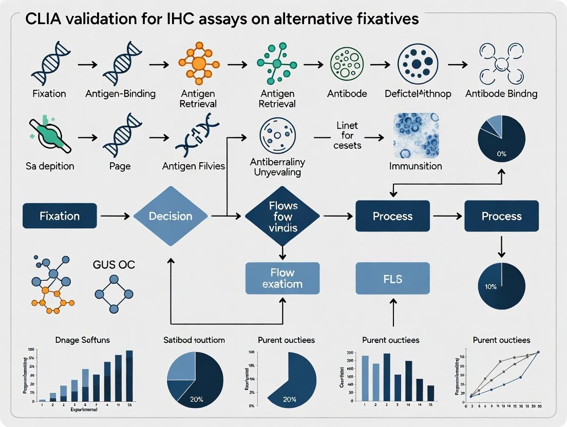
Visualizations
Title: Formalin's Molecular Impact on Assay Development
Title: Experimental Workflow for Fixative Comparison
The Scientist's Toolkit: Research Reagent Solutions
Table 3: Essential Materials for Fixative Limitation Studies
| Item | Function/Benefit | Key Consideration for CLIA Context |
|---|---|---|
| Automated Tissue Processor | Ensures consistent, reproducible dehydration and infiltration post-fixation. | Critical for standardizing pre-analytical variables during validation. |
| FFPE-Optimized RNA/DNA Extraction Kits (e.g., from Qiagen, Thermo Fisher) | Designed to reverse cross-links and recover fragmented nucleic acids. | Yield and quality directly impact downstream NGS assay validation. |
| PCR/RT-qPCR Assays for Multi-Size Amplicons | Measures extent of nucleic acid fragmentation functionally. | Provides data on acceptable amplicon size for companion diagnostic assays. |
| Validated Antibody Clones with CCC References | Antibodies with known clinical diagnostic performance in FFPE. | Essential baseline for comparing performance in alternative fixatives. |
| Automated IHC/ISH Stainer (e.g., Leica BOND, Ventana Benchmark) | Standardizes staining conditions, reducing technical variability. | Mandatory for reproducible, high-throughput assay development and validation. |
| Whole Slide Scanner & Image Analysis Software (e.g., Aperio, Vectra, QuPath) | Enables quantitative, objective scoring of IHC staining. | Supports development of reproducible scoring algorithms for validated assays. |
| Controlled Fixation Timer & pH Meter | Prevents over-fixation and ensures correct buffer pH for NBF. | Over-fixation is a major source of antigen masking variability. |
| Alternative Fixative Evaluations (e.g., Glyo-Fixx, PAXgene, Zinc-based) | Non-crosslinking or reduced cross-linking fixatives. | Potential to mitigate formalin's limitations; require full re-validation. |
| Antigen Retrieval Reagents (Citrate, EDTA, Tris-EDTA buffers, enzymatic) | Reverses methylene cross-links to unmask epitopes. | Optimization of AR is a major component of IHC assay development on FFPE. |
| Bioanalyzer/TapeStation System (Agilent) | Provides quantitative integrity numbers (RIN, DV200, DIN) for nucleic acids. | Establishes QC acceptance criteria for samples entering a validated assay pipeline. |
The transition from formalin fixation to alternative fixatives presents a critical challenge for clinical laboratory validation. For a CLIA (Clinical Laboratory Improvement Amendments)-validated IHC assay, the pre-analytical phase—specifically tissue fixation—must be rigorously standardized. This document provides application notes and detailed protocols for evaluating alternative fixatives, framed within the requirements for a robust CLIA validation study. The focus is on PAXgene, UMFIX, alcohol-based solutions, and other commercial fixatives, emphasizing their impact on macromolecular integrity and subsequent IHC performance.
Comparative Analysis of Alternative Fixatives
Table 1: Characteristics of Common Alternative Fixatives
| Fixative Type (Example Product) | Primary Chemistry | Fixation Duration (at RT) | Key Advantages for IHC/ISH | Key Considerations for CLIA Validation |
|---|---|---|---|---|
| PAXgene (PreAnalytiX) | Non-crosslinking, precipitating | 2-48 hours | Superior preservation of RNA/DNA; good morphology. | Requires specific processing; protocol deviation invalidates validation. |
| UMFIX (Sakura) | Methanol-based with additives | 4-72 hours | Good nucleic acid preservation; rapid tissue penetration. | May cause tissue brittleness; antigen retrieval may differ significantly from FFPE. |
| Alcohol-Based (FineFix, Glyo-Fixx) | Denaturing solvents (e.g., ethanol, methanol) | 1-24 hours | Fast, minimal crosslinking; preserves many epitopes. | Tissue shrinkage; potential for incomplete fixation of core. |
| Zinc-Based (Zinc Formalin, Z7, Z-Fix) | Mild crosslinking with zinc salts | 12-48 hours | Good morphology and antigenicity; improved nucleic acids vs. NBF. | Still involves crosslinking; requires optimization of retrieval. |
| HOPE (Herpes et al. Optimized Fixative) | Acetone-based with controlled humidity | 16-24 hours | Excellent preservation of proteins/nucleic acids for proteomics & PCR. | Complex, specialized fixation protocol; not routine for histology labs. |
Table 2: Quantitative Metrics for CLIA Validation Planning (Example Data)
| Analytical Performance Metric | Target Acceptance Criterion (for each antibody) | Example Result with PAXgene-fixed Tissue | Example Result with UMFIX-fixed Tissue |
|---|---|---|---|
| Signal Intensity (Score 0-3+) | ≥90% concordance with FFPE reference (for matched cases) | 95% concordance (n=20) | 92% concordance (n=20) |
| Background Staining | ≤10% of cases show unacceptable background | 5% (n=20) | 15% (n=20) |
| Cellular Localization Accuracy | 100% correct localization vs. known positive control | 100% met | 100% met |
| RNA Integrity Number (RIN) | Mean RIN >7.0 (if applicable) | Mean RIN = 8.2 | Mean RIN = 7.5 |
| Inter-assay Precision (%CV) | %CV <20% for quantitative IHC | 15% CV | 18% CV |
Detailed Protocols for Validation Experiments
Protocol 1: Parallel Fixation and Processing for Morphology & IHC Comparison Objective: To establish the optimal processing protocol for an alternative fixative and compare H&E morphology and IHC staining to standard FFPE. Materials: See "Scientist's Toolkit" below. Method:
- Tissue Sampling: Divide a fresh surgical specimen (>1 cm³) into matched sections (≈3-5 mm thick).
- Parallel Fixation: Immerse sections in:
- Container A: 10% NBF (20x tissue volume) for 24 hours.
- Container B: PAXgene fixative (20x volume) for 24 hours.
- Container C: UMFIX (20x volume) for 24 hours.
- Processing: Process each set through a dedicated tissue processor.
- For PAXgene: Use the proprietary PAXgene tissue processor protocol or a standard ethanol-based dehydration series.
- For UMFIX/Alcohol-based: Use a standard processor with ethanol-based dehydration (no xylene). Infiltrate with paraffin.
- Embedding, Sectioning, and H&E: Embed in paraffin, cut 4 µm sections, and perform H&E staining. Evaluate morphology (nuclear detail, cytoplasmic clearing, artifact).
- IHC Staining: Perform IHC for a panel of antibodies (e.g., Cytokeratin AE1/AE3, CD45, ER, Ki-67) on all sets. Use established FFPE protocols as a starting point, with and without antigen retrieval.
- Analysis: Score staining intensity, completeness, and specificity. Document any required changes to retrieval method, antibody dilution, or incubation time.
Protocol 2: Nucleic Acid Integrity Assessment Post-Fixation Objective: To quantify the preservation of RNA and DNA from tissues fixed in alternative solutions. Method:
- Fixation & Storage: Fix matched tissue pieces (≈20 mg) in NBF, PAXgene, and UMFIX for 6, 24, and 48 hours (n=3 per group). Store some PAXgene-fixed samples in stabilization solution per manufacturer instructions.
- Nucleic Acid Extraction: Following deparaffinization, use commercial kits optimized for FFPE (for all samples) or specific kits for PAXgene.
- Quantification & Quality Control:
- DNA: Assess yield via spectrophotometry (A260/A280). Perform PCR amplification of a housekeeping gene (e.g., GAPDH) at varying amplicon lengths (100 bp, 300 bp, 500 bp) to assess fragmentation.
- RNA: Measure yield and calculate RNA Integrity Number (RIN) using a Bioanalyzer or TapeStation.
- Downstream Application: Perform RT-qPCR for a target mRNA and compare Ct values across fixative groups.
Visualizations
Diagram 1: CLIA IHC Validation Workflow for Alt Fixatives
Diagram 2: IHC Challenge Path for Alternative Fixatives
The Scientist's Toolkit: Key Research Reagent Solutions
Table 3: Essential Materials for Alternative Fixative Research
| Item (Example Product) | Function in Validation Studies |
|---|---|
| PAXgene Tissue System (PreAnalytiX) | Integrated system of fixative, stabilizer, and dedicated processing reagents for optimal nucleic acid and morphology preservation. |
| UMFIX Fixative & Processing Kit (Sakura) | Methanol-based fixative with matching dehydration solutions for consistent processing without xylene. |
| HistoSpark Xylene-Free Dewaxer (Epredia) | Safe, effective dewaxing agent compatible with alcohol-fixed tissues and downstream molecular assays. |
| Antigen Retrieval Buffers (pH 6, pH 9, Tris-EDTA) (Leica, Dako) | Suite of buffers for optimizing epitope recovery across different fixative-antibody combinations. |
| RNAscope Kit for FFPE (ACD Bio-Techne) | RNA in situ hybridization platform validated for FFPE; requires optimization but works well on many alternative fixatives. |
| Automated IHC/ISH Slide Stainer (Ventana, Leica) | Essential for running high-precision, reproducible comparative staining during protocol optimization. |
| Digital Slide Scanner & Image Analysis SW (Aperio, HALO) | Enables quantitative, objective scoring of IHC staining intensity and distribution for precision data. |
| TapeStation/Bioanalyzer (Agilent) | Provides quantitative assessment of RNA (RIN) and DNA integrity from fixed tissues. |
| Multiplex IHC/IF Detection Kit (Akoya, Standard Bio) | For investigating co-expression markers, often more sensitive on less crosslinked alternative fixatives. |
Within the broader research thesis on CLIA validation for IHC assays on alternative fixatives, two key operational drivers emerge. The first is the need to enable integrated multi-omics analysis, specifically correlating genomic alterations with proteomic expression profiles from tissue samples. The second is the imperative to support lean, efficient lab workflows that reduce waste, time, and cost without compromising data quality for drug development. This application note details protocols designed to address these motivations using next-generation sequencing (NGS) and quantitative immunohistochemistry (IHC) on formalin-free fixative-preserved tissues.
Application Note: Integrated DNA/RNA Sequencing with Subsequent Protein Validation
Objective: To extract and sequence DNA and RNA from the same tissue specimen fixed in an alternative fixative (e.g., PAXgene), followed by targeted proteomic validation via IHC, enabling direct genotype-phenotype correlation.
Protocol 1: Co-Extraction of DNA and RNA from Alternative Fixative-Tissue
This protocol is optimized for FFPE-like tissue blocks fixed with non-formalin, molecular-preserving fixatives.
Materials:
- 2 x 10 µm curls or one 2.5 mm micro-punch from a PAXgene-fixed, paraffin-embedded (PFPE) block.
- Deparaffinization Solution (e.g., xylene substitute)
- Ethanol (100%, 90%, 70%)
- Proteinase K (≥20 mg/ml)
- Commercially available co-extraction kit (e.g., AllPrep DNA/RNA FFPE Kit, Qiagen)
- DNase I (RNase-free)
- Magnetic stand for 1.5 mL tubes
- Heated mixer or thermomixer
- NanoDrop or Qubit for quantification
Procedure:
- Deparaffinization:
- Place tissue in a 1.5 mL microcentrifuge tube.
- Add 1 mL of deparaffinization solution, vortex, and incubate at RT for 5 min.
- Centrifuge at full speed (≥13,000 x g) for 2 min. Carefully remove supernatant.
- Repeat steps once.
- Wash twice with 1 mL of 100% ethanol, centrifuging and removing supernatant each time.
- Air-dry pellet for 5-10 min.
Digestion:
- Add 150 µL of Proteinase K digestion buffer and 20 µL of Proteinase K to the pellet.
- Incubate on a heated mixer at 56°C for 60 min, then 80°C for 15 min to reverse crosslinks. Hold samples at 4°C.
Co-Extraction:
- Follow the manufacturer’s instructions for the chosen co-extraction kit. Briefly: a. Add appropriate buffer to the digest, vortex, and centrifuge. b. Apply lysate to a combined DNA/RNA spin column. RNA binds, DNA flows through. c. Perform on-column DNase I treatment for RNA fraction. d. Elute RNA in 30-50 µL RNase-free water. e. Process the flow-through for DNA binding on a separate column. e. Elute DNA in 50 µL elution buffer.
Quality Control (QC):
- Quantify DNA and RNA using fluorometric assays (Qubit).
- Assess DNA fragment size using a TapeStation Genomic DNA ScreenTape.
- Assess RNA Integrity Number (RIN) or DV200 (%) using a Bioanalyzer or TapeStation.
Table 1: Typical Yield and Quality from PFPE Tissue (2.5 mm punch)
| Nucleic Acid | Average Yield (ng) | QC Metric | Average Value | Acceptable Range for NGS |
|---|---|---|---|---|
| DNA | 450 - 1200 | DIN | 4.8 | ≥3.0 |
| RNA | 200 - 800 | DV200 | 65% | ≥30% |
Protocol 2: Focused NGS Panel Sequencing & Bioinformatics
For targeted sequencing of cancer-relevant genes from low-input, co-extracted nucleic acids.
Library Preparation:
- Use 20-50 ng of DNA for a targeted DNA panel (e.g., 150-gene oncology panel) using a hybrid-capture-based library prep kit.
- Use 20-50 ng of RNA for a targeted RNA fusion panel using an amplicon-based or hybrid-capture kit.
- Follow manufacturer protocols with no deviation for FFPE-derived inputs. Use 12-14 cycles of PCR amplification.
- Pool libraries and sequence on a mid-output flow cell (2x150 bp) to an average depth of 500x for DNA and 5M reads per sample for RNA.
Integrated Analysis Workflow:
- Primary Analysis: Demultiplexing (bcl2fastq), adapter trimming (Cutadapt).
- DNA Analysis: Align to hg38 (BWA), call variants (GATK Mutect2 for tumor-only with panel of normals), annotate (VEP).
- RNA Analysis: Align to hg38 (STAR), detect fusions (Arriba, STAR-Fusion), quantify gene expression (featureCounts).
- Integration: Create a unified report linking somatic mutations (DNA), gene fusions (RNA), and annotate with known or predicted protein expression changes.
Diagram 1: Integrated Genomics Workflow from PFPE Tissue
Application Note: Lean IHC Protocol for High-Throughput Protein Validation
Objective: To establish a validated, automated, and resource-efficient IHC protocol for validating proteomic targets identified via NGS, using lean principles to minimize reagent use and hands-on time.
Protocol 3: Automated, Lean IHC on Alternative Fixatives
A protocol optimized for Ventana BenchMark ULTRA autostainers, using minimal reagent volumes.
Materials:
- 4 µm sections of PFPE or other alternative fixative tissue on charged slides.
- Validated primary antibody (e.g., Anti-PD-L1, Anti-HER2, Anti-NTRK).
- OptiView or UltraView Universal DAB Detection Kit (Ventana).
- Reaction buffer, EZ Prep, and other standard Ventana reagents.
- Liquid Coverslips.
- Hematoxylin II and Bluing Reagent (for counterstain).
Procedure:
- Baking & Deparaffinization (On-Instrument):
- Load slides onto the autostainer.
- Select the "Alternative Fixative" protocol. The instrument will execute:
- Bake at 60°C for 8 minutes.
- Automated deparaffinization with EZ Prep at 72°C (multiple cycles).
- Cell Conditioning 1 (CC1) for antigen retrieval: 64-100°C for 32-64 min (condition-specific).
Primary Antibody Incubation (Lean Optimization):
- Apply 100-150 µL of primary antibody (prediluted in antibody diluent). This represents a 30-40% volume reduction versus standard 250 µL applications.
- Incubate at 36°C for 16-32 minutes (condition-specific).
- Rinse with reaction buffer (ultrafiltered water savings: ~50 mL/slide/run).
Detection & Counterstaining:
- Apply the UltraView DAB detection system per standard instructions (H2O2, DAB, Copper).
- Apply Hematoxylin II counterstain for 8 minutes, followed by bluing reagent for 4 minutes.
Post-Processing:
- Automatically remove slides from the instrument.
- Dehydrate through graded alcohols, clear in xylene, and coverslip.
Table 2: Lean IHC Protocol Savings vs. Standard Method
| Resource | Standard Protocol | Lean Protocol | Reduction |
|---|---|---|---|
| Primary Antibody | 250 µL/slide | 150 µL/slide | 40% |
| Reaction Buffer | 100 mL/run | 50 mL/run | 50% |
| Hands-On Time | 45 minutes | 15 minutes | 67% |
| Total Assay Time | 6.5 hours | 5 hours | 23% |
Protocol 4: Digital Quantification & CLIA-Validation Alignment
For objective scoring and data integration.
- Slide Scanning: Scan stained slides at 20x magnification using a digital whole slide scanner (e.g., Aperio).
- Image Analysis: Use FDA-cleared or validated digital pathology algorithms (e.g., VENTANA DP image analysis) for quantitative scoring (e.g., Tumor Proportion Score for PD-L1, membrane staining for HER2).
- Data Correlation: Integrate quantitative protein expression scores (from IHC) with corresponding genomic variant data (from NGS) in a structured database.
Diagram 2: Lean IHC & Digital Quantification Workflow
The Scientist's Toolkit: Research Reagent Solutions
Table 3: Essential Materials for Integrated Omics on Alternative Fixatives
| Item | Function & Rationale |
|---|---|
| PAXgene Tissue Fixative | Non-formalin, molecular-preserving fixative. Stabilizes RNA and DNA while maintaining morphology for IHC. Critical for integrated workflows. |
| AllPrep DNA/RNA FFPE Kit | Designed for simultaneous purification of genomic DNA and total RNA from FFPE-like tissues. Maximizes yield from precious samples. |
| Multiplexed Hybrid-Capture NGS Panels | Enable concurrent detection of SNVs, indels, CNVs, and fusions from low DNA/RNA inputs, supporting lean use of extracted nucleic acids. |
| Validated Rabbit Monoclonal Primary Antibodies | Essential for specific, reproducible IHC on alternative fixatives. Pre-validated clones reduce protocol development time. |
| UltraView Universal DAB Detection Kit | Low-background, high-sensitivity detection system for automated stainers. Optimized for use with reduced antibody volumes. |
| Digital Pathology Image Analysis Software | Provides reproducible, quantitative scoring of IHC, generating numerical data for correlation with genomic findings and regulatory submission. |
Within the broader research thesis on validating immunohistochemistry (IHC) assays for use with alternative fixatives (e.g., zinc-based, PAXgene), adherence to Clinical Laboratory Improvement Amendments (CLIA) regulations is the essential bridge from research to clinical utility. CLIA compliance is not an optional endpoint but the foundational framework that must be integrated into the experimental design from the outset. This document outlines application notes and protocols for generating CLIA-compliant validation data specific to IHC assays optimized for non-formalin fixatives.
Application Notes: Key Validation Parameters
For an IHC assay to be deployed in a CLIA-certified laboratory for patient testing, its validation must demonstrate robustness, reproducibility, and accuracy. The shift from Neutral Buffered Formalin (NBF) to an alternative fixative constitutes a major change requiring a full validation per CLIA and CAP guidelines. The following parameters, summarized in Table 1, must be quantitatively assessed.
Table 1: Core CLIA Validation Parameters for IHC Assays on Alternative Fixatives
| Validation Parameter | CLIA Requirement | Typical Target for IHC | Measurement Method |
|---|---|---|---|
| Accuracy | Agreement with a reference method. | ≥95% Positive/Negative Percent Agreement. | Comparison to established NBF results or orthogonal method (e.g., FISH, PCR). |
| Precision | Reproducibility of results. | ≥90% Intra-run and Inter-run agreement. | Testing of identical samples across runs, days, and operators. |
| Analytical Sensitivity | Detection limit of the assay. | Consistent staining at lowest expected antigen expression level. | Titration of primary antibody on low-expressing cell lines or tissues. |
| Analytical Specificity | Includes interference and cross-reactivity. | No staining in known negative tissues; staining blocked by peptide. | Testing on a panel of normal tissues; absorption with target peptide. |
| Reportable Range | Range of results the method can produce. | All possible staining intensities (0, 1+, 2+, 3+) are distinguishable. | Staining assessment across a calibrated tissue microarray with known expression levels. |
| Reference Range | Expected results in a normal population. | Definition of "negative" and "positive" staining patterns. | Analysis of relevant normal tissue types and non-targeted pathologies. |
Detailed Experimental Protocols
Protocol 1: Precision Testing (Reproducibility)
Objective: To determine intra-run, inter-run, and inter-operator precision for the IHC assay on tissues fixed in the alternative fixative. Materials: See "Scientist's Toolkit" below. Procedure:
- Sample Selection: Select 3-5 patient tissue cases representing negative, weak/low positive, and strong positive expression for the target antigen. Each tissue must be paired (same block) with sections fixed in NBF and the alternative fixative.
- Slide Preparation: Cut 30 serial sections from each alternative-fixative block. Randomize and label slides.
- Staining Runs: Perform IHC staining over 5 separate days (inter-run) by 2 trained operators (inter-operator).
- Each day, both operators stain one slide from each case (intra-run replicates of 2).
- Use the identical, optimized protocol (retrieval time, antibody dilution, detection system).
- Blinded Evaluation: A third-party pathologist scores all slides semi-quantitatively (e.g., 0, 1+, 2+, 3+) for intensity and/or percentage of positive cells.
- Data Analysis: Calculate percent agreement (exact and within ±1 score) for all comparisons. CLIA-compliant thresholds are typically ≥90%.
Protocol 2: Analytical Specificity (Interference and Cross-reactivity)
Objective: To confirm staining is specific to the target antigen and unaffected by the alternative fixative chemistry. Materials: See "Scientist's Toolkit" below. Procedure:
- Tissue Panel Staining: Perform the optimized IHC assay on a comprehensive normal tissue microarray (TMA) fixed with the alternative fixative. This assesses cross-reactivity with non-target tissues.
- Peptide Absorption Test:
- Pre-incubate the optimized dilution of the primary antibody with a 10-fold molar excess of the target immunizing peptide for 1 hour at room temperature.
- In parallel, incubate the primary antibody with a non-relevant peptide.
- Use both mixtures to stain a known positive control tissue fixed in the alternative fixative.
- Interpretation: Specific staining should be significantly reduced or abolished only in the tube with the target peptide. The normal TMA should show expected tissue-specific staining or no staining.
Visualizations
Title: CLIA Validation Pathway for Alternative Fixative IHC Assays
Title: Comparative Validation Workflow for Alternative Fixative IHC
The Scientist's Toolkit: Research Reagent Solutions
| Item | Function in Validation |
|---|---|
| Calibrated Tissue Microarray (TMA) | Contains cores with known antigen expression levels; essential for establishing reportable range and accuracy. |
| Immunizing Peptide (Target Specific) | Used in absorption experiments to confirm the analytical specificity of the primary antibody. |
| Cell Line Pellet Controls | Cell lines with known negative/positive expression, fixed in alternative fixative; critical for daily run monitoring and sensitivity assessment. |
| Alternative Fixative-Specific Antigen Retrieval Buffer | Optimized retrieval solution (e.g., high or low pH EDTA/Citrate) to reverse cross-links introduced by the non-NBF chemistry. |
| Validated Primary Antibody Clone | Antibody clone with documented performance in IHC, selected for its robustness and specificity on alternatively fixed tissues. |
| CLIA-Grade Detection System | A consistent, commercially prepared detection kit (polymer-based) to ensure minimal lot-to-lot variability and high sensitivity. |
| Digital Image Analysis Software | For quantitative, objective assessment of staining intensity and percentage, reducing scorer bias in precision studies. |
Blueprint for Success: Step-by-Step Protocol Development and Optimization for Alternative Fixative IHC
Within the critical path of CLIA validation for IHC assays on novel alternative fixatives, an initial feasibility assessment of antibody-antigen compatibility is paramount. Alternative fixatives (e.g., HOPE, PAXgene, Zinc-based formulations) induce distinct macromolecular cross-linking profiles compared to neutral buffered formalin (NBF). These alterations can mask, destroy, or modify key epitopes, leading to false-negative results or non-specific background. This Application Note details a systematic, tiered protocol for screening antibody performance and epitope compatibility on tissues fixed with candidate alternative reagents, providing the essential data required to select robust candidates for downstream, full assay validation.
Core Screening Workflow & Data Triage
The screening workflow follows a sequential, data-driven decision tree to efficiently triage antibodies. Key decision points are based on quantitative and qualitative metrics compared to NBF-positive control tissues.
Diagram Title: Antibody Screening Triage Workflow
Detailed Experimental Protocols
Protocol 3.1: Tier 1 – Multiplexed Epitope Retrieval Optimization
Objective: To identify the optimal antigen retrieval (AR) method for a given antibody on tissue fixed with an alternative fixative.
Materials: See "Research Reagent Solutions" table. Procedure:
- Sectioning: Cut serial 4 µm sections from FFPE blocks of NBF (control) and the alternative fixative (test). Mount on charged slides.
- Deparaffinization & Rehydration: Follow standard xylene and ethanol series.
- Multiplexed AR Setup: Using a slide rack system, subject serial test-fixative slides to different retrieval conditions in parallel:
- Condition A: Citrate Buffer (pH 6.0), 95-100°C, 20 min.
- Condition B: Tris-EDTA (pH 9.0), 95-100°C, 20 min.
- Condition C: Proteinase K (10 µg/mL), 37°C, 10 min.
- Condition D: No Retrieval (for epitopes sensitive to heat/ enzymatic unmasking).
- Immunostaining: Perform standardized IHC on all slides using the same primary antibody dilution, detection system (e.g., polymer-HRP), and DAB chromogen. Include a no-primary antibody control for each fixative/AR condition.
- Analysis: Visually assess under a microscope for specific membranous, cytoplasmic, or nuclear signal with minimal background. The optimal condition yields maximal specific signal intensity identical to the expected cellular localization.
Protocol 3.2: Tier 2 – Specificity and Semi-Quantitative Scoring
Objective: To confirm staining specificity and perform a semi-quantitative comparison to NBF standards.
Procedure:
- Using the optimal AR condition identified in Tier 1, stain a Tissue Microarray (TMA) containing cores of both NBF and alternative-fixative tissues. The TMA should include positive, negative, and normal tissue controls.
- Scoring: Two blinded, qualified pathologists score slides using a validated scale (e.g., 0-3+ for intensity, 0-100% for extent).
- Calculate Percent Recovery: For each positive control core, calculate the H-Score [(3 x % strong) + (2 x % moderate) + (1 x % weak)]. Determine the mean H-Score for the test fixative relative to the NBF mean.
- Assess Specificity: Check for non-specific stromal staining, cytoplasmic nuclear, or off-target patterns in negative tissues.
Protocol 3.3: Tier 3 – Quantitative Signal-to-Noise Ratio (SNR) Analysis
Objective: To obtain quantitative metrics of assay robustness for the alternative fixative.
Procedure:
- Perform IHC under Tier 1 & 2 optimized conditions on 10 replicate slides of a high-expressing positive tissue fixed in both NBF and the alternative fixative.
- Digital Image Analysis: Scan slides at 20x magnification. Using image analysis software (e.g., QuPath, HALO):
- Annotate 5-10 representative tumor regions per slide.
- Measure the mean optical density (OD) of the DAB signal within the annotated regions (Signal).
- Measure the mean OD in a non-reactive stromal or acellular region (Noise).
- Calculation: Calculate SNR for each replicate: SNR = Mean Signal OD / Mean Noise OD.
- Statistical Comparison: Perform an unpaired t-test to compare the mean SNR of the alternative fixative group versus the NBF group. Acceptable performance is defined as SNR ≥ 10 and no statistically significant (p < 0.05) reduction from the NBF control.
Data Presentation
Table 1: Representative Feasibility Screening Data for Anti-HER2 Antibody on Zinc-Formalin Fixed Tissues
| Antibody (Clone) | Fixative | Optimal AR | Mean H-Score (vs. NBF) | Specificity Confirmed? | Mean SNR (±SD) | Pass/Fail Tier 3 |
|---|---|---|---|---|---|---|
| HER2 (4B5) | NBF | pH 9.0 | 220 | Yes | 45 (±3.1) | N/A |
| HER2 (4B5) | Zinc-FF | pH 9.0 | 205 (93%) | Yes | 42 (±2.8) | PASS |
| HER2 (CB11) | NBF | pH 6.0 | 195 | Yes | 38 (±2.5) | N/A |
| HER2 (CB11) | Zinc-FF | pH 6.0 | 125 (64%) | No* | 15 (±4.2) | FAIL |
*Note: Clone CB11 on Zinc-FF showed aberrant cytoplasmic staining in negative controls.
Table 2: Key Metrics and Pass/Fail Criteria for Feasibility Assessment
| Screening Tier | Key Metrics Measured | Primary Pass/Fail Criteria | Data Output for CLIA Documentation |
|---|---|---|---|
| Tier 1: AR Opt. | Visual Signal Intensity, Localization | Identification of a retrieval method yielding specific, localized signal. | Photomicrographs, AR condition log. |
| Tier 2: Specificity | H-Score, Staining Pattern Concordance | Specific pattern; Mean H-Score ≥ 70% of NBF control. | H-Score tables, pattern comparison images. |
| Tier 3: Robustness | Signal-to-Noise Ratio (SNR), Statistical P-value | SNR ≥ 10; No significant decrease vs. NBF (p ≥ 0.05). | SNR data table, statistical test result. |
The Scientist's Toolkit: Research Reagent Solutions
Table 3: Essential Materials for Compatibility Screening
| Item/Category | Example Product(s) | Function in Screening |
|---|---|---|
| Alternative Fixatives | Zinc-formalin (Zinc-FF), PAXgene, HOPE solution, CyMol | Test reagents for tissue fixation, creating cross-linking profiles distinct from NBF for epitope compatibility testing. |
| Multiplex AR Buffers | Citrate Buffer (pH 6.0), Tris-EDTA/EGTA (pH 9.0), TRS (pH 1-10 range) | Key reagents for unmasking epitopes altered by novel fixation chemistries in a systematic screen. |
| Validated IHC Controls | Full-face FFPE blocks or TMAs of cell lines/tissues with known antigen expression (positive/negative) | Essential for establishing baseline NBF performance and evaluating specificity/ recovery on test fixatives. |
| Polymer-Based Detection | EnVision (Dako/Agilent), MACH (Biocare), ImmPRESS (Vector) | High-sensitivity, low-background detection systems critical for achieving a quantifiable SNR in Tier 3. |
| Chromogen | DAB (3,3'-Diaminobenzidine), Metal-enhanced DAB | Produces a stable, permanent, and quantifiable (via optical density) signal precipitate. |
| Digital Pathology Platform | Aperio/Leica, Vectra/Polaris (Akoya), HALO, QuPath | Enables whole-slide imaging, precise region annotation, and quantitative analysis of signal intensity and noise. |
| Image Analysis Software | Indica Labs HALO, Visiopharm, QuPath (open-source) | Provides tools for quantifying mean optical density, cell segmentation, and calculating H-Scores and SNR metrics. |
Within the rigorous framework of CLIA validation for IHC assays utilizing alternative fixatives, the pre-analytical phase is paramount. This phase introduces significant, often uncontrollable, variability that can compromise assay reproducibility and clinical reliability. This document provides detailed application notes and protocols to systematically evaluate and control three critical pre-analytical variables: fixation time, tissue processing, and sectioning. Standardization of these steps is a foundational prerequisite for any subsequent analytical validation.
Table 1: Impact of Formalin Fixation Time on Antigen Retrieval Efficacy and IHC Signal Intensity
| Target Antigen (Cell Location) | Optimal Fixation Time (10% NBF) | Under-fixed (<12h) Effect | Over-fixed (>72h) Effect | Recommended Retrieval Method for Extended Fixation |
|---|---|---|---|---|
| Ki-67 (Nuclear) | 6-24 hours | Diffuse, weak staining | Marked signal loss | Heat-Induced Epitope Retrieval (HIER), Citrate pH 6, extended time |
| HER2 (Membrane) | 6-48 hours | Artifactual internalization | Masking, requires strong HIER | HIER, EDTA pH 9.0 |
| CD3 (Membrane/Cytoplasmic) | 18-72 hours | Potential false-negative | Moderate signal loss | HIER, Citrate pH 6 |
| p53 (Nuclear) | 8-48 hours | Non-specific background | Significant attenuation | HIER, Tris-EDTA pH 9.0 |
Table 2: Comparison of Alternative Fixative Properties Relevant to CLIA Validation
| Fixative (Common Name) | Primary Mechanism | Optimal Fixation Time | Key Advantages for IHC | Major Limitations for Validation |
|---|---|---|---|---|
| Ethanol (70-100%) | Protein dehydration/denaturation | 18-24 hours (at 4°C) | Excellent antigen preservation; minimal masking | Poor morphology; tissue hardening |
| Zinc Formalin (Z7) | Cross-linking with zinc salts | 24-48 hours | Superior morphology; reduced epitope masking vs. NBF | Less predictable cross-linking; requires protocol re-optimization |
| PAXgene (Non-crosslinking) | Acid precipitation | 24-72 hours | Excellent nucleic acid and protein preservation | Cost; specialized processing required |
| Glyoxal-based solutions | Cross-linking (different adducts than formaldehyde) | 6-24 hours | Reduced health hazard; faster penetration | Potential autofluorescence; novel retrieval needed |
Table 3: Tissue Processor Program Variables and Their Impact on Sectioning Quality
| Processing Step | Standard (Paraffin) Duration | Aggressive (Fast) Shortening Risk | Gentle (Long) Protocol Benefit | Critical for Alternative Fixatives? |
|---|---|---|---|---|
| Dehydration (Ethanol) | 70% to 100%, 1h each | Incomplete dehydration, poor infiltration | Ensures complete water removal | Yes - Ethanol-fixed tissues are more prone to over-hardening. |
| Clearing (Xylene) | 2-3 changes, 1h each | Cloudy tissue, sectioning ribbons shred | Clear, translucent tissue | Yes - PAXgene tissues require adjusted timing. |
| Infiltration (Paraffin) | 3 changes, 1h each at 60°C | Soft blocks, sections wrinkle | Uniform, firm blocks | Critical - May need lower wax melting point for alcohol-fixed tissues. |
| Total Cycle Time | ~12-14 hours | High risk of artifacts | Optimal morphology | Must be re-validated per fixative. |
Experimental Protocols
Protocol 3.1: Systematic Fixation Time Course for CLIA Validation Studies
Objective: To empirically determine the optimal fixation window for a specific antigen-fixative pair and establish the allowable tolerance as part of the assay validation. Materials: See Scientist's Toolkit (Section 5). Procedure:
- Tissue Selection: Obtain fresh, surgically resected tissue specimen (e.g., tonsil, carcinoma). With informed consent and IRB approval, slice into congruent sections (≥3mm thick, ≤4cm area) using a standardized tissue slicer within 5 minutes of resection.
- Fixative Allocation: Immerse tissue slices in a 20:1 volume-to-tissue ratio of the target fixative (e.g., 10% NBF, Zinc Formalin, Ethanol).
- Time Course Setup: For each fixative, allocate slices to fixation intervals: 1, 6, 12, 24, 48, 72, and 168 hours (1 week). Maintain temperature at 25°C ± 2°C (room temp) for formalin-based; 4°C for alcohol-based.
- Processing: After each time point, transfer tissue to standardized cassettes and process using a validated, consistent processing schedule (see Table 3, Gentle protocol).
- Embedding and Sectioning: Embed in paraffin wax. Section each block at 4µm using a microtome with a fresh, high-profile blade. Float sections on a 42°C water bath containing DEPC-treated water to minimize RNase activity (if needed). Mount on charged slides.
- IHC Staining: Subject all slides from all time points to the same IHC run for the target antigen(s), including appropriate positive and negative controls.
- Analysis: Perform digital image analysis (DIA) to quantify staining intensity (optical density), percentage of positive cells, and membrane completeness (for HER2-like targets). Assess morphological preservation by a certified pathologist.
Protocol 3.2: Evaluation of Sectioning-Induced Antigen Loss (Recut Stability)
Objective: To validate the stability of the antigen-antibody interaction in stored blocks and define the maximum allowable recut interval for assay re-runs. Materials: Prepared paraffin blocks, microtome, charged slides. Procedure:
- Block Selection: Use blocks from Protocol 3.1 fixed for the determined optimal time.
- Sequential Sectioning: Face the block appropriately. Collect ten consecutive 4µm sections. Store the block at room temperature.
- Time-Course Recuts: At intervals of 1 day, 1 week, 1 month, 3 months, and 6 months, return the block to the microtome, face it lightly (remove ~50µm), and collect two new consecutive sections.
- Staining and Analysis: Stain all sections (initial and recuts) in a single IHC batch to eliminate run-to-run variation. Use DIA to compare the staining intensity of the recut sections to the average of the initial ten sections (baseline).
- Acceptance Criterion: Establish a cutoff (e.g., ≤15% loss in mean optical density) to define the valid "recut period" for the validated assay.
Pathway and Workflow Visualizations
Title: Pre-Analytical Variables Workflow
Title: Protocol Selection Logic for CLIA Validation
The Scientist's Toolkit: Essential Research Reagent Solutions
| Item / Reagent Solution | Function in Pre-Analytical Roadmap Studies |
|---|---|
| Neutral Buffered Formalin (10%, pH 7.2-7.4) | Gold-standard cross-linking fixative; serves as the primary benchmark for comparing all alternative fixatives in validation studies. |
| Precision Tissue Slicer Matrix | Enables creation of multiple, morphologically congruent tissue sections from one specimen for parallel fixation time-course experiments. |
| Validated Automated Tissue Processor | Provides reproducible and programmable dehydration, clearing, and infiltration; critical for isolating fixation as the sole variable. |
| High-Profile Microtome Blades | Produces thin, consistent sections with minimal compression and chatter, reducing variable antigen exposure and technical artifacts. |
| Positively Charged/Adhesive Slides | Prevents tissue section detachment during stringent retrieval protocols, especially critical for alcohol-fixed or delicate tissues. |
| Certified Antigen Retrieval Buffers (Citrate pH 6.0, Tris/EDTA pH 9.0) | Standardized solutions for HIER; essential for unmasking epitopes cross-linked by aldehyde-based fixatives. |
| Digital Image Analysis (DIA) Software | Provides objective, quantitative measurement of IHC staining intensity (H-score, optical density) for comparing fixation variables. |
| Controlled Temperature Fixation Chamber | Maintains precise temperature during fixation (e.g., 4°C for alcohols, 25°C for formalin), eliminating a key environmental variable. |
Within the broader thesis on CLIA validation for IHC assays on alternative fixatives (e.g., HOPE, PAXgene), this application note details the imperative adaptation of antigen retrieval (AR) and detection methodologies. Transitioning from formalin-fixed, paraffin-embedded (FFPE) tissues to novel fixative chemistries necessitates a systematic re-evaluation of retrieval protocols and detection system compatibility to ensure optimal antigenicity, specificity, and sensitivity for clinical-grade assays.
Core Challenge: Fixative Chemistry Dictates Retrieval Strategy
Alternative fixatives employ distinct chemical mechanisms for tissue preservation, directly impacting protein conformation and antigen masking. The standard heat-induced epitope retrieval (HIER) protocols optimized for FFPE's methylene crosslinks are often suboptimal.
Table 1: Comparison of Fixative Chemistry and Retrieval Demands
| Fixative Type | Primary Chemistry | Crosslink Nature | Typical AR Intensity Required | Common Challenge |
|---|---|---|---|---|
| Neutral Buffered Formalin (FFPE) | Formaldehyde | Methylene bridges | Standard HIER (pH 6-10) | Over-retrieval can damage morphology. |
| HOPE (HEPES-glutamic acid buffer mediated Organic solvent Protection Effect) | Acetone-based, organic solvents | Less protein crosslinking, more precipitation | Mild HIER or enzymatic retrieval | Antigens are sensitive; over-retrieval leads to loss. |
| PAXgene | Non-formaldehyde, proprietary precipitating fixatives | Minimal crosslinking, precipitation-based | Variable; often requires fine-tuned HIER at lower temperature/time | Inconsistent results with standard FFPE protocols. |
| Zinc-based Fixatives | Zinc salts in buffer | Ionic stabilization, no crosslinks | Often minimal or no HIER required | Detection may require optimization to reduce background. |
Application Notes & Protocols
Note 1: Systematic Retrieval Optimization Protocol
Objective: To empirically determine the optimal AR method for a target antigen (e.g., HER2) on an alternative fixative (e.g., HOPE-fixed tissue).
Materials: See "Scientist's Toolkit" below.
Workflow:
- Sectioning: Cut 4μm sections from HOPE-fixed, paraffin-embedded blocks onto charged slides.
- Deparaffinization & Rehydration: Use a non-xylene, alcohol-based series compatible with the fixative.
- Antigen Retrieval Matrix Setup:
- Prepare three retrieval buffers: Citrate (pH 6.0), Tris-EDTA (pH 9.0), and a low-concentration enzymatic solution (e.g., 0.05% pepsin in 0.01N HCl).
- For each buffer, test a time/temperature gradient: 95°C for 10min, 15min, 20min; and 100°C for 10min, 20min.
- Retrieval Execution: Perform HIER using a calibrated water bath or steamer. For enzymatic retrieval, use a humidified incubator at 37°C for 5-15 minutes.
- Immunostaining: Proceed with a standardized detection protocol (see Protocol 2) immediately after cooling and washing.
- Analysis: Score slides for signal intensity (0-3+), background, and morphological preservation. The optimal condition yields maximum specific signal with minimal background and preserved morphology.
Note 2: Detection System Compatibility Testing
Objective: To evaluate the performance of different detection chemistries (e.g., HRP vs. AP, polymer vs. ABC) on an alternative fixative.
Rationale: Altered tissue chemistry can affect enzyme stability and polymer penetration.
Protocol:
- After optimal AR, block slides with appropriate protein block (casein-based blocks often perform better than serum on precipitated proteins).
- Apply primary antibody at the titrated concentration.
- Detection Comparison Arm:
- Arm A: HRP-labeled polymer system (e.g., EnVision+).
- Arm B: Alkaline Phosphatase (AP)-labeled polymer system.
- Arm C: Traditional Avidin-Biotin Complex (ABC) with HRP.
- Develop with compatible chromogens (DAB for HRP, Fast Red for AP).
- Counterstain, dehydrate, and mount.
- Quantitative Analysis: Use image analysis software to calculate signal-to-noise ratio (SNR). The system with the highest SNR and cleanest background is optimal.
Table 2: Example Optimization Results for p53 on PAXgene Tissue
| Retrieval Method | Detection System | Mean Signal Intensity (AU) | Background (AU) | SNR | Morphology Score (1-5) |
|---|---|---|---|---|---|
| Citrate pH 6.0, 95°C, 10min | HRP Polymer | 1250 | 210 | 5.95 | 4 |
| Tris-EDTA pH 9.0, 95°C, 15min | HRP Polymer | 1850 | 450 | 4.11 | 3 |
| Tris-EDTA pH 9.0, 95°C, 10min | HRP Polymer | 1650 | 180 | 9.17 | 5 |
| Tris-EDTA pH 9.0, 95°C, 10min | AP Polymer | 1200 | 150 | 8.00 | 5 |
| No Retrieval | HRP Polymer | 300 | 100 | 3.00 | 5 |
AU = Arbitrary Units; SNR = Signal-to-Noise Ratio.
The Scientist's Toolkit: Key Research Reagent Solutions
| Item | Function & Rationale for Alternative Fixatives |
|---|---|
| pH-stable HIER Buffers (Citrate, Tris-EDTA, Borate) | Essential for testing the pH dependence of antigen unmasking on novel crosslinks. |
| Low-concentration Enzymatic Retrieval Solutions (Pepsin, Trypsin) | Crucial for delicate antigens on low-crosslink fixatives (e.g., HOPE) where heat may destroy epitopes. |
| Non-ionic Detergent (e.g., Tween-20, Triton X-100) | Added to wash buffers to reduce non-specific binding on precipitated proteins. |
| Casein-based Blocking Solution | Often superior to serum-based blocks for reducing background on non-formalin tissues. |
| Polymer-based Detection Systems (HRP/AP) | Recommended over ABC systems due to reduced endogenous biotin interference and more consistent penetration. |
| Alternative Chromogens (Fast Red, VIP, Vector Blue) | Provides options if endogenous enzyme activity (e.g., peroxidases in HOPE tissue) is a concern. |
| Controlled Temperature Water Bath | Preferred over pressure cookers for fine-tuning retrieval time/temperature during optimization. |
Visualizations
Title: Optimization Workflow for New Fixative IHC Protocols
Title: Chemical Basis for Tailoring Antigen Retrieval
Within the framework of CLIA validation for IHC assays, the adoption of novel alternative fixatives necessitates rigorous initial protocol establishment. This document details the critical application notes and protocols for determining optimal primary antibody titrations and establishing appropriate controls when transitioning from formalin-fixed, paraffin-embedded (FFPE) tissues to a new fixative matrix. The performance of immunohistochemical (IHC) markers is highly dependent on the fixation chemistry, making systematic titration and control strategies paramount for assay reliability and subsequent validation.
Core Principles of Titration in Alternative Fixatives
The primary goal of titration is to identify the antibody concentration that yields the strongest specific signal with the lowest non-specific background. In alternative fixatives, epitope presentation and stability may differ significantly from FFPE. Therefore, a checkerboard titration, varying both antibody concentration and antigen retrieval conditions, is recommended.
Experimental Protocol: Checkerboard Titration for Primary Antibody
Materials and Equipment
- Tissue microarrays (TMAs) containing known positive and negative tissues, fixed in the new fixative matrix and processed to paraffin blocks.
- Serial sections of the TMAs at 4-5 µm thickness.
- Primary antibody of interest.
- Negative control reagent (IgG from same host species as primary antibody or antibody diluent).
- Compatible IHC detection system (e.g., polymer-based HRP or AP).
- Antigen retrieval solutions (e.g., citrate buffer pH 6.0, EDTA/TRIS buffer pH 9.0, enzymatic retrieval).
- Automated IHC stainer or humidified chamber for manual staining.
- Light microscope for evaluation.
Methodology
- Sectioning: Cut serial sections from the TMA blocks fixed in the new matrix.
- Antigen Retrieval Optimization: Choose two retrieval methods (e.g., heat-induced epitope retrieval (HIER) at low pH and high pH). Include a no-retrieval condition.
- Antibody Dilution Series: Prepare a minimum of four serial dilutions of the primary antibody (e.g., 1:50, 1:100, 1:200, 1:400) based on the manufacturer's recommendation for FFPE.
- Staining Procedure:
- Deparaffinize and hydrate sections.
- Perform antigen retrieval as per the selected conditions.
- Block endogenous peroxidase and apply protein block.
- Apply the primary antibody dilutions to sequential sections. Include a negative control for each retrieval condition.
- Apply the detection system and chromogen (e.g., DAB).
- Counterstain, dehydrate, clear, and mount.
- Evaluation: Score slides for intensity (0-3+), proportion of positive cells, and background staining. The optimal condition is the highest dilution with maximal specific signal and minimal background.
Data Presentation: Titration Results
Table 1: Checkerboard Titration Results for Anti-CD20 (Clone L26) in Novel Alcohol-Based Fixative Matrix
| Retrieval Condition | Antibody Dilution | Intensity Score (0-3+) | % Positive Cells | Background Score (0-3+) | Overall Suitability |
|---|---|---|---|---|---|
| Citrate pH 6.0 | 1:50 | 3+ | 95% | 2+ | Poor |
| Citrate pH 6.0 | 1:100 | 3+ | 95% | 1+ | Good |
| Citrate pH 6.0 | 1:200 | 2+ | 90% | 0 | Optimal |
| Citrate pH 6.0 | 1:400 | 1+ | 85% | 0 | Suboptimal |
| EDTA pH 9.0 | 1:50 | 2+ | 90% | 3+ | Poor |
| EDTA pH 9.0 | 1:100 | 2+ | 90% | 2+ | Poor |
| EDTA pH 9.0 | 1:200 | 1+ | 80% | 1+ | Poor |
| None | 1:100 | 0 | 0% | 0 | Ineffective |
Essential Control Strategies for CLIA Validation Context
A robust control strategy is non-negotiable for validation. The following controls must be incorporated during initial staining condition establishment.
Protocol: Control Slide Setup for Each IHC Run
- Positive Tissue Control: A slide containing a tissue known to express the target antigen, fixed in the new fixative matrix, must be included in every run.
- Negative Tissue Control: A slide containing tissue known not to express the target antigen, fixed in the new fixative matrix.
- Reagent Negative Control (Primary Antibody Omission): Replace the primary antibody with antibody diluent or isotype-matched irrelevant IgG on the test tissue. This controls for non-specific binding from the detection system.
- Process Control (Optional but Recommended): A slide stained for a ubiquitously expressed antigen (e.g., beta-actin) to confirm overall tissue and protocol integrity with the new fixative.
The Scientist's Toolkit: Research Reagent Solutions
Table 2: Essential Materials for IHC Titration on Alternative Fixatives
| Item | Function & Importance |
|---|---|
| Tissue Microarray (TMA) | Contains multiple tissue cores on one slide, enabling high-throughput, parallel comparison of staining conditions under identical protocol steps. Essential for efficient titration. |
| Alternative Fixative-Processed Tissues | Tissues fixed and processed in the novel fixative matrix. The cornerstone of assay development; FFPE controls are insufficient for establishing new conditions. |
| Modular IHC Detection System | A polymer-based detection system (e.g., HRP/DAB) with secondary antibody and label pre-complexed. Offers high sensitivity and low background, crucial for optimizing signal-to-noise ratio. |
| pH-varied Antigen Retrieval Buffers | Critical for unmasking epitopes altered by the new fixation chemistry. Testing a range (e.g., citrate pH 6.0, EDTA/TRIS pH 8.0-9.0) is mandatory. |
| Automated IHC Stainer | Provides superior reproducibility by standardizing incubation times, temperatures, and wash volumes across all titration slides, reducing variable introduction. |
| Digital Slide Scanner & Image Analysis Software | Enables objective, quantitative assessment of staining intensity (optical density) and percentage positivity, moving beyond subjective scoring for validation-ready data. |
Visualization: Experimental Workflow and Relationship Logic
Title: IHC Antibody Titration Workflow for New Fixatives
Title: IHC Run Control Logic for CLIA Validation
Solving Common Pitfalls: Troubleshooting Staining Issues and Enhancing Assay Robustness
Application Notes
Within the framework of CLIA validation for IHC assays, particularly when investigating alternative tissue fixatives, diagnosing poor staining is paramount. The performance characteristics of an assay—its sensitivity, specificity, and robustness—are directly quantified during validation. Suboptimal staining artifacts, such as weak signal, high background, and non-specific binding, compromise these metrics and can lead to failed validation runs or inaccurate clinical interpretations. This document outlines systematic troubleshooting approaches, integrating current best practices and quantitative data to guide assay optimization and validation.
The transition from formalin-based fixatives to alternatives (e.g., alcohol-based, non-aldehyde) can significantly alter epitope availability, tissue morphology, and non-specific protein interactions. These changes can manifest as the staining issues described herein. Therefore, a rigorous diagnostic protocol is essential to isolate the variable—be it fixation, pre-analytical steps, or the assay itself—and to establish a validated, reliable protocol.
Key Quantitative Data on Staining Issues and Fixatives
Table 1: Impact of Common Variables on Staining Artifacts
| Variable | Weak Signal Likelihood | High Background Likelihood | Common Root Cause in Alternative Fixatives |
|---|---|---|---|
| Over-fixation (Neutral Buffered Formalin >72h) | High | Low | Excessive cross-linking, epitope masking. |
| Under-fixation (Alcohol-based, <1h) | Medium | High | Poor morphology, increased non-specific protein binding. |
| Antigen Retrieval pH (Low: 6.0) | Low for some targets | Low | Optimal for many phosphorylated epitopes. |
| Antigen Retrieval pH (High: 9.0) | Low for some targets | Medium | Optimal for nuclear/transcription factors; can increase background. |
| Primary Antibody Concentration (High) | Low | High | Excess unbound antibody binds non-specifically. |
| Blocking Time (<10 min) | Low | High | Inadequate suppression of endogenous sites. |
| Detection System Amplification (Excessive) | Low | High | Over-amplification of low-level non-specific signal. |
Table 2: CLIA Validation Metrics Affected by Staining Artifacts
| Performance Characteristic | Impact of Weak Signal | Impact of High Background | Acceptable Range (Typical CLIA) |
|---|---|---|---|
| Analytical Sensitivity | Severely Reduced | Falsely Elevated | ≥95% detection of known positives. |
| Analytical Specificity | May be unaffected | Severely Reduced | ≥90% (minimal cross-reactivity). |
| Signal-to-Noise Ratio | Very Low | Very Low | ≥3:1 for positive vs. negative cells. |
| Inter-Assay Precision (CV) | Increased | Increased | ≤15% for quantitative IHC. |
Experimental Protocols
Protocol 1: Systematic Diagnostic for Staining Failures
Purpose: To methodically identify the root cause of poor staining within an IHC assay undergoing CLIA validation. Materials: Tissue microarrays (TMAs) containing known positive and negative controls, fixed with both standard NBF and the alternative fixative under investigation.
Control Verification:
- Stain control slides with a well-validated, robust antibody (e.g., anti-Vimentin) using the standard protocol.
- Expected Result: Strong, specific staining in positive control tissues.
- Diagnosis: If staining fails here, the issue is core reagents or instrumentation, not the fixative/antibody in question.
Fixation Comparison:
- Stain paired serial sections from NBF-fixed and alternative-fixed tissues with the target antibody under standard protocol.
- Quantify: Use image analysis to measure mean optical density (OD) of signal in target regions and adjacent background.
- Diagnosis: Weak signal only in alternative-fixed samples suggests epitope masking or altered conformation. High background only in alternative fixative suggests insufficient cross-linking and increased non-specific binding.
Antigen Retrieval Titration:
- For the problematic fixative, test a series of retrieval conditions: Citrate pH 6.0, Tris-EDTA pH 9.0, and a protease-induced epitope retrieval (PIER) for 5, 10, and 15 minutes.
- Diagnosis: Improved signal indicates initial retrieval was suboptimal. Increased background suggests retrieval is too aggressive.
Antibody and Blocking Optimization:
- Perform a checkerboard titration of the primary antibody (e.g., 1:50, 1:100, 1:200, 1:500) against varying blocking times (10, 30, 60 min) with a protein block (e.g., 5% normal serum).
- Diagnosis: Identification of the concentration that maximizes signal-to-noise ratio.
Protocol 2: Quantitative Background Assessment
Purpose: To objectively measure and compare non-specific binding. Materials: Negative control tissue (known absence of target), isotype control or primary antibody omission control.
- Stain negative control tissue sections with the full IHC protocol.
- Apply the same protocol but substitute the primary antibody with an irrelevant immunoglobulin of the same species and isotype at the same concentration (Isotype Control).
- Apply the protocol with diluent only in place of the primary antibody (No Primary Control).
- Scan slides and use image analysis software to measure the OD in 5-10 representative fields per slide.
- Calculate the average background OD for each control. The highest of these values represents the assay background floor. The specific signal OD must significantly exceed this floor (e.g., S/N ≥ 3).
Visualization
Title: IHC Staining Problem Diagnostic Workflow
Title: How Alternative Fixatives Cause Staining Issues
The Scientist's Toolkit
Table 3: Essential Research Reagent Solutions for IHC Troubleshooting
| Item | Function in Diagnosis/Optimization | Key Consideration for CLIA Validation |
|---|---|---|
| Tissue Microarray (TMA) with known positive/negative cores | Provides internal controls on a single slide for direct comparison of staining performance under different conditions. | Essential for assessing precision and reproducibility across multiple tissue types. |
| Polymer-based Detection System | Amplifies signal with high sensitivity while reducing non-specific binding common with traditional avidin-biotin systems. | Must be validated as part of the total test system. Lot-to-lot consistency is critical. |
| Automated Staining Platform | Ensures precise, reproducible timing and application of reagents, a key variable in standardization. | Required for high-complexity CLIA testing to minimize operator-induced variability. |
| Serum/Protein Blocking Solution | Saturates non-specific protein binding sites to reduce background. Choice of serum (e.g., normal goat, rabbit) should match secondary antibody host. | Blocking time and concentration must be standardized and documented in the SOP. |
| Antigen Retrieval Buffers (pH 6.0 Citrate & pH 9.0 Tris-EDTA) | Unmasks epitopes cross-linked by fixation. The optimal buffer and time are target- and fixative-dependent. | Retrieval method is a critical pre-analytical variable requiring strict control. |
| Isotype Control Antibody | An irrelevant immunoglobulin matching the primary antibody's host species, isotype, and concentration. Distinguishes specific from non-specific antibody binding. | Mandatory negative control for assay specificity assessment during validation. |
| Whole Slide Image Analysis Software | Enables quantitative measurement of signal intensity (OD) and background in defined regions of interest. | Provides objective, quantitative data for establishing positivity thresholds and calculating S/N ratios. |
Application Notes
Within the framework of CLIA validation for IHC assays on alternative fixatives (e.g., ethanol-based, HOPE, Zinc), rigorous optimization of pre-analytical and analytical variables is critical. This process ensures robustness, reproducibility, and analytical sensitivity equivalent to or exceeding standard NBF-fixed tissue. Key optimization levers—Antigen Retrieval (AR) pH, retrieval time/temperature, and primary antibody incubation—are interdependent and must be systematically calibrated for each fixative-antibody pair. Alternative fixatives alter protein cross-linking patterns, necessitating tailored retrieval conditions to optimally expose epitopes while preserving tissue morphology. The primary goal is to establish a standardized, validated protocol that meets CLIA requirements for precision, accuracy, and reportable range.
Table 1: Effect of Retrieval pH on IHC Staining Intensity (H-Score) Across Fixatives
| Target | Fixative | Retrieval pH 6.0 | Retrieval pH 8.0 | Retrieval pH 9.0 | Optimal pH |
|---|---|---|---|---|---|
| ER (SP1) | NBF | 180 | 220 | 195 | 8.0 |
| ER (SP1) | Ethanol-based | 95 | 160 | 210 | 9.0 |
| Ki-67 (MIB-1) | NBF | 200 | 220 | 180 | 8.0 |
| Ki-67 (MIB-1) | Zinc | 210 | 195 | 170 | 6.0 |
| p53 (DO-7) | HOPE | 110 | 185 | 240 | 9.0 |
Table 2: Optimization of Retrieval Time and Temperature for Ethanol-Fixed Tissue
| Retrieval Method | Temperature | Time (min) | Stain Intensity | Background | Morphology |
|---|---|---|---|---|---|
| Pressure Cooker | ~121°C | 5 | Low | Low | Excellent |
| Pressure Cooker | ~121°C | 10 | High | Moderate | Good |
| Water Bath | 97°C | 20 | Moderate | Low | Excellent |
| Water Bath | 97°C | 40 | High | High | Fair |
Table 3: Primary Antibody Incubation Optimization for CLIA Validation
| Antibody Clone | Fixative | Standard Conc. (μg/mL) | Optimized Conc. (μg/mL) | Incubation Time (RT) | Signal-to-Noise Ratio |
|---|---|---|---|---|---|
| HER2 (4B5) | NBF | 1.0 | 1.0 | 32 min | 8.5 |
| HER2 (4B5) | Ethanol-based | 1.0 | 2.0 | 60 min | 7.8 |
| PD-L1 (22C3) | NBF | 1.0 | 1.0 | 32 min | 6.0 |
| PD-L1 (22C3) | Zinc | 1.0 | 0.5 | 20 min | 7.2 |
Experimental Protocols
Protocol 1: Systematic Antigen Retrieval pH Optimization
- Tissue Sectioning: Cut 4μm sections from FFPE blocks of alternative fixatives (e.g., ethanol, Zinc, HOPE) and NBF controls.
- Deparaffinization & Rehydration: Bake slides at 60°C for 1 hour. Process through xylene (3 changes, 5 min each) and graded ethanol (100%, 95%, 70%, 5 min each) to distilled water.
- pH Buffer Preparation: Prepare citrate-based buffer (pH 6.0), Tris-EDTA buffer (pH 8.0), and Tris-EDTA buffer (pH 9.0).
- Retrieval Procedure: Using a pre-heated decloaking chamber or water bath, perform heat-induced epitope retrieval (HIER) at 97°C for 20 minutes in each pH buffer.
- Cooling & Washing: Cool slides for 30 minutes at room temperature in the buffer. Rinse in distilled water and wash in Tris-buffered saline with 0.025% Tween-20 (TBST) for 5 minutes.
- Staining: Proceed with standardized IHC staining protocol (peroxide block, protein block, primary antibody, detection, chromogen, hematoxylin).
- Analysis: Score slides using H-score or equivalent quantitative image analysis. Determine optimal pH for each fixative-antibody pair.
Protocol 2: Titration of Primary Antibody Incubation Time and Concentration
- Slide Preparation: Prepare serial sections from a tissue microarray (TMA) containing known positive and negative tissues fixed in the alternative fixative of interest.
- Standard AR: Perform AR using the optimal pH and time/temperature conditions determined in Protocol 1.
- Antibody Dilution Series: Prepare a 2-fold dilution series of the primary antibody (e.g., 4.0, 2.0, 1.0, 0.5, 0.25 μg/mL) in antibody diluent.
- Incubation Time Matrix: For each concentration, test three incubation times: 20 minutes (room temperature), 32 minutes (room temperature), and 60 minutes (room temperature). Perform all steps on an automated IHC stainer for consistency.
- Detection & Analysis: Complete staining with a polymer-based detection system. Quantify staining intensity and background using digital pathology software. Calculate the signal-to-noise ratio for each condition.
- Validation: Select the condition (concentration/time) that provides the highest signal-to-noise ratio and meets the CLIA validation criteria for positive and negative controls.
Protocol 3: CLIA Validation for Precision (Repeatability and Reproducibility)
- Sample Set: Select 20 cases fixed in the alternative fixative, spanning the assay's reportable range (negative, weak, moderate, strong).
- Inter-Run Precision: Stain one slide from each case in three separate staining runs (different days, different lots of detection reagents). Use the optimized protocol.
- Intra-Run Precision: Stain three replicate slides from each case in a single run.
- Reader Precision: Have three qualified pathologists score all slides independently using the validated scoring algorithm.
- Analysis: Calculate the coefficient of variation (CV%) for quantitative scores (e.g., H-score) and the percent agreement for categorical scores. CLIA validation requires CV% < 15% for continuous data and >90% concordance for categorical data.
Diagrams
IHC Optimization Levers & Objectives
CLIA Validation Workflow for IHC
The Scientist's Toolkit: Research Reagent Solutions
Table 4: Essential Materials for IHC Optimization on Alternative Fixatives
| Item/Category | Specific Example/Product | Function in Optimization |
|---|---|---|
| Fixatives | Neutral Buffered Formalin (NBF), Ethanol-based fixatives (e.g., FineFIX, UMFIX), Zinc Formalin, HOPE fixative. | Serve as the test variable. Alternative fixatives require tailored protocols to match NBF performance. |
| Antigen Retrieval Buffers | Citrate Buffer (pH 6.0), Tris-EDTA Buffer (pH 8.0 & 9.0), EDTA Buffer (pH 8.0). | Unmask epitopes cross-linked by different fixatives. pH is a critical optimization lever. |
| Retrieval Device | Decloaking Chamber (Pressure Cooker), Water Bath, Automated Slide Stainer with retrieval capability. | Provide controlled application of heat and time for consistent, reproducible epitope retrieval. |
| Validated Primary Antibodies | RTU clones for biomarkers (ER, PR, HER2, Ki-67, PD-L1, etc.) with known performance in NBF. | The analyte of interest. Concentration and incubation time must be re-optimized for alternative fixatives. |
| Detection System | Polymer-based HRP or AP detection systems (e.g., EnVision, Ultravision). | Amplify signal. Must be kept constant during optimization to isolate variable effects. |
| Chromogen | DAB (3,3'-Diaminobenzidine), AEC (3-Amino-9-Ethylcarbazole). | Visualizes antibody binding. Choice can affect contrast and permanence. |
| Automated IHC Stainer | BenchMark Ultra, BOND-III, Autostainer Link 48. | Essential for CLIA validation, ensuring standardized reagent application, timing, and temperature across runs. |
| Digital Pathology System | Scanner (e.g., Aperio, VENTANA DP) and Image Analysis Software (e.g., HALO, QuPath). | Enables quantitative, objective scoring of staining intensity (H-score, % positivity) for precise optimization and validation data. |
| Control Tissues | Multi-tissue TMAs containing known positive and negative tissues for each target, fixed in both NBF and alternative fixatives. | Critical for determining assay sensitivity, specificity, and establishing the reportable range during validation. |
Within the framework of a broader thesis on CLIA (Clinical Laboratory Improvement Amendments) validation for Immunohistochemistry (IHC) assays using alternative fixatives, controlling pre-analytical variables is paramount. A validated IHC assay requires demonstration of robustness, reproducibility, and accuracy. Fixation delay and incomplete penetration are two critical, yet often uncontrolled, pre-analytical factors that directly impact antigen preservation and staining reproducibility. Standardizing these variables is essential for generating reliable data suitable for drug development and clinical research.
Table 1: Effect of Fixation Delay on Antigen Immunoreactivity Scores (IRS) for Common Biomarkers
| Biomarker (Example) | Tissue Type | Delay Time (hrs, RT) | Average IRS (0-12) | % Signal Loss vs. Immediate Fixation | Key Morphological Impact |
|---|---|---|---|---|---|
| ER (Estrogen Receptor) | Breast | 0 (Immediate) | 10.2 ± 0.8 | 0% | Sharp nuclear detail |
| 1 | 9.5 ± 1.1 | 6.9% | Mild cytoplasmic blurring | ||
| 3 | 7.1 ± 1.5 | 30.4% | Significant nuclear smudging | ||
| 6 | 4.3 ± 2.0 | 57.8% | Poor nuclear definition | ||
| HER2 | Breast | 0 (Immediate) | 9.8 ± 0.7 | 0% | Crisp membrane staining |
| 2 | 8.0 ± 1.3 | 18.4% | Focal membrane discontinuity | ||
| 4 | 5.5 ± 1.7 | 43.9% | Weak, fragmented staining | ||
| Ki-67 | Lymph Node | 0 (Immediate) | 8.5 ± 1.0 | 0% | Clear nuclear staining |
| 1 | 7.8 ± 1.2 | 8.2% | Slight background increase | ||
| 2 | 6.2 ± 1.4 | 27.1% | Diffuse cytoplasmic staining | ||
| p53 | Colon | 0 (Immediate) | 9.1 ± 0.9 | 0% | Strong specific nuclear stain |
| 3 | 6.4 ± 1.6 | 29.7% | Increased non-specific background |
Note: RT = Room Temperature (22°C). IRS is a hypothetical composite score for illustration. Actual validation data must be generated per CLIA guidelines.
Table 2: Fixative Penetration Rate in Dense Tissues
| Fixative | Tissue Type | Approx. Penetration Rate (mm/hour) | Time to Fully Fix 5mm Biopsy (hours) | Key Consideration |
|---|---|---|---|---|
| 10% NBF | Dense Breast | 0.5 - 1.0 | 5 - 10 | Core requires sectioning for uniform fixation |
| Fatty Breast | 1.0 - 2.0 | 2.5 - 5 | Fat impedes penetration | |
| Liver | 1.5 - 2.5 | 2 - 3.3 | Uniform fixation achievable | |
| Ethanol-Based | Dense Breast | 2.0 - 3.0 | 1.7 - 2.5 | Faster but may cause brittleness |
| PAXgene | Dense Breast | ~1.0 | ~5 | Designed for molecular integrity |
Detailed Experimental Protocols
Protocol 3.1: Systematic Evaluation of Fixation Delay
Objective: To quantify the impact of graded ischemia time on antigen immunoreactivity for CLIA validation of an IHC assay.
Materials:
- Fresh tissue specimen (surgical or biopsy).
- Pre-chilled 10% Neutral Buffered Formalin (NBF) or alternative fixative (e.g., PAXgene, Ethanol-based).
- Timers, labeled cassettes, cold storage facility (4°C).
- Standardized tissue processing, embedding, and microtomy setup.
- Validated IHC staining platform with defined antibodies and detection system.
- Digital slide scanner and image analysis software (e.g., HALO, QuPath).
Procedure:
- Tissue Acquisition & Partitioning: Immediately upon resection, place specimen on a chilled pad. Using a sterile blade, divide the tissue into at least 5 equal segments (e.g., ~5x5x3 mm each).
- Delay Induction: Assign each segment to a delay group (0, 0.5, 1, 2, 4, 6 hours). Place segments in a dry, sterile container at room temperature (simulating warm ischemia) for their designated delay time.
- Fixation: At the precise time point, immerse each tissue segment in a 20:1 volume ratio of pre-chilled fixative. Fix for a standardized time (e.g., 24 hours for NBF).
- Processing & Embedding: Process all samples in an identical, standardized protocol. Embed in paraffin, ensuring similar orientation.
- Sectioning & Staining: Cut sequential 4µm sections from each block. Perform IHC staining for target antigens in a single batch using a validated, automated protocol to minimize inter-run variability.
- Analysis: Scan slides. Using image analysis, quantify staining intensity (e.g., H-score, Allred score for ER/PR), percentage positive cells, and staining localization. Record morphological artifacts.
Protocol 3.2: Assessing Fixative Penetration and Completeness
Objective: To verify uniform fixative penetration throughout a tissue specimen as part of assay validation.
Materials:
- Tissue specimen of known thickness.
- Fixative of choice (e.g., 10% NBF, alternative fixative).
- pH indicator strips or solution (for NBF).
- Scalpel blade.
Procedure (Dye-Indicator Method):
- Fixation: Fix the intact tissue specimen using the standard protocol.
- Sectioning & Assessment: After fixation and processing, cut the paraffin block deeply to expose the tissue center. Take a 4µm section from the center of the block.
- Staining: Perform H&E staining on the central section.
- Microscopic Evaluation: Assess the H&E slide for a clear, uniform eosinophilic staining pattern. Incompletely fixed central areas will appear basophilic, softer, and show poor cellular detail compared to the well-fixed periphery.
- Validation Criterion: For assay validation, specify that tissue samples exceeding a certain thickness (e.g., >3mm) must be bisected or sliced upon receipt to ensure the center-to-periphery morphology and staining are indistinguishable.
Visualizations
Title: Impact of Fixation Delay on IHC Workflow and Antigen Integrity
Title: Protocol to Assess Fixative Penetration Completeness
The Scientist's Toolkit: Key Research Reagent Solutions
Table 3: Essential Materials for Standardizing Pre-Analytical Fixation
| Item / Reagent Solution | Function in Protocol | Key Specification for Standardization |
|---|---|---|
| Pre-Chilled Neutral Buffered Formalin (10% NBF) | Standard cross-linking fixative. Buffering prevents acid artifact. | pH 7.2-7.4. Use fresh, chilled (4°C) to slow autolysis during initial penetration. |
| Alternative Fixatives (e.g., PAXgene Tissue, Glyo-Fixx, Ethanol-based) | May offer superior antigen/molecular preservation for specific assays. | Must be validated against NBF per CLIA guidelines. Penetration rate differs. |
| RNA/DNA Stabilization Solution (e.g., RNAlater) | Preserves nucleic acids during delay for parallel NGS analysis. | Does not fix for morphology; tissue must be later fixed or processed frozen. |
| Tissue Processing and Embedding Cassettes | Holds tissue during delay, fixation, and processing. | Use permeable, non-reactive cassettes. Label with solvent-resistant ink/bar codes. |
| Digital Timer/Lab Tracking Software | Precisely records ischemia and fixation time for each sample. | Critical for audit trail in CLIA validation. Integrates with LIMS. |
| Tissue Sectioning Aid (e.g., Cold Plate, Ice Pack) | Maintains tissue integrity during grossing for delay experiments. | Minimizes additional heat-induced degradation during partitioning. |
| Validated Primary Antibody Clone and Detection Kit | For IHC staining of target antigen in delay/penetration studies. | Clone must be robust to expected pre-analytical variability. Use same lot for study. |
| Morphometric Image Analysis Software | Quantifies IHC staining intensity and distribution objectively. | Required for generating quantitative data for validation (e.g., H-score, % positivity). |
Selecting and Sourcing Appropriate Controls for Alternative Fixative Protocols
Within the framework of CLIA validation for immunohistochemistry (IHC) assays, the adoption of alternative fixatives (e.g., alcohol-based, HOPE, Zinc-based) introduces significant pre-analytical variability. Rigorous validation requires meticulous selection and sourcing of controls to ensure assay specificity, sensitivity, and reproducibility. These controls must account for the distinct antigen retrieval and epitope presentation characteristics imposed by non-formalin fixatives.
Core Control Categories for Validation
The following control types are essential for a comprehensive validation study.
Table 1: Essential Control Types for Alternative Fixative IHC Validation
| Control Category | Purpose | Key Considerations for Alternative Fixatives |
|---|---|---|
| Positive Tissue Control | Demonstrates assay functionality; verifies staining protocol. | Must be fixed in the same alternative fixative as test samples. Should exhibit known, homogeneous antigen expression. |
| Negative Tissue Control | Assesses non-specific background staining. | Tissue with known absence of the target antigen, processed identically to test samples. |
| Reagent Control (No Primary Antibody) | Identifies background from detection system or endogenous enzymes. | Uses buffer instead of primary antibody on a test sample. Critical for assessing alternative fixative-induced enzyme activity. |
| Biological Negative Control | Confirms antibody specificity within the relevant tissue context. | Tissue with adjacent negative cell types (e.g., stromal cells in a tumor section). |
| Titration Control Set | Determines optimal antibody dilution for the alternative fixative. | A series of slides with antibody dilutions on control tissue. Optimal dilution often differs from FFPE protocols. |
| FFPE Reference Control | Provides a benchmark against the standard of care. | A serial section from the same tissue block processed in parallel as FFPE. Directly highlights fixation-induced variance. |
| Multi-level Tissue Control | For quantitative/semi-quantitative assays, ensures linearity. | Tissue with varying levels of antigen expression (low, medium, high), all fixed identically. |
Sourcing Strategies for Control Tissues
Sourcing appropriate tissues is a primary challenge. Key strategies include:
Table 2: Sourcing Strategies for Control Tissues
| Source | Advantages | Challenges & Mitigations |
|---|---|---|
| Residual Patient Tissue | Biologically relevant, diverse pathology. | Limited availability; ethical/regulatory constraints. Solution: Establish an IRB-approved biobank with explicit consent for research use. |
| Commercial Tissue Microarrays (TMAs) | High-throughput, multi-tissue on one slide. | Often FFPE-only. Solution: Source from biobanks specializing in custom alternative-fixative TMAs (e.g., Pantomics, US Biomax). |
| Cell Line Xenografts | Unlimited, homogeneous material. | May not replicate native tissue architecture. Solution: Use multiple lines with known antigen expression; fix immediately post-excision. |
| Normal/Non-Diseased Organ Tissues | Readily available (e.g., surgical margins). | May lack target antigen. Best used as negative or background assessment controls. |
| Synthetic or Peptide Controls | Highly standardized, no biohazard risk. | Lack cellular and morphological context. Use as a supplementary specificity control. |
Experimental Protocols
Protocol 4.1: Establishing a Positive Control Tissue Bank for Alternative Fixatives
Objective: To create a characterized repository of tissues fixed in alternative fixatives for use as positive controls. Materials: Fresh tissue specimens, alternative fixative (e.g., 70% Ethanol, Zinc Acetate solution), cassettes, processor, paraffin. Procedure:
- Obtain fresh tissue with informed consent and IRB approval.
- Immediately upon collection, slice tissue into ≤ 4 mm thick sections.
- Immerse sections in a minimum of 10x volume of the chosen alternative fixative. Fixation time varies (e.g., 18-24h for ethanol at 4°C; 24h for Zinc at RT).
- Process tissues through graded alcohols, clearing agent, and paraffin embedding using a protocol optimized for the fixative (typically slower than FFPE).
- Cut 4 µm sections and stain with H&E for morphological validation.
- Perform IHC for a panel of common antigens (e.g., Cytokeratin, Vimentin, CD45) to characterize antigen preservation.
- Document staining intensity, homogeneity, and background. Archive slides and block with full metadata.
Protocol 4.2: Parallel FFPE vs. Alternative Fixative Control Staining
Objective: To directly compare antibody performance and establish optimal dilution for the alternative fixative. Materials: Adjacent tissue slices, Neutral Buffered Formalin (NBF), alternative fixative, target primary antibody. Procedure:
- Divide a fresh tissue specimen into three adjacent slices.
- Fix slice A in NBF for 24h (standard FFPE). Fix slice B in the alternative fixative per protocol. Fix slice C in the alternative fixative but with a different duration/time as an additional variable.
- Process all slices through embedding identically post-fixation.
- Cut serial sections from all blocks.
- For the target antibody, perform antigen retrieval optimized for each fixative type (e.g., pH 6 citrate for FFPE vs. pH 9 EDTA for alcohol-fixed).
- Run IHC with a dilution series (e.g., 1:50, 1:100, 1:200, 1:500) of the primary antibody on all fixative types.
- Compare signal-to-noise ratio, cellular localization, and intensity. Select the dilution giving optimal specific staining with minimal background for the alternative fixative.
The Scientist's Toolkit: Research Reagent Solutions
Table 3: Essential Materials for Control Selection & Validation
| Item | Function | Example/Notes |
|---|---|---|
| Custom TMA Services | Provides tailored control tissues fixed in alternative formats. | Supplier: [Pantomics]. Allows inclusion of characterized positive/negative cores. |
| Antigen Retrieval Buffer Kit (pH 6-10) | Systematically determines optimal epitope recovery for alternative fixatives. | Product: Citrate (pH 6.0), Tris-EDTA (pH 9.0), high-pH retrieval solutions. |
| Multiplex IHC/IF Validation Panels | Validates multiple targets on scarce control tissue. | Antibody panels for co-localization (e.g., cytokeratin + p53 + immune cell marker). |
| Digital Slide Scanner & Image Analysis Software | Enables quantitative comparison of staining intensity across fixative types. | Tools: Aperio, Vectra, HALO. Critical for objective CLIA validation data. |
| Control Cell Line Pellet Array | Standardized positive control material. | Create pellets from cell lines with known antigen status, fix in alternative fixative, embed in a single block. |
| Endogenous Enzyme Blocking Kits | Mitigates increased peroxidase/alkaline phosphatase activity in some alternative fixatives. | Essential for alcohol-fixed tissues. Use higher concentrations or longer incubation times. |
| Reference Standard Antibody Sets | Well-characterized antibodies for common targets to benchmark fixation effects. | Resources: HPA (Human Protein Atlas) validated antibodies, FDA-cleared IVD antibodies. |
Visualizations
Title: Control Selection Workflow for IHC Validation
Title: Control Comparison Pathway: FFPE vs. Alternative Fixative
Demonstrating Analytical Performance: The CLIA Validation Framework and Comparative Benchmarking
The transition of immunohistochemistry (IHC) assays from formalin-fixed, paraffin-embedded (FFPE) tissue to alternative fixatives (e.g., alcohol-based, zinc-based, non-crosslinking) represents a significant methodological shift. For successful translation into Clinical Laboratory Improvement Amendments (CLIA)-regulated environments, such as companion diagnostics or clinical trial assay development, rigorous and standardized validation is non-negotiable. This document provides detailed application notes and protocols for defining the core CLIA validation parameters—Accuracy, Precision, Sensitivity, and Specificity—specifically for IHC assays optimized for alternative fixatives. This work forms a critical chapter in a broader thesis establishing a comprehensive validation framework to ensure assay reliability, reproducibility, and clinical utility in drug development and diagnostic research.
Accuracy: Comparison to a Reference Standard
Accuracy measures the agreement between the test result and an established reference standard. For alternative fixative IHC, this involves parallel testing against FFPE, the current clinical mainstay.
Experimental Protocol: Accuracy (Method Comparison)
- Tissue Selection: Select 30-60 archival cases encompassing the expected range of target antigen expression (negative, weak, moderate, strong) and relevant tumor/tissue types.
- Paired Sample Preparation: For each case, obtain adjacent tissue sections. Fix one section in 10% Neutral Buffered Formalin (NBF) for 24-72 hours and process to FFPE. Fix the adjacent section in the target alternative fixative (e.g., FineFIX, Z7, PAXgene) using the vendor's optimized protocol and process to paraffin.
- IHC Staining: Stain all slides (FFPE and alternative fixative pairs) in the same batch using the optimized protocol for the alternative fixative. Include appropriate controls.
- Blinded Evaluation: Have at least two qualified pathologists score all slides in a blinded manner using the intended clinical scoring system (e.g., H-score, % positivity, 0-3+).
- Data Analysis: Calculate the concordance rate (%, e.g., within one scoring step). Use statistical methods like Cohen's kappa (for categorical scores) or Bland-Altman analysis (for continuous scores like H-score).
Table 1: Accuracy Data Summary for a Hypothetical HER2 IHC Assay on Alcohol-Based Fixative
| Case # | FFPE (Reference) Score | Alternative Fixative Score | Within +/-1 Score Agreement | Concordance Status |
|---|---|---|---|---|
| 1-20 | 0, 1+, 2+, 3+ (distribution) | 0, 1+, 2+, 3+ (distribution) | Yes/No | Concordant/Discordant |
| Overall (n=50) | -- | -- | 48/50 (96%) | κ = 0.92 (Excellent) |
Precision: Reproducibility and Repeatability
Precision assesses the closeness of agreement between independent test results under specified conditions. Intra-run, inter-run, inter-instrument, and inter-operator precision must be evaluated.
Experimental Protocol: Precision (Reproducibility)
- Sample Set: Select 5-8 cases spanning the assay dynamic range (negative, low positive, high positive). Create a tissue microarray (TMA) or use whole slides from tissues fixed in the alternative fixative.
- Experimental Design:
- Repeatability (Intra-run): One operator stains the set three times in one run.
- Intermediate Precision (Inter-run/Inter-day): Two operators stain the set over three separate days, using the same lot of reagents.
- Reproducibility (Inter-instrument): Perform staining on two different validated autostainers.
- Evaluation: All slides are scored by the same pathologist. For quantitative scoring (H-score), calculate the coefficient of variation (CV%). For categorical scoring, calculate the percent agreement.
Table 2: Precision Data Summary for a PD-L1 IHC Assay on Zinc-Based Fixative
| Precision Type | Sample Level | Mean H-score | Standard Deviation | CV% | % Agreement (Categorical) |
|---|---|---|---|---|---|
| Intra-run (n=3) | Low Positive | 45 | 2.1 | 4.7% | 100% |
| Inter-day (n=9) | High Positive | 180 | 8.5 | 4.7% | 100% |
| Inter-operator (n=6) | Negative | 5 | 0.8 | 16.0%* | 100% |
| Acceptable due to low signal near zero. |
Analytical Sensitivity: Lower Limit of Detection (LLOD)
Analytical sensitivity defines the lowest amount of target antigen that can be reliably detected. For alternative fixatives, epitope presentation may differ, necessitating re-establishment of LLOD.
Experimental Protocol: Analytical Sensitivity (LLOD)
- Cell Line Model: Use isogenic cell lines with known, graded expression levels of the target antigen or a cell line titration model (e.g., mixing positive and negative cells).
- Sample Preparation: Fix and process cell pellets in both NBF and the alternative fixative. Create TMA blocks with serial dilutions of positive cells in a negative background.
- Staining and Analysis: Stain the TMA with the optimized protocol. The LLOD is defined as the lowest concentration of positive cells that yields a stain intensity distinguishable from the negative control (zero cells) with 95% confidence. This can be determined using statistical software (e.g., linear regression of signal vs. concentration).
Table 3: Sensitivity Comparison: FFPE vs. Alcohol-Based Fixative
| Fixative | Target | Assay | LLOD (% Positive Cells) | Staining Intensity at LLOD |
|---|---|---|---|---|
| NBF (FFPE) | ER | Clone SP1 | 1-2% | Weak but distinct nuclear |
| Alcohol-Based | ER | Clone SP1 (opt.) | 5% | Weak but distinct nuclear |
Specificity: Assessment of Non-Specific Binding
Specificity ensures the assay detects only the target antigen. This includes analytical specificity (cross-reactivity) and clinical specificity (negative status in non-target tissues).
Experimental Protocol: Specificity
- Blocking/Neutralization: Pre-incubate the primary antibody with a 10-fold molar excess of the target peptide antigen. Loss of signal confirms specificity.
- Cross-Reactivity Panel: Stain a TMA containing tissues known to express homologous proteins or related epitopes.
- Non-Target Tissue Panel: Stain a comprehensive normal tissue microarray (e.g., 30+ tissues) fixed with the alternative fixative to check for unexpected off-target staining.
- Isotype Control: Run parallel stains with a concentration-matched irrelevant primary antibody of the same isotype.
The Scientist's Toolkit: Key Research Reagent Solutions
| Item | Function in Alternative Fixative IHC Validation |
|---|---|
| Alternative Fixatives (e.g., FineFIX, Z7 Fixative, PAXgene Tissue System) | Non-formalin fixatives that preserve morphology and potentially enhance antigenicity for specific targets. |
| Epitope Retrieval Buffers (pH 6, pH 8, pH 9, EDTA, Citrate) | Critical for reversing the unique cross-links or precipitation effects of alternative fixatives to expose target epitopes. |
| Validated Primary Antibody Clones (Optimized Titers) | Antibodies must be selected and titrated specifically for the fixative-protocol combination. |
| Multiplex IHC Detection Systems (e.g., Opal, MACSima) | Enable simultaneous evaluation of multiple markers on scarce alternative-fixed samples for co-localization studies. |
| Automated IHC Stainers (e.g., Ventana, Leica, Dako) | Essential for running standardized, high-precision reproducibility experiments. Protocols must be re-programmed. |
| Digital Pathology & Image Analysis Software (e.g., HALO, QuPath) | Provides objective, quantitative scoring for H-scores, % positivity, and intensity analysis crucial for validation data. |
| Tissue Microarray (TMA) Builder | Allows efficient analysis of hundreds of tissue cores across different fixatives in a single experiment. |
Visualizations
Diagram 1: CLIA Validation Workflow for Alternative Fixative IHC
Diagram 2: Key Analytical Comparisons in Validation
Diagram 3: Precision Testing Design Matrix
In the context of CLIA validation for immunohistochemistry (IHC) assays on novel alternative fixatives, establishing a comparator is a regulatory and scientific imperative. Formalin-Fixed, Paraffin-Embedded (FFPE) tissue remains the universally accepted archival standard due to its unparalleled preservation of morphological detail and macromolecular integrity over decades. The primary objective of correlation studies is to demonstrate that IHC assays performed on tissues fixed with experimental alternatives yield quantitatively and qualitatively equivalent results to those obtained from matched FFPE samples. This establishes FFPE as the "gold standard" comparator. Success is defined by a high degree of concordance in staining intensity, cellular localization, and diagnostic scoring, ensuring that historical clinical data and established diagnostic thresholds remain relevant.
Experimental Protocols
Protocol 1: Paired Sample Tissue Microarray (TMA) Construction & Staining
Objective: To enable high-throughput, parallel comparison of IHC staining across matched tissue samples fixed in FFPE and alternative fixatives. Materials: Surgically resected tissue specimens (neoplastic and normal adjacent), neutral buffered formalin (NFF), experimental fixative(s), paraffin, microtome, TMA constructor, charged slides. Methodology:
- Sample Procurement & Division: Immediately following resection, divide each specimen into multiple congruent segments (≥3mm³).
- Parallel Fixation: Immerse matched segments concurrently in:
- Comparator: 10% NFF for 24-48 hours at room temperature.
- Test: Experimental fixative(s) for its optimized duration.
- Processing & Embedding: Process all samples through identical dehydration and clearing cycles. Embed in paraffin blocks.
- TMA Construction: Core each donor block (FFPE and matched alternative-fixative) with a 1.0mm needle. Insert cores into a recipient TMA block in a predefined, mirrored pattern for direct comparison.
- Sectioning: Cut 4-5µm sections from the TMA block and mount on charged slides.
- IHC Staining: Perform IHC on serial TMA sections using automated stainers under identical conditions (antigen retrieval, primary antibody incubation, detection system) for a panel of biomarkers (e.g., ER, HER2, Ki-67, PD-L1, p53).
Protocol 2: Digital Image Analysis & Quantitative Correlation
Objective: To obtain objective, quantitative data on staining concordance. Materials: Whole-slide scanner, digital image analysis software (e.g., HALO, QuPath), statistical software. Methodology:
- Scanning: Scan all IHC-stained TMA slides at 20x magnification.
- Annotation: Annotate identical regions of interest (ROI) for each matched tissue core pair.
- Quantification: For each core, use appropriate algorithms:
- Membrane markers (HER2): Measure membrane staining intensity (0-3+ scale) and completeness.
- Nuclear markers (Ki-67, p53): Calculate positive nuclear count and labeling index (%).
- H-Score: Apply algorithm: (3 * percentage of strongly stained cells) + (2 * percentage of moderately stained cells) + (1 * percentage of weakly stained cells). Range: 0-300.
- Statistical Analysis: Perform Pearson or Spearman correlation analysis comparing the quantitative scores from alternative-fixative samples to their FFPE counterparts. Aim for a correlation coefficient (r) > 0.90.
Data Presentation
Table 1: Quantitative Correlation of IHC Biomarkers Between Experimental Fixative A and FFPE
| Biomarker (Target) | N (Matched Pairs) | FFPE Mean H-Score (±SD) | Fixative A Mean H-Score (±SD) | Correlation Coefficient (r) | 95% Confidence Interval |
|---|---|---|---|---|---|
| Estrogen Receptor (Nuclear) | 50 | 210 (± 45) | 205 (± 48) | 0.98 | 0.96 - 0.99 |
| HER2 (Membrane) | 50 | 2.1* (± 0.8) | 2.0* (± 0.9) | 0.95 | 0.91 - 0.97 |
| Ki-67 (Nuclear) | 50 | 25.5% (± 12.1) | 24.8% (± 11.7) | 0.97 | 0.95 - 0.98 |
| PD-L1 (CPS) | 30 | 15.2 (± 10.5) | 14.8 (± 10.1) | 0.93 | 0.86 - 0.97 |
Average semi-quantitative score (0-3+). *Combined Positive Score.
Visualizations
Title: Workflow for FFPE Correlation Studies
Title: Correlation Study's Role in CLIA Validation
The Scientist's Toolkit: Essential Research Reagent Solutions
| Item | Function in Correlation Studies |
|---|---|
| Tissue Microarray (TMA) Builder | Enables high-throughput assembly of hundreds of matched tissue cores from FFPE and test blocks into a single slide for parallel staining. |
| Automated IHC Stainer | Ensures absolute protocol consistency (timing, temperature, reagent volumes) across all slides, eliminating a major source of pre-analytical variability. |
| Validated Primary Antibody Clones | Antibodies with proven performance on FFPE are prerequisites; clones sensitive to fixation changes may require re-optimization or replacement. |
| Multiplex IHC Detection Kit | Allows simultaneous detection of multiple biomarkers on a single slide, conserving precious matched samples and ensuring perfect co-localization. |
| Whole Slide Scanner & DIA Software | Provides high-resolution digital images for objective, quantitative analysis of staining intensity, distribution, and co-localization. |
| Antigen Retrieval Buffer (pH 6 & pH 9) | Critical for unmasking epitopes; testing both pH conditions is essential when validating an antibody on a new fixative. |
| RNA/DNA Co-extraction Kit | For correlative molecular studies, assessing nucleic acid quality from the same tissue block used for IHC validates the fixative's multi-omic utility. |
| Reference Standard Tissues | Commercially available FFPE cell line pellets or tissues with known biomarker expression levels provide an external control across staining runs. |
Within the broader thesis on CLIA (Clinical Laboratory Improvement Amendments) validation for immunohistochemistry (IHC) assays on alternative fixatives, the development of rigorous documentation is the critical final step. This phase translates experimental research on novel fixatives (e.g., ethanol-based, HOPE, zinc-formalin) into actionable, compliant laboratory procedures. The Validation Report provides the evidentiary backbone, while the Procedure Manual (Standard Operating Procedure - SOP) ensures consistent, day-to-day application. This article details the creation of these documents, framed as essential Application Notes and Protocols for bringing a validated IHC assay into a CLIA-regulated environment.
Key Components of the Validation Report
The Validation Report is a comprehensive record of all experimental data and analyses that demonstrate the assay's performance meets pre-defined acceptance criteria.
2.1. Structured Data Presentation (Summary Tables) All quantitative validation data must be consolidated into clear tables. For an IHC assay on alternative fixatives, key parameters include:
Table 1: Summary of Validation Performance Criteria & Results
| Performance Parameter | Acceptance Criteria | Result (e.g., Ethanol-Based Fixative) | Result (10% NBF Control) | Met Criteria? |
|---|---|---|---|---|
| Accuracy (vs. Reference) | ≥ 95% Concordance | 96.7% | 99.1% | Yes |
| Precision (Repeatability) | CV ≤ 15% | 8.2% | 6.5% | Yes |
| Precision (Reproducibility) | CV ≤ 20% | 12.1% | 10.3% | Yes |
| Analytical Sensitivity | Detection at 1:128 dilution | 1:256 | 1:512 | Yes |
| Analytical Specificity | No cross-reactivity with negative tissues | Pass | Pass | Yes |
| Reportable Range | Linear from 1+ to 3+ intensity | Linear | Linear | Yes |
| Robustness (Fixation Time) | 18-72 hours acceptable | 24-48 hours optimal | 6-72 hours | Pass (Note) |
Table 2: Comparative Staining Intensity Scores (Example Antigen: CD3)
| Tissue Type | Alternative Fixative Mean Score (n=10) | NBF Mean Score (n=10) | p-value (t-test) |
|---|---|---|---|
| Tonsil | 2.9 ± 0.3 | 3.0 ± 0.0 | 0.15 |
| Lymph Node | 2.7 ± 0.5 | 2.8 ± 0.4 | 0.22 |
| Spleen | 2.5 ± 0.5 | 2.6 ± 0.5 | 0.31 |
2.2. Detailed Experimental Protocols from Validation Protocol 2.2.1: Precision (Reproducibility) Testing Objective: Assess inter-operator and inter-day variability of the IHC assay using the alternative fixative. Materials: See "Scientist's Toolkit" below. Method:
- Select three tissue blocks fixed in the alternative fixative (e.g., ethanol-based) with known low, medium, and high antigen expression.
- Cut 30 serial sections from each block.
- Three distinct, trained technologists will stain 10 slides each (distributed across 5 non-consecutive days) using the identical, optimized protocol.
- All slides are scored independently by two pathologists using a semi-quantitative scale (0-3+).
- Calculate the Coefficient of Variation (%CV) for scores across all operators and days. Analysis: A %CV ≤ 20% for each expression level is typically acceptable for CLIA validation.
Protocol 2.2.2: Analytical Specificity (Cross-Reactivity) Objective: Verify staining is specific to the target antigen. Method:
- Assemble a tissue microarray (TMA) containing a panel of tissues known to be negative for the target antigen but potentially sharing structural similarities.
- Process the TMA with the validated IHC protocol.
- Include controls where the primary antibody is: a) omitted, b) replaced with an isotype control, c) pre-absorbed with a blocking peptide.
- Evaluate all cores for any non-specific staining. Analysis: Absence of staining in negative tissues and in negative control conditions confirms specificity.
Development of the Procedure Manual (SOP)
The SOP is the actionable guide derived from the Validation Report. It must be explicit, unambiguous, and followed precisely.
3.1. Core SOP Structure:
- Purpose and Scope
- Principal Responsibilities
- Materials and Reagents (Refer to Toolkit)
- Safety Considerations
- Step-by-Step Procedure
- Quality Control Steps
- Troubleshooting Guide
- References and Associated Documents (Link to Validation Report)
3.2. Diagram: IHC SOP Workflow for Alternative Fixatives
Diagram Title: IHC SOP Workflow for Alternative Fixative Tissues
The Scientist's Toolkit: Key Research Reagent Solutions
Table 3: Essential Materials for IHC Validation on Alternative Fixatives
| Item | Function & Critical Note |
|---|---|
| Alternative Fixative (e.g., Ethanol-based, Z-Fix) | Preserves tissue architecture and antigenicity differently than NBF; optimization of time/pH is crucial. |
| Validated Primary Antibody Clone | Specific clone proven to work with the alternative fixative; lot number must be documented. |
| Polymer-based Detection System | Amplifies signal; chosen for low background and compatibility with the fixation method. |
| pH-specific Epitope Retrieval Buffer | Critical for unmasking antigens altered by alternative fixation (e.g., high-pH for ethanol-fixed tissues). |
| Multitissue Control Block | Block containing both alternative-fixed and NBF-fixed tissues for run-to-run QC. |
| Automated IHC Stainer | Ensures reproducibility; protocol parameters (times, temps) must be locked. |
| Digital Slide Scanner & Image Analysis SW | Enables quantitative, objective scoring of staining intensity for validation data. |
| CLIA-Compliant QC Documentation Log | Tracks all reagent lots, equipment maintenance, and daily QC results for audit readiness. |
Diagram: Documentation Development Pathway
Diagram Title: From Research to CLIA: Documentation Pathway
1. Introduction & Thesis Context This application note details the implementation of post-validation quality assurance (QA) protocols for a novel immunohistochemistry (IHC) assay validated for use with alternative fixatives (e.g., HOPE, Zinc-based, PAXgene), as per CLIA requirements. The broader thesis posits that robust, ongoing QA is critical to demonstrate assay stability and reliability in a clinical research or drug development setting after initial validation. This document provides specific, actionable protocols for routine QC and proficiency testing (PT).
2. Key Quality Indicators & Acceptance Criteria (Quantitative Summary) The following table summarizes the quantitative benchmarks derived from the initial CLIA validation study, which now serve as ongoing QA acceptance criteria.
Table 1: Ongoing QA Acceptance Criteria for Alternative Fixative IHC Assay
| Quality Indicator | Metric | Acceptance Criterion | Frequency |
|---|---|---|---|
| Positive Control Reactivity | Staining Intensity (0-3+ scale) | Score ≥ 2+ (vs. established benchmark) | Per run |
| Negative Control Reactivity | Staining Intensity (0-3+ scale) | Score = 0 | Per run |
| Background Staining | Semi-quantitative Score (0-3) | Score ≤ 1 | Per run |
| Assay Precision | Coefficient of Variation (CV) | Intra-run CV < 15%; Inter-run CV < 20% | Quarterly review |
| Proficiency Testing | Concordance Rate | ≥ 90% with reference result or consensus | Biannually |
3. Detailed Experimental Protocols
Protocol 3.1: Routine Daily QC Procedure Objective: To monitor assay performance for each staining run. Materials: Positive control tissue (known reactivity), negative control tissue (known non-reactive), alternative fixative-specific retrieval buffer, validated primary antibody, detection system. Procedure:
- Section control tissues (positive and negative) processed with the target alternative fixative.
- Include controls on every slide or in each staining batch as per validation specifications.
- Process slides through the fully validated IHC protocol (deparaffinization, retrieval, blocking, primary antibody incubation, detection, counterstaining).
- Two qualified personnel independently score slides using the validated scoring system (e.g., H-score, % positivity x intensity).
- Record scores in the QC log. The run is accepted only if all control results meet the criteria in Table 1.
Protocol 3.2: Quarterly Precision Monitoring (Inter-run) Objective: To assess assay reproducibility over time. Materials: A set of 3-5 patient samples representing low, medium, and high expression levels, fixed in the alternative fixative and embedded in a single block. Procedure:
- Cut sections from the precision block in a single session to minimize pre-analytical variation.
- Stain one slide from this set in three separate assay runs over a two-week period.
- Ensure all other reagents and instrumentation are as used routinely.
- A single pathologist scores all slides in a blinded manner.
- Calculate the inter-run Coefficient of Variation (CV) for each sample's quantitative score (e.g., H-score).
- Compare CVs to acceptance criteria (Table 1). Investigate any outliers.
Protocol 3.3: Biannual Proficiency Testing (PT) Objective: To ensure continued analyst and assay competency. Materials: A set of 10 previously characterized cases (PT panel) fixed in the alternative fixative, provided by an external PT program or internally developed from a residual tissue bank. Procedure:
- PT slides are embedded within routine workflow by the lab manager without analyst knowledge.
- Analysts process and score PT slides according to standard operating procedures.
- Scores are compared to the established reference values (consensus diagnosis or orthogonal method result).
- Calculate concordance rate. A rate ≥90% is required for satisfactory performance. Perform root cause analysis and remedial training for any failure.
4. Visualization: QA Workflow and Relationships
Diagram Title: Ongoing QA Workflow Cycle for IHC Assay
5. The Scientist's Toolkit: Essential Research Reagent Solutions Table 2: Key Reagents for QA in Alternative Fixative IHC
| Item | Function in QA Protocols | Critical Specification |
|---|---|---|
| Characterized Control Tissues | Serves as positive/negative controls for daily runs and precision blocks. | Fixed in the same alternative fixative as test samples. |
| Alternative Fixative-Specific Antigen Retrieval Buffer | Unmasks epitopes optimized for the alternative fixative chemistry. | pH and buffer composition must match validation protocol exactly. |
| Validated Primary Antibody Clone | Specific binding to target antigen. | Clone, dilution, and incubation conditions locked per validation. |
| Multispecies HRP Polymer Detection System | Amplifies signal with high sensitivity and low background. | Must be compatible with the alternative fixative; lot-to-lot consistency is key. |
| Reference Proficiency Testing Panel | Provides ground truth for biannual competency assessment. | Should be comprised of samples fixed in the alternative fixative with consensus scores. |
| Automated Staining Platform | Standardizes all incubation and wash steps. | Protocol parameters (times, temps, volumes) must be unchangeable post-validation. |
Conclusion
The successful CLIA validation of IHC assays for alternative fixatives represents a strategic advancement in modern pathology and translational research, enabling laboratories to leverage superior biomolecule preservation without sacrificing regulatory compliance or assay reliability. This journey requires a systematic approach: a clear understanding of the fixative's properties (Intent 1), a meticulously developed and optimized protocol (Intent 2), proactive troubleshooting to ensure robustness (Intent 3), and a rigorous, comparative analytical validation meeting all CLIA requirements (Intent 4). Moving forward, as novel fixatives and multiplexed assays continue to emerge, the principles outlined here will form the cornerstone of a flexible yet rigorous validation paradigm. This evolution is critical for supporting personalized medicine, where integrated analysis of proteins and nucleic acids from a single, optimally preserved specimen will drive more accurate diagnostics and therapeutic decisions.