Bridging the Gaps: Advanced Strategies for Complete Viral Genome Sequencing in Research and Drug Development
This article provides a comprehensive, current guide for researchers and drug development professionals tackling sequencing coverage gaps in viral genomes.
Bridging the Gaps: Advanced Strategies for Complete Viral Genome Sequencing in Research and Drug Development
Abstract
This article provides a comprehensive, current guide for researchers and drug development professionals tackling sequencing coverage gaps in viral genomes. We first establish the critical importance of complete genomes for evolutionary tracking, vaccine design, and antiviral development. We then detail state-of-the-art methodological approaches, including probe-based enrichment and long-read sequencing, for gap closure. The guide offers practical troubleshooting and optimization protocols for common pitfalls like high GC regions and subgenomic RNAs. Finally, we present a framework for validating complete assemblies and comparing sequencing platforms. Our synthesis equips scientists with a holistic strategy to obtain high-fidelity viral genomes, accelerating biomedical discovery.
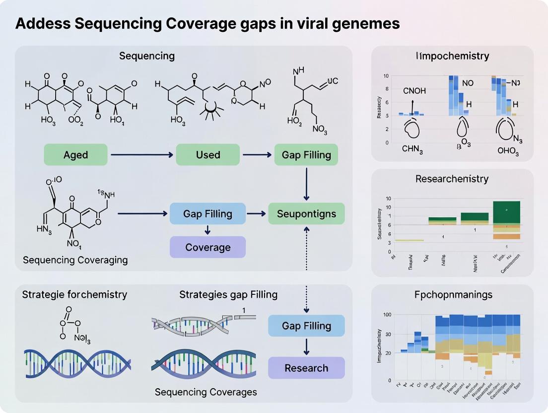
Why Coverage Gaps Matter: The Critical Impact on Viral Evolution, Pathogenesis, and Therapeutic Development
Coverage gaps are regions within a sequenced viral genome where the number of aligned reads (depth of coverage) is insufficient for confident base calling, assembly, or variant identification. These gaps compromise data completeness and can obscure critical genetic information, posing significant challenges for genomic surveillance, therapeutic target identification, and vaccine development.
Troubleshooting Guides & FAQs
FAQ 1: Why do I suddenly have zero-coverage regions in my Illumina data for an amplicon-based SARS-CoV-2 panel?
- Problem: Primer mismatches due to new viral mutations.
- Solution: Use updated primer schemes or switch to a tiled amplicon approach. Validate in silico primer binding with current strain sequences before wet-lab work.
FAQ 2: My nanopore sequencing of HIV-1 shows consistent dropouts in specific GC-rich regions. How can I resolve this?
- Problem: Systematic biases against high or low GC-content regions in some sequencing technologies.
- Solution: Balance your sequencing library with a mix of technologies (e.g., combine Oxford Nanopore with Illumina for hybrid assembly). Use PCR additives like Q5 High-GC Enhancer during amplification.
FAQ 3: During assembly of a novel flavivirus, I have gaps in repetitive terminal regions. What should I do?
- Problem: Long Terminal Repeats (LTRs) and other repeats confuse assemblers.
- Solution: Employ specialized assemblers (e.g., VICUNA, SPAdes) designed for viral repeats. Supplement with RACE (Rapid Amplification of cDNA Ends) or Sanger sequencing to close terminal gaps.
FAQ 4: How can I verify if a low-coverage region is a technical artifact or a genuine genomic deletion?
- Problem: Differentiating signal from noise.
- Solution: Perform orthogonal validation using a different assay (e.g., droplet digital PCR for copy number variation) or a different sequencing chemistry. Triangulate data from multiple aligned reads, checking for split reads or paired-end discordance.
Experimental Protocols
Protocol 1: Tiled Amplicon Sequencing for Highly Variable Viruses
Objective: Generate overlapping amplicons to minimize primer dropout.
- Primer Design: Using a reference genome, design primer pairs to generate 400-800 bp amplicons with 50-150 bp overlaps.
- Multiplex PCR: Perform two pools of multiplex PCR using a high-fidelity polymerase.
- Pool Normalization: Quantify amplicons by fluorometry, then normalize and pool equimolarly.
- Library Prep: Use a tagmentation-based (e.g., Nextera) or ligation-based library kit.
- Sequencing: Sequence on an Illumina MiSeq (2x250 bp) to ensure overlap.
Protocol 2: Hybrid Assembly for Complex Viral Genomes
Objective: Combine long and short-read data to resolve repeats and gaps.
- Sample Prep: Extract viral DNA/RNA from the sample.
- Long-Read Library: Prepare an unamplified library for Oxford Nanopore MinION sequencing.
- Short-Read Library: Prepare a PCR-free library for Illumina sequencing (e.g., 2x150 bp).
- Basecalling & QC: Basecall nanopore reads (Guppy) and QC all reads (FastQC).
- Assembly: Perform initial assembly using the long reads with Canu or Flye.
- Polishing: Polish the assembly 3-4 times using the high-accuracy Illumina reads with Medaka and Pilon.
Table 1: Common Sources of Coverage Gaps by Sequencing Approach
| Sequencing Method | Primary Gap Sources | Typical Genome Regions Affected |
|---|---|---|
| Amplicon-Based (Illumina) | Primer mismatch, amplicon size bias | Spike protein gene (SARS-CoV-2), Hypervariable regions (HIV-1) |
| Metagenomic (Shotgun) | Host DNA dominance, low viral titer | Entire genome, but especially low-copy regions |
| Long-Read (Nanopore/PacBio) | DNA/RNA degradation, basecalling errors | Homopolymer tracts, high-GC regions |
Table 2: Impact of Coverage Thresholds on Variant Calling
| Minimum Coverage Depth | Variant Calling Confidence | Risk of Missing True Variants | Risk of Calling False Variants |
|---|---|---|---|
| 10x | Low | Very High | High |
| 30x | Moderate | Moderate | Low |
| 100x | High | Low | Very Low |
| 200x+ | Very High | Very Low | Very Low |
The Scientist's Toolkit: Research Reagent Solutions
| Reagent / Kit | Function in Addressing Coverage Gaps |
|---|---|
| Q5 High-GC Enhancer (NEB) | Improves amplification efficiency through GC-rich regions prone to dropout. |
| RNase H | Degrades RNA in cDNA hybrids to improve 2nd strand synthesis and coverage uniformity. |
| PCR-Cleanup Size Selection Beads (SPRI) | Removes primer dimers and selects optimal amplicon size to improve library complexity. |
| Target-Specific Probe Panels (Hybrid Capture) | Enriches for viral reads from complex backgrounds without primer bias. |
| dUTP / UDG System | Controls carryover contamination in amplification-heavy protocols. |
| DNA Damage Repair Mix (e.g., NEB FFPE) | Repairs nicked/degraded DNA common in archived samples before long-read library prep. |
Visualizations
Title: Primary Causes Leading to Viral Genome Coverage Gaps
Title: Hybrid Sequencing Workflow to Resolve Coverage Gaps
Technical Support Center: Troubleshooting Incomplete Viral Genomes
Frequently Asked Questions (FAQs)
Q1: My NGS run yielded a viral genome with <90% coverage. What are the primary technical causes? A: Incomplete coverage often stems from: 1) Low viral load in the sample, leading to insufficient template. 2) PCR amplification bias during library prep, especially for high-GC regions. 3) Primer mismatches in amplicon-based protocols due to unknown viral diversity. 4) Sequence dropouts from homopolymeric regions or secondary structures that challenge polymerases. 5) Suboptimal read depth or uneven sequencing coverage.
Q2: How does incomplete data specifically impact phylogenetic inference and transmission cluster resolution? A: Missing data breaks phylogenetic signal. A 2023 study showed that genomes with >20% missing sites reduced the accuracy of inferred transmission clusters by up to 65% compared to full genomes. Incomplete data inflates branch length uncertainties and can lead to incorrect topological placements, obscuring the directionality of transmission chains.
Q3: What are the best practices to salvage and analyze datasets with unavoidable coverage gaps? A: Implement a tiered approach: 1) Mask uncertain sites rather than infer them. 2) Use phylogenetic models that account for missing data (e.g., ascertainment bias correction). 3) For transmission networks, integrate epidemiological metadata to constrain possible linkages where genetic data is incomplete. 4) Clearly report the proportion and location of gaps in all publications.
Q4: My outbreak surveillance pipeline is flagging too many partial genomes as "new variants." How can I reduce false positives? A: This is a common issue. Adjust your variant-calling threshold: only call a new variant if polymorphisms are supported by ≥10x read depth and present in ≥90% of reads in the covered region. Ignore mutations in regions with <10x coverage. Implement a coverage-based filter before phylogenetic placement; genomes below a defined coverage completeness threshold (e.g., <80%) should be annotated as "low confidence" for lineage assignment.
Q5: Are there specific genome regions where coverage gaps are most detrimental for functional inference? A: Yes. Gaps in key functional regions cripple interpretation. The table below summarizes high-impact zones:
Table 1: Impact of Coverage Gaps in Critical Viral Genomic Regions
| Genomic Region | Key Function | Consequence of Incomplete Data |
|---|---|---|
| Spike Protein (SARS-CoV-2) | Host cell receptor binding, neutralization epitopes | Inability to assess antigenic drift, vaccine escape mutations. |
| Polymerase (RdRp) | Viral replication, drug target (e.g., Remdesivir) | Missed mutations conferring antiviral resistance. |
| Envelope Glycoproteins (HIV, Influenza) | Host tropism, immune evasion | Blinded surveillance for shifts in host range or pathogenicity. |
| Promoter/Regulatory Regions | Transcription control (e.g., HIV LTR) | Unpredictable impact on viral replicative capacity. |
Troubleshooting Guides
Issue: Consistently Low Coverage in Specific Genome Regions (e.g., High GC Content)
- Step 1: Verify with QC Tools. Run
FastQCorMosdepthto visualize coverage distribution. Confirm the drop is localized. - Step 2: Optimize Library Prep. For amplicon panels, design tiled primers with degeneracy to handle variance. For hybrid capture, increase probe concentration/tiling density for difficult regions.
- Step 3: Adjust Sequencing Chemistry. Use a sequencing mix optimized for high-GC content (e.g., Q5 High-Fidelity GC Rich PCR kits).
- Step 4: In Silico Correction. Use tools like
NCBI-BLASTto map missing regions against a curated, closely related reference, clearly annotating these as in silico inferences.
Issue: Reconstructing Transmission Chains from Mixed Complete/Partial Genomes
- Step 1: Standardize Data. Align all genomes to the same reference. Replace missing sites with "N" (ambiguity), not gaps, for consistent alignment length.
- Step 2: Model-Aware Tree Building. Use a software like
IQ-TREEwith theASC(ascertainment bias correction) model to avoid overestimating genetic distances from missing data. - Step 3: Statistical Testing. Employ a tool like
TreeTimeto assess the confidence of specific node placements and cluster definitions. Clusters supported by <90% bootstrap values due to missing data should be considered tentative. - Step 4: Integrate Metadata. Use a Bayesian framework like
BEASTto incorporate sample collection dates and locations, which can help resolve uncertainties when genetic data is incomplete.
Experimental Protocol: Hybrid Capture for Enriching Low-Titer Viral Genomes from Host Background
Objective: To obtain complete viral genome sequences from clinical samples with high host nucleic acid background and low viral load.
Materials: See "The Scientist's Toolkit" below. Procedure:
- Extraction: Perform total nucleic acid extraction using a silica-membrane column kit. Include carrier RNA if viral load is suspected to be very low.
- Library Preparation: Convert RNA to cDNA. Fragment DNA via ultrasonication to ~200bp. Perform end-repair, A-tailing, and adapter ligation using a double-stranded DNA library prep kit. Amplify with 6-8 PCR cycles.
- Hybrid Capture: a. Denature: Heat 1 µg of library DNA at 95°C for 5 minutes, then immediately chill on ice. b. Hybridize: Incubate with a panel of biotinylated, 80-base RNA probes (tiled across the target viral genome) at 65°C for 16-24 hours in a hybridization buffer. c. Capture: Bind the probe-DNA hybrids to streptavidin-coated magnetic beads. Wash sequentially with high- and low-stringency buffers to reduce off-target binding. d. Elute & Amplify: Elute the captured DNA with NaOH, neutralize, and perform a second round of PCR (12-14 cycles).
- Sequencing: Pool and sequence on an Illumina platform (MiSeq/NextSeq) to a target depth of >1000x mean coverage.
- Analysis: Map reads to the viral reference genome using
BWA-MEMorBowtie2. Call consensus with a threshold of 10x coverage and 90% agreement.
Diagram: Hybrid Capture Workflow for Viral Enrichment
Diagram: Impact of Incomplete Data on Phylogenetic Inference
The Scientist's Toolkit: Research Reagent Solutions
Table 2: Essential Reagents for Addressing Coverage Gaps
| Item | Function | Example Product/Brand |
|---|---|---|
| Target-Specific Hybrid Capture Probes | Enrich viral sequences from complex background; crucial for low-titer samples. | MyBaits Expert (Arbor Biosciences), xGen (IDT). |
| High-Fidelity PCR Mix for GC-Rich Targets | Reduces polymerase dropouts in difficult genomic regions. | Q5 High-Fidelity GC-Rich (NEB), KAPA HiFi HotStart (Roche). |
| Carrier RNA | Improves recovery of low-concentration viral RNA during extraction. | poly(A) RNA, MS2 bacteriophage RNA. |
| Methylated Adapters & Duplicate Removal Beads | Enables accurate PCR duplicate removal, improving coverage evenness. | Unique Dual Indexes (UDIs) (Illumina), AMPure XP beads (Beckman Coulter). |
| Synthetic Control Genome | Spike-in control to monitor enrichment efficiency and coverage gaps. | RNA Virus Control (Seracare), custom gBlocks (IDT). |
FAQs & Troubleshooting Guides
Q1: During viral genome assembly, I encounter many short contigs and cannot achieve a complete genome. What are the primary causes and solutions? A: This is typically caused by low sequencing coverage and high variability regions. Solutions include:
- Increase Sequencing Depth: For Illumina, aim for >1000x coverage for highly variable viruses (e.g., HIV, influenza). For nanopore/ PacBio, >100x is often sufficient for scaffolding.
- Hybrid Assembly: Combine short-read (Illumina) and long-read (ONT/PacBio) data. Use short reads for accuracy and long reads to span repetitive or low-complexity regions.
- Targeted Enrichment: Use sequence capture probes (e.g., Twist Pan-Viral Panel) to enrich viral material from host or environmental samples before sequencing.
Q2: How do I determine if a gap in my assembled viral genome is biologically real (e.g., an un-translatable region) versus an artifact of poor sequencing? A: Follow this diagnostic workflow:
- Map Reads Back: Visualize BAM/CRAM files in IGV. A true biological gap will have no mapped reads across all samples. An artifact often has low, fragmented coverage.
- Check Parallel Samples: Does the gap appear consistently in multiple independent isolates/sequencing runs of the same virus?
- Consult Reference Databases: Use BLAST against NCBI Virus, VIPR, or GISAID to see if the region is consistently absent in all known strains.
- PCR/Sanger Verification: Design primers flanking the putative gap for wet-lab validation.
Q3: What are the best practices for functional annotation of viral genes when the reference genome is incomplete or has low-quality annotations? A: Implement a multi-source annotation pipeline:
- Primary Annotation: Use tools like Prokka or VAPiD, but do not rely solely on them.
- Homology Search: Perform sensitive homology searches using HMMER (against PFAM, VOGDB) and HHpred for distant relationships.
- De Novo Prediction: Use tools like DeepTFactor or Phyre2 for protein structure/function prediction when homology is weak.
- Orthology Assignment: Use OrthoFinder or eggNOG-mapper to infer function from conserved protein families.
- Manual Curation: Always curate results against recent literature. Table 1 summarizes key resources.
Table 1: Key Resources for Viral Functional Annotation
| Resource Name | Type | Primary Use | URL/Reference |
|---|---|---|---|
| VOGDB | Database | Protein families & functional annotation of viral proteins | https://vogdb.org |
| VIPR | Database | Repository of annotated viral sequences & tools | https://www.viprbrc.org |
| PHROG | Database | Prophage (viral) protein families | https://phrogs.lmge.uca.fr |
| HMMER | Tool | Sensitive protein profile searches | http://hmmer.org |
| InterProScan | Tool | Integrates multiple protein signature databases | https://www.ebi.ac.uk/interpro |
Q4: My research focuses on viral pathogenesis. How can sequencing gaps directly hinder the identification of virulence factors? A: Gaps can obscure critical genomic elements:
- Promoters/Regulatory Elements: These non-coding regions are often AT-rich and prone to assembly gaps. Missing them can prevent understanding of gene regulation during infection.
- Frameshifts & Readthrough Events: Gaps can mask programmed ribosomal frameshifts or stop-codon readthroughs essential for expressing virulence factors (e.g., gag-pol fusion in retroviruses).
- Recombination Breakpoints: These hotspots are often in complex, repetitive regions that are difficult to sequence. Missing them prevents understanding of viral evolution and emergence.
- RNA Modification Sites: Direct RNA sequencing (ONT) can detect modifications (m6A) that affect pathogenesis. Gaps in RNA-seq coverage miss these epitranscriptomic signals.
Experimental Protocol: Hybrid Sequencing for Gap Closure in a Novel Herpesvirus
Objective: Generate a complete, high-accuracy genome sequence from a clinical isolate.
Materials: See "Research Reagent Solutions" table below.
Methodology:
- Nucleic Acid Extraction: Use the QIAamp MinElute Virus Spin Kit for high-purity viral DNA.
- Library Preparation & Sequencing:
- Illumina: Prepare library using Nextera XT DNA Library Prep Kit (2x150bp). Sequence on MiSeq to achieve >500x predicted coverage.
- Oxford Nanopore: Prepare library using Ligation Sequencing Kit V14 (SQK-LSK114). Load on R10.4.1 flow cell and sequence on GridION for 48 hours.
- Hybrid Assembly:
- Trim Illumina reads with Trimmomatic (
ILLUMINACLIP:TruSeq3-PE-2.fa:2:30:10). - Base-call and trim nanopore reads with Guppy (
--config dna_r10.4.1_e8.2_400bps_sup.cfg). - Perform hybrid assembly using Unicycler (
--mode hybrid).
- Trim Illumina reads with Trimmomatic (
- Polishing & Validation:
- Polish the Unicycler assembly with the Illumina reads using Polypolish.
- Assess assembly completeness with QUAST and CheckV.
- Validate critical gaps via PCR with LongAmp Taq and Sanger sequencing.
Research Reagent Solutions
| Item | Function | Example Product |
|---|---|---|
| Viral DNA/RNA Extraction Kit | Isolate high-purity, inhibitor-free nucleic acid from complex samples. | QIAamp MinElute Virus Spin Kit |
| Ultra II FS DNA Library Prep Kit | Prepare Illumina-compatible libraries from low-input viral DNA. | NEBNext Ultra II FS DNA Library Prep Kit |
| Ligation Sequencing Kit | Prepare DNA libraries for nanopore sequencing, enabling long reads. | Oxford Nanopore SQK-LSK114 |
| Pan-Viral Enrichment Probe Set | Enrich viral sequences from high-background (host, microbiome) samples. | Twist Pan-Viral Panel |
| Long-Range PCR Master Mix | Amplify across large gaps or repeats for validation. | NEB LongAmp Taq Master Mix |
| SMRTbell Prep Kit | Prepare libraries for PacBio HiFi sequencing (high-accuracy long reads). | PacBio SMRTbell Prep Kit 3.0 |
Viral Genome Assembly & Gap Resolution Workflow
Decision Tree for Characterizing Genomic Gaps
Troubleshooting & FAQs for Viral Genome Sequencing Coverage Gaps
This support center addresses common experimental challenges in achieving complete viral genome sequences, which is critical for accurate drug and vaccine target identification.
FAQ 1: Why do I consistently have low or zero coverage in specific regions of the viral genome (e.g., GC-rich areas or secondary structures)?
- Answer: This is often due to biases in library preparation protocols (especially amplicon-based methods) and polymerase stalling. GC-rich regions can cause inefficient amplification, while strong secondary structures in viral RNA can cause reverse transcriptase or polymerase enzymes to dissociate.
- Protocol Solution (Hybrid Capture for Problematic Regions):
- Probe Design: Design biotinylated DNA or DNA/RNA hybrid probes (e.g., using xGen Lockdown Probes) targeting the low-coverage regions identified in your initial sequencing run. Include flanking sequences.
- Library Prep: Prepare a standard short-read sequencing library (e.g., Illumina DNA Prep) from your viral cDNA.
- Hybridization: Denature the library and hybridize with the probe pool (65°C for 16-24 hours in a thermocycler with heated lid).
- Capture: Bind hybridized fragments to streptavidin beads, wash stringently to remove off-target fragments.
- Amplification & Sequencing: Perform a limited-cycle PCR to amplify captured fragments and proceed with sequencing. This enriches for hard-to-sequence regions.
FAQ 2: How can I identify true resistance mutations versus sequencing artifacts introduced by coverage gaps or errors?
- Answer: Artifacts often appear at very low frequencies (<2%) and are not reproducible across technical replicates or different sequencing platforms. True resistance mutations are often present at higher frequencies and correlate with phenotypic assay data.
- Protocol Solution (Triangulation Validation):
- Multi-Protocol Sequencing: Sequence the same sample using at least two independent methods (e.g., amplicon sequencing AND hybrid capture OR metagenomic sequencing).
- Duplicate Analysis: Perform library preparation and sequencing in independent technical duplicates.
- Variant Calling: Use a robust, multi-algorithm variant caller (e.g., combining LoFreq, iVar, and GATK). Only call mutations that are:
- Present in >5% of reads (platform-dependent threshold).
- Called by at least 2 variant-calling algorithms.
- Reproduced across both sequencing methods or technical duplicates.
FAQ 3: My epitope mapping is incomplete due to fragmented viral surface protein reads. How can I obtain full-length, high-quality sequences for structural analysis?
- Answer: Short-read sequencing often fragments long open reading frames (ORFs). Utilizing long-read sequencing (Oxford Nanopore or PacBio) for these specific genes is the recommended solution.
- Protocol Solution (Targeted Long-Read Sequencing of Viral Glycoprotein Genes):
- Targeted Amplification: Design primers to generate amplicons spanning the entire glycoprotein gene(s) (may be 2-5kb). Use a high-fidelity, long-range polymerase (e.g., PrimeSTAR GXL).
- Size Selection: Purify the full-length amplicon using magnetic beads (e.g., SPRIselect) with a size cutoff to remove primer dimers and shorter fragments.
- Library Prep for Nanopore: Use the Ligation Sequencing Kit (SQK-LSK114). Do not fragment the DNA. Attach sequencing adapters directly to the amplicon.
- Sequencing & Analysis: Load onto a MinION flow cell (R10.4.1 preferred for accuracy). Basecall with Dorado (using super-accuracy model) and perform consensus polishing (e.g., with Medaka) to achieve high (>Q30) accuracy for the full-length gene.
Table 1: Impact of Sequencing Method on Coverage Uniformity for a Model Virus (HIV-1)
| Sequencing Method | Average Coverage Depth | % Genome Covered >20x | Problematic Regions (e.g., pol secondary structure) Coverage |
|---|---|---|---|
| Amplicon (V3-V4) | 10,000x | 98.5% | <10x or 0x |
| Metagenomic (Shotgun) | 150x | 65.2% | Inconsistent, low depth |
| Hybrid Capture | 850x | 99.1% | >100x |
| Long-Read (Nanopore) | 50x | 99.8% | Full-length reads |
Table 2: False Positive Mutation Rate by Validation Strategy
| Validation Strategy | Mean False Positive Calls per 10kb Viral Genome | Key Requirement |
|---|---|---|
| Single Protocol, Single Caller | 4.7 | N/A |
| Single Protocol, Dual Caller Concordance | 1.2 | Use of orthogonal algorithms (e.g., LoFreq + iVar) |
| Dual Protocol (Amplicon + Capture) | 0.3 | Independent library prep |
| Dual Protocol + Phenotypic Correlation | ~0.1 | Access to neutralization/IC50 data |
Visualizations
Title: Workflow for Addressing Viral Sequencing Gaps
Title: Decision Tree for Validating Viral Mutations
The Scientist's Toolkit: Research Reagent Solutions
| Item | Function | Key Consideration |
|---|---|---|
| xGen Hybridization Capture Probes | Biotinylated probes to enrich for specific low-coverage viral genomic regions. | Design probes against known "gap" regions from public databases (GISAID). |
| PrimeSTAR GXL DNA Polymerase | High-fidelity polymerase for long-range PCR of viral glycoprotein genes (>5kb). | Minimizes amplification errors critical for epitope analysis. |
| SPRIselect Magnetic Beads | Size-selection and clean-up of cDNA/amplicon libraries. | Critical for removing primers and selecting optimal fragment sizes. |
| Oxford Nanopore Ligation Sequencing Kit (SQK-LSK114) | Prepares native DNA libraries for long-read sequencing on MinION/PromethION. | Enables full-length viral gene sequencing without fragmentation. |
| Pan-viral Enrichment Probe Sets (e.g., ViroCap) | Capture probes for a broad range of known viruses from complex samples. | Useful for detecting co-infections that may interfere with target identification. |
| Unique Molecular Identifiers (UMIs) | Short random barcodes ligated to each cDNA molecule before amplification. | Allows bioinformatic correction of PCR/sequencing errors for accurate low-frequency variant calling. |
Closing the Gaps: A Toolkit of Modern Sequencing and Enrichment Techniques for Complete Viral Genomes
Technical Support Center: Troubleshooting & FAQs
Q1: My hybridization-based capture panel shows high on-target rates but consistently misses coverage for specific viral genotypes. What could be the cause and how can I resolve it?
A: This is a common issue stemming from probe-template mismatches due to high viral genetic diversity. To resolve:
- Diagnose: Map your missed regions against the reference used for probe design. Use tools like BLAST to check for local sequence divergence (>10-15% is problematic).
- Solution - Probe Optimization: Redesign probes for the variable region using a tiled, degenerate probe strategy.
- Protocol - Degenerate Probe Design:
- Gather all available sequences for the target virus genotype/clade from databases (NCBI Virus, GISAID).
- Perform multiple sequence alignment (MSA) using MAFFT or Clustal Omega.
- Identify conserved blocks flanking the variable region.
- Design overlapping 80-120nt probes that tile across the region. At variable positions, incorporate mixed bases (e.g., W for A/T) or universal bases (e.g., inosine) following synthesis compatibility.
- Increase probe tiling density (e.g., 3x overlap) over these regions.
- Protocol - Degenerate Probe Design:
- Alternative - Wet-Lab Adjustment: Lower the hybridization temperature by 2-5°C in the capture step to allow for mismatch hybridization. Re-optimize wash stringency empirically.
Q2: I am using a tiling amplicon approach for viral genome sequencing, but I'm getting dropout in high-GC (>70%) regions. How can I improve uniformity?
A: Dropout in high-GC regions is often due to inefficient primer binding and polymerase stalling.
- Diagnose: Check the raw amplicon sizes on a bioanalyzer. Smearing or smaller fragments indicate incomplete amplification.
- Solution - Reagent & Protocol Optimization:
- Protocol - High-GC Amplicon Generation:
- PCR Formulation: Use a PCR mix specifically engineered for high-GC content (e.g., with additives like betaine, DMSO, or TMAC).
- Thermocycling Profile: Implement a slow ramp rate (e.g., 0.5°C/sec) from annealing to extension to facilitate primer binding and polymerase initiation.
- Two-Step PCR: Design a secondary, nested primer set internal to the first, targeting the same region but with a lower overall GC content in its binding sites if possible.
- Polymerase Choice: Switch to a polymerase blend with high processivity and GC-neutral bias.
- Protocol - High-GC Amplicon Generation:
- Primer Redesign: If possible, redesign tiling primers so their 3' ends anchor in adjacent, more moderate-GC sequence stretches.
Q3: My CRISPR-based enrichment (e.g., for PAC-MAN or CARMEN) shows low efficiency. What are the critical steps to optimize gRNA design and cleavage?
A: Low efficiency often traces back to gRNA activity and target accessibility.
- Diagnose: Check the pre- and post-enrichment target abundance via qPCR. If low, the issue is likely gRNA-related.
- Solution - gRNA Design & Validation Protocol:
- Design Rules: Use current algorithms (CRISPRscan, ChopChop) to pick gRNAs with high predicted efficiency. Prioritize a PAM site (e.g., NGG for SpCas9) close to the center of your target region. Avoid sequences with significant secondary structure.
- Validation Workflow:
- In vitro Transcription: Synthesize candidate gRNAs.
- Test Cleavage: Set up a cleavage reaction using recombinant Cas9/gRNA complex and a synthetic double-stranded DNA target containing the viral sequence. Use a plasmid-based target as a control.
- Analyze: Run products on a Bioanalyzer or gel. Select gRNAs with >80% cleavage efficiency in vitro before proceeding to library enrichment.
- Critical Parameter: Ensure your input library is not denatured. Cas9 requires double-stranded DNA targets. Use 100-500ng of intact, double-stranded library as input.
Q4: For a broad viral family panel (e.g., all Flaviviruses), how do I balance probe comprehensiveness against off-target human host binding?
A: This is a key challenge in clinical/metagenomic samples with high host background.
- Diagnose: Check sequencing metrics: a very low fraction of reads mapping to the viral target (<1%) with high alignment to the host genome (e.g., human hg38) indicates host off-target capture.
- Solution - In Silico Probe Filtering & Blocking Agents:
- Protocol - Host Depletion Probe Design:
- After generating candidate probes from viral alignments, perform an in silico subtraction.
- Blast all candidate 80-120mer probes against the host reference genome(s). Discard any probe with >80% identity over >50bp.
- For the remaining probes, calculate melting temperature (Tm) and cross-hybridization potential. Tools like
BLATorbowtie2in sensitive mode can be used.
- Experimental Suppression: Add unlabeled host blocking oligonucleotides (e.g., Cot-1 DNA, sheared human DNA, or synthetic oligos blocking repetitive elements) in excess during hybridization to sequester host-derived reads.
- Protocol - Host Depletion Probe Design:
Table 1: Comparison of Targeted Enrichment Modalities for Viral Sequencing
| Parameter | Hybridization Capture Panels | Tiling Amplicon PCR | CRISPR-based Enrichment |
|---|---|---|---|
| Typical Input DNA (ng) | 50-500 | 1-100 | 50-200 |
| Hands-on Time | 12-24 hours | 3-6 hours | 6-12 hours |
| Design Flexibility | High (post-synthesis) | High (per-run) | Moderate (requires new gRNA) |
| Tolerance to SNPs | Moderate (Degrades with mismatch) | Low (Primer dropout) | High (Tolerates mismatches outside seed/PAM) |
| Uniformity of Coverage | Good (Depends on probe design) | Variable (Prone to GC bias) | Good (Depends on gRNA spacing) |
| Best For | Diverse strains, large genomes | Low input, high sensitivity | Specific variant discrimination, portable |
Table 2: Troubleshooting Guide: Common Metrics and Thresholds
| Problem | Key Metric to Check | Acceptable Threshold | Corrective Action |
|---|---|---|---|
| Low On-Target Rate (Capture) | % Reads on Target | >20% for complex panels | Increase probe tiling; Add blocking agents. |
| High Duplicate Rate (Amplicon) | % PCR Duplicates | <30% | Reduce PCR cycles; Increase input diversity. |
| Coverage Dropout (GC-rich) | Fold-80 Base Penalty | <2 | Optimize PCR with additives; Redesign primers. |
| Inefficient CRISPR Cleavage | In vitro cleavage efficiency | >80% | Re-design and re-test gRNA activity. |
| Poor Uniformity (Panels) | Coverage CV (Coefficient of Variation) | <0.5 (lower is better) | Increase probe overlap/tiling density. |
Experimental Protocols
Protocol 1: Designing and Validating a Tiled, Degenerate Probe Set
Objective: To generate hybridization probes that recover highly variable viral regions. Steps:
- Sequence Curation: Download all relevant viral sequences from public repositories. Curate to remove low-quality entries.
- Alignment & Conservation Plotting: Perform MSA. Generate a conservation score plot (e.g., with
plotconfrom EMBOSS). - Probe Definition: In conserved regions (>90% identity), design 100mer probes with 2x tiling density. In variable regions (50-90% identity), increase density to 3-5x and incorporate degeneracy (mixed bases).
- Off-Target Filtering: Blast all probe sequences against the host genome. Remove probes with significant hits (E-value < 1e-10, length >50bp).
- Synthesis & Pooling: Order probes as a pooled oligonucleotide library. Amplify and convert to double-stranded biotinylated capture baits per manufacturer's protocol (e.g., Agilent SureSelectXT).
Protocol 2: CRISPR-Cas9 Enrichment for Viral Targets (Adapted from CAST-seq)
Objective: To enrich for specific viral sequences from a fragmented NGS library using Cas9. Steps:
- gRNA Design & Preparation: Design two gRNAs flanking the target region (200-500bp apart). Synthesize guide RNAs via in vitro transcription and purify.
- Cas9-gRNA RNP Complex Formation: Incubate 10pmol of Cas9 nuclease with a 1.5x molar ratio of each gRNA in NEBuffer 3.1 at 25°C for 10 minutes.
- Library Digestion: Mix 100-200ng of your double-stranded, adapter-ligated NGS library with the RNP complex. Incubate at 37°C for 60 minutes.
- Target Isolation: Use streptavidin beads with biotinylated oligonucleotides complementary to the overhang sequences created by Cas9 cleavage (requires custom design) to pull down the linearized target fragments.
- Wash & Elute: Wash beads stringently. Elute the captured fragments in low-EDTA TE buffer.
- Amplification: Perform a limited-cycle PCR (8-12 cycles) with index primers to generate the final sequencing library.
Visualizations
Title: Workflow for Addressing Viral Sequencing Coverage Gaps
Title: CRISPR-based Enrichment Protocol Workflow
The Scientist's Toolkit: Research Reagent Solutions
| Item | Function / Application | Example Vendor/Brand |
|---|---|---|
| Hybridization Capture Kit | Provides buffers, beads, and protocol for solution-based target enrichment. | Agilent SureSelectXT, IDT xGen |
| High-GC PCR Additive Mix | Betaine, DMSO, or TMAC to reduce secondary structure and improve polymerase processivity in high-GC regions. | Thermo Fisher, QIAGEN |
| Recombinant Cas9 Nuclease | High-purity, high-activity nuclease for CRISPR-based enrichment workflows. | New England Biolabs, IDT |
| Blocking Oligos (Cot-1 DNA) | Unlabeled host DNA to pre-bind repetitive elements and reduce off-target capture in host-rich samples. | Thermo Fisher, Roche |
| PCR Polymerase for High Fidelity | Enzyme blends designed for uniform amplification of complex templates with minimal bias. | KAPA HiFi, Q5 High-Fidelity |
| Streptavidin Magnetic Beads | For solid-phase capture of biotinylated targets (e.g., baits or cleaved fragments). | Dynabeads, Sera-Mag |
| Universal Blocking Oligos (TSO) | Oligos blocking adapter sequences to prevent primer dimer formation in low-input protocols. | IDT, Sigma-Aldrich |
Troubleshooting Guides & FAQs
Q1: During PacBio HiFi library prep for a diverse viral pool, my yield is consistently low. What are the primary causes and solutions? A: Low yield often stems from input DNA quality or size selection inefficiency.
- Cause 1: Degraded or sheared viral genomic DNA. Solution: Use fresh phenol-chloroform or magnetic bead-based extraction, minimizing vortexing. Check integrity on a pulse-field or standard gel.
- Cause 2: Inefficient removal of short fragments during size selection. Solution: Optimize bead-to-sample ratio (e.g., using 0.45x left-side followed by 0.3x right-side SPRI selection). For very low inputs (<100ng), consider carrier RNA during precipitation.
- Protocol: High-Integrity Viral DNA Extraction for HiFi
- Concentrate viral particles from culture supernatant using PEG-8000 precipitation (10% w/v, 4°C overnight).
- Resuspend pellet in DNase/RNase-free PBS with proteinase K (0.5 mg/ml).
- Lyse with SDS (1% final), incubate at 56°C for 1 hour.
- Perform phenol:chloroform:isoamyl alcohol (25:24:1) extraction twice.
- Precipitate DNA with isopropanol and glycogen, wash with 70% ethanol.
- Resuspend in low TE buffer. Quantify using Qubit dsDNA HS Assay.
Q2: My Oxford Nanopore (ONT) sequencing run of a coronavirus genome shows a high proportion of reads failing basecalling (\"Reads Pore\"). How can I improve this? A: This typically indicates issues with the motor protein or adapter-ligation.
- Cause 1: Contaminants (e.g., salts, organics) co-precipitated with DNA. Solution: Perform additional ethanol washes (80%) and ensure pellet is fully dried. Use AMPure XP bead clean-up post-ligation.
- Cause 2: Damaged or overused flow cell pores. Solution: Perform a "Flow Cell Check" in MinKNOW before loading sample. If pore availability is <800 for R10.4.1, precondition with nuclease flush.
- Protocol: ONT Flow Cell Preconditioning & Loading for Viral RNA
- Perform a Flow Cell Check in MinKNOW. Note pore availability.
- If <800, prepare a nuclease flush mix: 480 µl nuclease-free water, 20 µl Flow Cell Flush Tether (FLT-FLK111), 500 µl Flow Cell Flush Buffer (FLB-FLK111).
- Load 800 µl of mix via the priming port, wait 5 minutes, reload.
- Prime flow cell with 800 µl of fresh flush buffer, then load library as per protocol.
Q3: When assembling a complex herpesvirus genome (with large repeats) from HiFi data, my assembler (e.g., Flye, hifiasm) produces fragmented contigs. How do I resolve this? A: This suggests the assembler is breaking at long, homogeneous repeats. The solution is to adjust assembly parameters or use a specialized workflow.
- Cause: Default assembly parameters may not tolerate the high identity and length of viral terminal/inverted repeats. Solution: Increase the overlap error rate tolerance and disable repeat trimming.
- Protocol: Herpesvirus Assembly with HiFi Reads using Flye
- Subsample reads to ~50x coverage:
seqtk sample reads.fastq 50000 > subreads.fastq. - Run Flye with repeat-sensitive settings:
- Polish the assembly once with the full read set using
medaka_consensus.
- Subsample reads to ~50x coverage:
Q4: For ONT direct RNA sequencing of HIV genomes, my read lengths are far shorter than expected. What step is likely problematic? A: RNA degradation or incorrect handling of the reverse transcription step in the cDNA-PCR protocol is common.
- Cause 1: RNA fragmentation during extraction or storage. Solution: Always include RNAse inhibitors, work on ice, and use Trizol-LS with glycogen carrier. Avoid freeze-thaw cycles.
- Cause 2: Over-denaturation during the reverse transcription step for cDNA-PCR sequencing. Solution: Do not exceed 70°C for 3 minutes during RNA primer annealing.
- Protocol: Full-Length Viral RNA Preservation for ONT
- To viral supernatant, add 3x volume of Trizol LS. Incubate 5 min RT.
- Add glycogen (5 µl of 20mg/ml), mix, then add 0.3x volume chloroform. Shake vigorously.
- Centrifuge at 12,000g, 15 min, 4°C. Transfer aqueous phase.
- Precipitate with isopropanol, wash with 80% ethanol (not 70%).
- Resuspend in nuclease-free water with 1 U/µl RNAse inhibitor. Store at -80°C in aliquots.
Table 1: Performance Metrics for Resolving Viral Repetitive Regions
| Metric | PacBio HiFi (Sequel II/IIe) | Oxford Nanopore (R10.4.1, Duplex) | Ideal for Viral Research Gap When... |
|---|---|---|---|
| Read Length (N50) | 15-25 kb | 10-50 kb (simplex), >100 kb (duplex possible) | Targeting very long (>10 kb) homologous repeats (e.g., poxvirus inverted terminal repeats). |
| Raw Read Accuracy | >99.9% (Q30) | ~99% (simplex Q20), >99.9% (duplex Q30) | Detecting low-frequency variants (<1%) within a viral quasispecies. |
| Required DNA Input | 1-5 µg (standard), 100-500 ng (low input) | 50-1000 ng (ligation), 10-50 ng (PCR-cDNA) | Sample is extremely limited (e.g., direct from clinical specimen). |
| Typical Coverage for Closure | 30-50x | 50-100x (due to lower single-pass accuracy) | Budget is constrained and high multiplexing is needed. |
| Best Suited Repeat Type | Moderate-length tandem repeats, GC-rich regions | Long homopolymer runs, methylated repeats (epigenetic context) | The biological question involves epigenetic regulation of viral latency. |
Table 2: Troubleshooting Common Library Preparation Failures
| Symptom | PacBio HiFi Likely Cause | ONT Likely Cause | Immediate Diagnostic Step |
|---|---|---|---|
| No sequencing output | SMRTbell template nicked; failed polymerase binding | Flow cell pores blocked; motor protein inactive | Check BioAnalyzer/TapeStation profile for library size. Run Flow Cell Check in MinKNOW. |
| Extremely short reads | Over-shearing of input DNA; severe DNA degradation | Contaminants inhibiting motor protein; incorrect buffer | Run a genomic DNA control sample. Check fluorometric quantification vs. fragment analyzer. |
| High adapter dimer peak | Inefficient purification post-ligation; insufficient size selection | Ligation time/temp incorrect; AMPure bead ratio wrong | Re-run size selection with adjusted bead ratios. Analyze library with HS D5000/HS BioAnalyzer assay. |
| Low multiplexing yield | Inaccurate sample quantification leading to imbalance | PCR bias during barcoding; some samples degraded | Re-quantify samples with fragment-aware assay (Qubit + fragment analyzer). Re-pool based on molarity. |
Experimental Workflow Diagrams
Diagram Title: PacBio HiFi Viral Genome Sequencing Workflow
Diagram Title: ONT Direct RNA Viral Sequencing Workflow
The Scientist's Toolkit: Research Reagent Solutions
Table 3: Essential Materials for Long-Read Viral Genomics
| Item | Function & Rationale | Example Product/Brand |
|---|---|---|
| Glycogen (Molecular Grade) | Carrier for ethanol precipitation of low-concentration viral nucleic acids; increases visible pellet and recovery. | GlycoBlue (Thermo Fisher), Molecular Grade Glycogen (Roche). |
| SPRIselect Beads | Size-selective purification of DNA fragments; critical for removing short fragments and adapter dimers in HiFi/ONT lib prep. | SPRIselect / AMPure XP (Beckman Coulter). |
| Proteinase K (RNA-free) | Degrades nucleases and viral capsid proteins during extraction; essential for obtaining high-molecular-weight DNA/RNA. | Proteinase K, molecular biology grade (Roche). |
| RNase Inhibitor | Protects viral RNA from degradation during all steps post-cell lysis; critical for full-length transcript recovery. | Superase-In (Thermo Fisher), RNasin (Promega). |
| Low-TE Buffer (pH 8.0) | Resuspension buffer for extracted DNA; EDTA chelates Mg2+ to inhibit nuclease activity, Tris stabilizes pH. | Invitrogen UltraPure 1X TE Buffer, diluted 10-fold. |
| Flow Cell Flush Kit | Rejuvenates Oxford Nanopore flow cells by clearing blocked pores; extends usable life for viral sequencing runs. | ONT Flow Cell Flush Kit (EXP-WSH004). |
Technical Support Center
Troubleshooting Guides & FAQs
Q1: During hybrid assembly, my consensus genome has an unusually high number of ambiguous bases (N's) at the junctions between short-read contigs and long-read scaffolds. What is the likely cause and solution?
A: This typically indicates a lack of sufficient overlap or conflicting data between the two datasets during the scaffolding step.
- Cause: The most common cause is suboptimal mapping of short reads to the long-read scaffold, often due to high error rates in the raw long reads or significant differences in coverage depth between datasets.
- Solution:
- Polish the scaffold: Use the high-accuracy short reads to polish the long-read scaffold before the final hybrid merging step. Tools like
PilonorNextPolishare designed for this. - Adjust mapping parameters: Loosen mapping stringency (e.g., decrease the
--score-minparameter inminimap2) for the initial alignment of short reads to long reads to ensure overlaps are detected. - Verify coverage: Check that coverage is relatively uniform. A table of recommended coverage for viral genome assembly is below.
- Polish the scaffold: Use the high-accuracy short reads to polish the long-read scaffold before the final hybrid merging step. Tools like
| Sequencing Type | Recommended Minimum Coverage for Viral Genomes | Optimal Coverage Range for Hybrid Assembly |
|---|---|---|
| Short-Read (Illumina) | 100x | 200x - 500x |
| Long-Read (ONT) | 50x | 100x - 200x |
| Long-Read (PacBio HiFi) | 20x | 30x - 100x |
Protocol: Hybrid Assembly with Polishing
- Initial Assembly: Generate a primary assembly from long reads using
FlyeorCanu. - Polish with Short Reads: Map short reads to the draft assembly using
bwa mem. RunPilon(java -Xmx16G -jar pilon.jar --genome draft.fasta --frags aligned.bam --output polished --changes). - Scaffold Integration: Use the polished long-read assembly as a trusted scaffold. Map short-read-only contigs (from
SPAdesin--metamode) to it usingminimap2to fill remaining gaps.
Q2: I am not recovering the terminal repeats/ends of my linear viral genome, leading to coverage gaps. How can I address this with a hybrid approach?
A: This is a common challenge in viral genomics due to the difficulty of sequencing through hairpin loops or identical repeats.
- Cause: Short reads cannot span the repeated regions, and long reads may have dropouts or systematic errors at sequence ends.
- Solution: Employ a targeted enrichment or PCR-free ligation protocol for long-read libraries combined with circular consensus sequencing (CCS) if using PacBio.
- Experimental Protocol: Terminal End Recovery
- Sample Prep: For Nanopore, use a PCR-free ligation sequencing kit (SQK-LSK114) to avoid amplification bias against terminal structures.
- Sequencing: Sequence the same sample on both platforms. For PacBio, target ≥20kb inserts and generate HiFi reads.
- Hybrid Analysis: Use TAR-VIR (Terminal Repeat Finder for Viral genomes) or manually inspect long reads that are longer than the expected genome length in
GeneiousorIGV. These reads will contain the complete terminal repeat. Use these as a scaffold to anchor short-read contigs.
Q3: My hybrid assembly results in chimeric contigs or misassemblies in low-complexity regions. How can I validate and correct these?
A: This often stems from repetitive regions or homologous sequences within the viral genome or host DNA.
- Cause: Misalignment of both short and long reads in repetitive zones.
- Solution: Implement an orthogonal validation step.
- Validation by Mapping: Map all raw reads (both short and long) back to the final hybrid assembly using
minimap2(long) andbwa(short). Visually inspect the alignment inIGVfor coverage drops (>50% drop) or mis-oriented reads at putative chimeric junctions. - Experimental Validation: Design PCR primers flanking the suspected misassembly. Perform Sanger sequencing on the amplicon to confirm the correct sequence.
- Re-assembly with Constraints: If a misassembly is confirmed, use the validated sequence as a "trusted anchor" to guide the assembler. In
Unicycler, you can provide the corrected sequence as a "trusted contig" to constrain the assembly graph.
- Validation by Mapping: Map all raw reads (both short and long) back to the final hybrid assembly using
The Scientist's Toolkit: Research Reagent Solutions
| Item | Function in Hybrid Sequencing for Viral Genomes |
|---|---|
| PCR-Free Library Prep Kits (ONT SQK-LSK114, PacBio SMRTbell) | Minimizes amplification bias, crucial for recovering GC-rich or structured terminal repeats in viral genomes. |
| Magnetic Bead Cleanup Kits (SPRI beads) | For precise size selection of long-read libraries to remove short fragments and optimize read length. |
| Host Depletion Kits (e.g., NEBNext Microbiome DNA Enrichment) | Enriches viral sequences by selectively removing methylated host (e.g., human) DNA, improving viral coverage. |
| Direct cDNA or Direct RNA Sequencing Kits (ONT) | Allows for sequencing of viral RNA genomes without amplification, preserving base modifications and simplifying coverage of ends. |
| High-Fidelity PCR Mix (for validation) | Used for generating accurate amplicons from hybrid assemblies for Sanger sequencing validation of problematic regions. |
Workflow & Relationship Diagrams
Title: Hybrid Sequencing Viral Genome Workflow
Title: Root Causes of Coverage Gaps & Hybrid Solutions
Troubleshooting Guides & FAQs
FAQ Section: Sample Preparation & Input Quality
Q1: My viral RNA yields from clinical swabs are consistently low and degraded. What are the critical steps to improve this? A: Low yields often stem from inefficient lysis and nuclease activity. Implement the following:
- Immediate Stabilization: Place swabs in nucleic acid stabilization buffer (e.g., RNAshield) immediately after collection. Flash-freeze in liquid nitrogen if processing is delayed >1 hour.
- Enhanced Lysis: Use a combination of proteinase K and a chaotropic salt-based lysis buffer. For tough envelopes, include a brief mechanical homogenization step (e.g., bead beating).
- Carrier RNA: Add 1 µg of yeast tRNA or poly-A RNA per extraction to improve binding efficiency of low-concentration viral RNA during silica-column purification.
- Inhibition Removal: Perform a post-elution cleanup using kits designed for difficult samples (e.g., OneStep PCR Inhibitor Removal Kit). Validate with a spike-in control.
Q2: How do I accurately assess the quality of my input viral nucleic acid when quantities are minimal? A: Standard spectrophotometry is unreliable for low-concentration samples. Use fluorescence-based assays:
- For DNA/RNA Integrity: Agilent Bioanalyzer or TapeStation with High Sensitivity assays. A DV200 value (percentage of RNA fragments >200 nucleotides) >30% is a suggested minimum for successful viral sequencing.
- For Concentration: Use Qubit with RNA HS or DNA HS assays. This is specific for nucleic acids and ignores contaminants.
- Key Metric Table:
| Metric | Assay/Instrument | Target for Viral Sequencing | Purpose |
|---|---|---|---|
| Concentration | Qubit Flurometer | >0.1 ng/µL (minimum) | Quantifies amplifiable nucleic acid. |
| Integrity Number | Bioanalyzer (RINe) | >7 (if detectable) | Assesses eukaryotic RNA degradation. Less informative for viral RNA alone. |
| DV200 | Bioanalyzer/TapeStation | >30% (for viral RNA) | Better metric for fragmented viral RNA in host background. |
| A260/A280 | Spectrophotometer | 1.8-2.0 | Purity check (protein/organic contamination). Unreliable at low conc. |
| A260/A230 | Spectrophotometer | 2.0-2.2 | Purity check (salt/carbohydrate contamination). Unreliable at low conc. |
Q3: My amplicon-based sequencing shows drastic coverage drop-offs or complete failures at certain genome regions. What causes this? A: This is a classic sign of amplification bias, primarily due to:
- Primer Mismatch: Sequence divergence in primer binding sites, especially in highly variable viruses.
- Secondary Structure: High GC content or stem-loop structures in the template that polymerases cannot traverse.
- Amplicon Length: Longer amplicons (>1500 bp) amplify less efficiently, causing lower coverage.
- PCR Inhibition: Residual inhibitors from extraction co-purification.
Troubleshooting Guide: Minimizing Amplification Bias
Problem: Uneven or incomplete genome coverage with amplicon panels. Solution: Employ a multi-primer amplification strategy.
- Protocol: Tiling PCR with Overlapping Amplicons
- Design: Design two or more sets of primers (primer sets A and B) that tile across the viral genome with 50-100 bp overlaps. The primer binding sites for Set B should be internal to the amplicons generated by Set A.
- First-Strand Synthesis: For RNA viruses, perform random hexamer and/or targeted primer reverse transcription.
- Multiplex PCR 1: Perform the first multiplex PCR using Primer Set A.
- Multiplex PCR 2: Dilute Product A 1:50. Use it as the template for a second multiplex PCR using Primer Set B.
- Pool and Purify: Equimolar pool of Products A and B, then purify using SPRI beads.
- Rationale: If a primer site in Set A is mutated and fails to amplify, the overlapping region may be successfully generated by Set B, thereby bridging the gap.
Problem: Loss of low-abundance variants due to early PCR cycle bottlenecks. Solution: Optimize PCR conditions and cycle number.
- Protocol: Limited-Cycle, High-Input PCR
- Input: Use maximum recommended input volume/concentration for your library prep kit (e.g., up to 1 µg of cDNA).
- Polymerase: Use a high-fidelity, high-processivity polymerase (e.g., Q5 Hot Start, PrimeSTAR GXL).
- Cycling: Reduce total amplification cycles. For library amplification post-enrichment, limit to 10-14 cycles. Perform pre-library PCR in as few cycles as possible (often 20-25) to generate sufficient template.
- Replicates: Perform 3-4 independent reverse transcription and amplification reactions per sample. Pool them before library purification to average out stochastic early-cycle bias.
The Scientist's Toolkit: Research Reagent Solutions
| Item | Function & Rationale |
|---|---|
| RNase Inhibitor (e.g., murine, recombinant) | Critical for RNA viral workflows. Inactivates RNases during cell lysis and RT to preserve sample integrity. |
| DTT or β-Mercaptoethanol | Reducing agent added to lysis buffers to disrupt viral capsid proteins and inhibit RNases. |
| SPRI (Solid Phase Reversible Immobilization) Beads | Size-selective purification of nucleic acids. Used for cleanup, size selection, and library normalization. |
| dUTP/Uracil-Specific Excision Reagent (USER Enzyme) | Incorporation of dUTP in PCR2 during library prep allows enzymatic removal of previous PCR products, dramatically reducing cross-contamination and index hopping. |
| Target-Specific Reverse Transcriptase Primers | For known viruses, using a pool of primers targeting the viral genome increases the chance of full-length cDNA synthesis versus random hexamers alone. |
| PCR Additives (e.g., Betaine, DMSO) | Reduces secondary structure in high-GC regions, improving polymerase processivity and yield for problematic amplicons. |
| Synthetic Spike-In Controls (e.g., ERCC RNA) | Added pre-extraction to monitor technical variability, efficiency, and bias across the entire wet-lab workflow. |
| High-Fidelity DNA Polymerase | Essential for minimizing polymerase-introduced errors that can be mistaken for true viral diversity. |
Visualizations
Title: Viral Genome Sequencing Wet-Lab Workflow
Title: Causes and Mitigation of Amplification Bias
Solving Common Pitfalls: Expert Troubleshooting for Stubborn Coverage Gaps and Low-Titer Samples
Troubleshooting Guides & FAQs
FAQ 1: How does high GC content affect viral genome sequencing, and how can I mitigate it? High GC regions (>70%) cause polymerase stalling during amplification, leading to low or no coverage in critical viral genomic regions, such as the Herpesviridae terminase complex. This results in assembly gaps and incomplete genomes.
Solution:
- Reagent Selection: Use a PCR polymerase or reverse transcriptase specifically engineered for high GC content (e.g., GC-rich buffers, additives like betaine or DMSO).
- Protocol Adjustment: Implement a two-step PCR with a touchdown or slow-ramping thermal cycling profile to improve primer binding specificity.
- Sequencing Technology: Consider using a sequencing platform less susceptible to GC bias, such as single-molecule technologies (e.g., PacBio).
Experimental Protocol: Amplification of High-GC Viral Regions
- Prepare a 50 µL PCR reaction mix:
- 1X GC-rich specific polymerase buffer.
- 200 µM of each dNTP.
- 0.5 µM forward and reverse primers.
- 1 M betaine.
- 5% DMSO.
- 10-100 ng of viral cDNA/DNA.
- 1.25 U of GC-rich optimized polymerase.
- Run the following thermal cycler program:
- Initial Denaturation: 98°C for 2 min.
- 35 cycles of: Denaturation at 98°C for 10 sec, Annealing at 65-72°C (gradient recommended) for 30 sec, Extension at 72°C for 1 min/kb.
- Final Extension: 72°C for 5 min.
FAQ 2: Why do secondary structures in RNA viruses cause sequencing failures, and how can they be resolved? Stable RNA secondary structures (hairpins, stem-loops) in viruses like Flaviviridae or Coronaviridae cause reverse transcriptase (RT) to dissociate, resulting in truncated cDNA, low yield, and 5' coverage drop-off.
Solution:
- Denaturants: Include DMSO (5-10%) or formamide (5%) in the RT reaction to disrupt base pairing.
- Elevated Temperature: Perform reverse transcription at a higher temperature (50-55°C) using thermostable RT enzymes.
- Primer Design: Design primers outside predicted structured regions using tools like mFold or RNAfold.
Experimental Protocol: Reverse Transcription of Structured Viral RNA
- Combine on ice:
- 1-500 ng viral RNA.
- 1 µL random hexamers (50 ng/µL) or gene-specific primer.
- 1 µL 10 mM dNTP mix.
- Nuclease-free water to 13 µL.
- Heat mixture to 65°C for 5 min, then immediately place on ice.
- Add 4 µL of 5X RT buffer, 1 µL RNase inhibitor, 1 µL DMSO, and 1 µL thermostable reverse transcriptase.
- Incubate at 55°C for 45-60 min.
- Inactivate at 85°C for 5 min.
FAQ 3: What issues do homopolymeric regions pose in viral sequencing, and what are the best practices? Long homopolymeric tracts (e.g., poly-A tails in influenza, repetitive regions in Herpesviruses) cause sequencing frameshifts and indels in short-read technologies, misassembling repeat-containing genes crucial for virulence and drug targeting.
Solution:
- Long-Read Sequencing: Utilize PacBio HiFi or Oxford Nanopore sequencing to span entire repetitive regions, enabling correct assembly.
- Overlapping Amplicons: Design tiling amplicons that begin and end in unique sequences flanking the homopolymer.
- Error-Correction: Use computational polishing (e.g., Medaka, Nanopolish) with long-read data to correct residual errors.
Experimental Protocol: Tiling Amplicon Approach for Homopolymers
- Using a reference genome, design primers to generate 1.5-2.5 kb amplicons with 200-500 bp overlaps.
- Perform PCR as described in FAQ 1's protocol, optimizing for the target region's characteristics.
- Purify amplicons using a magnetic bead-based clean-up system.
- Quantify, pool amplicons equimolarly, and proceed to library preparation for long-read sequencing.
Table 1: Impact of Additives on High-GC Region Amplification Yield
| Additive | Concentration | Mean Yield (ng/µL) | Coverage Uniformity (% CV)* | Recommended Viral Family |
|---|---|---|---|---|
| None (Standard Buffer) | N/A | 12.3 | 45.2 | Low GC viruses (e.g., Poxviridae) |
| Betaine | 1.0 M | 45.7 | 22.1 | Herpesviridae, Adenoviridae |
| DMSO | 5% | 38.9 | 25.8 | Papillomaviridae |
| Betaine + DMSO | 1.0 M + 5% | 52.4 | 18.3 | High GC regions in Coronaviridae |
*% CV: Percent Coefficient of Variation across amplicon depth.
Table 2: Sequencing Technology Comparison for Challenging Regions
| Platform | Read Length | Homopolymer Error Rate | GC Bias | Best Suited Challenge |
|---|---|---|---|---|
| Illumina | Short (150-300bp) | Very Low | High | General purpose, low complexity |
| PacBio HiFi | Long (10-25 kb) | Low (<0.1%) | Low | Homopolymers, Secondary Structure |
| Oxford Nanopore | Very Long (>100 kb) | Moderate | Very Low | Large repeats, Structural Variants |
| Sanger | Long (500-1000bp) | Low | Moderate | High GC, Validation |
Visualizations
Title: Troubleshooting Path for Viral Sequencing Challenges
Title: High-Temp RT Protocol for Structured RNA
The Scientist's Toolkit: Research Reagent Solutions
Table 3: Essential Reagents for Addressing Sequencing Challenges
| Reagent/Chemical | Function | Example Product/Type |
|---|---|---|
| Betaine (5M stock) | PCR enhancer; equalizes DNA melting temperatures by reducing base stacking, crucial for high GC regions. | Sigma-Aldrich B0300 |
| Dimethyl Sulfoxide (DMSO) | Disrupts secondary structure in both DNA and RNA; improves primer annealing in GC-rich templates. | Molecular biology grade, sterile-filtered. |
| GC-Rich Optimized Polymerase | Polymerase with high processivity and stability in high GC and structured templates. | KAPA HiFi HotStart, PrimeSTAR GXL. |
| Thermostable Reverse Transcriptase | Operates at high temperatures (50-60°C) to melt RNA secondary structures during cDNA synthesis. | SuperScript IV, ThermoScript. |
| 7-deaza-dGTP | Analog nucleotide that incorporates into DNA, reducing secondary structure formation in downstream sequencing. | Roche Applied Science. |
| Magnetic Bead Clean-up Kits | For size selection and purification of long amplicons or libraries, removing primers and enzymes. | SPRIselect beads (Beckman Coulter). |
| Long-Read Sequencing Kit | Library preparation kits optimized for maintaining integrity of long, complex fragments. | PacBio SMRTbell, Nanopore LSK-114. |
Troubleshooting Guides & FAQs
Q1: After host rRNA depletion, my viral RNA yield is extremely low, preventing library construction. What went wrong?
A: This is common when starting material is limited. Depletion protocols can non-specifically remove or degrade target RNA. First, verify the input RNA Integrity Number (RIN) is >7. If using probe-based depletion (e.g., Pan-Human/Ribo-Zero), ensure the hybridization temperature and time are precisely controlled. Excessive digestion with RNase H can degrade viral RNA. Consider adding carrier RNA (e.g., yeast tRNA) during depletion to improve recovery. For very low inputs (<10 ng total RNA), switch to a targeted enrichment approach (e.g., probe capture) instead of depletion.
Q2: My targeted viral capture enrichment failed, showing high off-target (host) reads. What are the key optimization steps?
A: High host background post-capture usually indicates suboptimal hybridization. Key checks:
- Blocking Reagents: Ensure you used sufficient Cot Human DNA and/or oligonucleotide blockers to suppress repetitive host sequences.
- Hybridization Time/Temp: Follow manufacturer guidelines exactly. Under-hybridization reduces on-target binding. For custom panels, a temperature gradient (65-75°C) may be needed.
- Probe Design: If using custom probes, check for probe self-complementarity and ensure they are designed against the correct viral strain consensus. Update probes if the virus has high mutation rates.
- Post-Capture PCR Cycles: Minimize amplification cycles (8-12) to reduce bias and duplication.
Q3: During metagenomic sequencing of plasma, I get no viral reads despite clinical evidence of infection. What strategies can improve detection?
A: This indicates overwhelming host nucleic acid masking the viral signal. Implement a combined depletion and enrichment workflow:
- Pre-process: Use nuclease (e.g., Benzonase) treatment to digest unprotected nucleic acids from broken host cells, enriching for encapsidated viral nucleic acids.
- Deplete: Use a combined rRNA and globin mRNA depletion kit.
- Concentrate: For DNA viruses, use virus-like particle (VLP) concentration via ultrafiltration (e.g., 100kDa filters).
- Amplify: Employ a whole genome amplification (WGA) method with random primers for DNA viruses or SISPA/SMART-seq for RNA viruses, though this may introduce bias.
Q4: How do I choose between probe-based capture (hybridization) and amplicon sequencing for recovering complete viral genomes?
A: The choice depends on viral load and diversity. See the comparison table below.
| Parameter | Probe-Based Capture (Hybridization) | Amplicon Sequencing (Multiplex PCR) |
|---|---|---|
| Best For Viral Load | Low to moderate (e.g., >100 copies/µg host DNA) | Moderate to high (e.g., >1000 copies/µg host DNA) |
| Host Contamination | Moderate (5-40% on-target rate) | Very Low (>95% on-target rate) |
| Variant Calling | Excellent for discovery of novel variants & recombination | Prone to amplification bias; may miss novel/primer-mismatch strains |
| Coverage Uniformity | Good, but can be uneven across genome | Excellent (if primers are well-designed) |
| Development Time | Long (probe design/synthesis) | Short (primer design) |
| Cost per Sample | High | Low |
| Risk | Cross-hybridization with host genome | Primer mismatch leading to dropout |
Q5: My amplicon sequencing for HIV/SARS-CoV-2 has consistent "dropouts" (gaps) in coverage. How can I resolve this?
A: Coverage gaps are typically due to primer mismatches from viral diversity. Solutions:
- Use Degenerate Primers: Redesign tiling amplicon primers with inosine or wobble bases at highly variable positions.
- Multiplex Primer Pools: Use two or more overlapping primer sets targeting the same region but designed from different clade consensus sequences.
- Switch to Probe Capture: For highly variable regions, long biotinylated probes (120mer) tolerate more mismatches than PCR primers.
- Software Pipeline: Use a reference-based assembler (e.g., SPAdes, IVA) that is tolerant to low coverage regions, not just a mapper.
Detailed Experimental Protocols
Protocol 1: Combined Depletion and Hybridization Capture for Low Titer DNA Viruses from Blood
Objective: Enrich viral DNA from plasma with high human background. Key Materials: See "Research Reagent Solutions" below. Workflow:
- Input: 1 mL of plasma or serum.
- Nuclease Treatment: Add 2 µL of Benzonase (25 U/µL) and 5 µL of MgCl2 (1M) to sample. Incubate at 37°C for 1 hour to digest free-floating host DNA/RNA.
- Viral Concentration: Transfer to 100kDa molecular weight cutoff ultrafiltration column. Centrifuge at 4,000 x g for 30 min. Recover retentate (~50 µL).
- DNA Extraction: Use a silica-membrane column kit with carrier RNA added to lysis buffer. Elute in 20 µL.
- Library Prep: Use a low-input, dual-indexed library prep kit (e.g., Nextera XT). 12 PCR cycles recommended.
- Hybridization Capture:
- Denature library at 95°C for 10 min.
- Combine with 5 µL of custom xGen pan-viral probe pool (e.g., ViroCap), 5 µg Cot Human DNA, and xGen Hybridization Buffer.
- Hybridize at 65°C for 16 hours in a thermal cycler with heated lid.
- Capture Recovery: Bind to streptavidin beads, wash stringently, and perform post-capture PCR for 10-12 cycles.
- Sequencing: Pool and sequence on Illumina platform (2x150 bp), aiming for 20-50 million reads per sample.
Protocol 2: rRNA Depletion and Random-Primed Amplification for Unknown RNA Viruses
Objective: Recover RNA viral genomes from tissue with high host RNA. Workflow:
- Input: 100 ng - 1 µg of total RNA (RIN > 7).
- rRNA Depletion: Use a probe-based kit (e.g., Illumina Ribo-Zero Plus). Critical Step: Do not exceed the recommended RNase H digestion time (30 min).
- RNA Cleanup: Purify with RNA SPRI beads. Elute in 11 µL.
- First-Strand Synthesis: Use SuperScript IV with random hexamers (50 µM final) and dNTPs (10 mM each). Incubate: 65°C for 5 min, 4°C for 1 min, 25°C for 10 min, 50°C for 50 min.
- Second-Strand Synthesis: Use RNase H and DNA Pol I with dNTPs. Incubate at 16°C for 2.5 hours.
- Library Construction: Use a DNA library prep kit from the double-stranded cDNA. Use ¼ reaction volumes and minimize PCR cycles (8-10).
- Sequencing & Analysis: Sequence. For bioinformatics, first map to host genome (e.g., GRCh38) and subtract aligning reads. Assemble remaining reads de novo.
Diagrams
Diagram 1: Decision Workflow for Viral Enrichment Strategy
Diagram 2: Hybridization Capture Wet-Lab Protocol Steps
The Scientist's Toolkit: Research Reagent Solutions
| Reagent / Kit | Function | Key Consideration |
|---|---|---|
| Benzonase Nuclease | Degrades all unprotected DNA & RNA (linear, circular, ds/ss). Used to digest free host nucleic acids, enriching for encapsidated viral genomes. | Requires Mg²⁺. Must be thoroughly inactivated (heat) before downstream steps. |
| Ribo-Zero Plus / Pan-Human rRNA Depletion Kit | Removes cytoplasmic and mitochondrial ribosomal RNA from human total RNA samples, increasing proportion of viral RNA. | Compatible with low inputs (down to 10 ng). RNase H step is critical; over-digestion harms yield. |
| MyOne Streptavidin C1/T1 Beads | Magnetic beads used to capture biotinylated probes hybridized to target sequences during enrichment workflows. | T1 beads have higher capacity. Use in ratio recommended by probe manufacturer. |
| xGen Lockdown Pan-Viral Probe Pool | A set of biotinylated 120mer DNA oligonucleotides designed to tile across known viral genomes for hybridization capture. | Broad but not exhaustive. Update probes for newly emerging viruses. Requires Cot Human DNA blockers. |
| Cot Human DNA | Highly sheared, sonicated human genomic DNA used as a blocking agent during hybridization to suppress binding of probes to repetitive human sequences. | Essential for reducing host background in capture. Must be fresh and properly diluted. |
| KAPA HyperPrep Kit (Low Input) | Library preparation kit optimized for low-input and degraded DNA/RNA samples. Includes efficient adapter ligation and limited-cycle PCR. | Minimize PCR cycles to retain complexity and reduce duplicates. |
| QIAamp Viral RNA Mini Kit / MagMAX Viral Kit | Solid-phase extraction kits for purifying viral nucleic acids from plasma, serum, or other body fluids. Include carrier RNA to improve low-concentration yield. | Carrier RNA is crucial for recovery of <1000 copy/mL samples. |
Troubleshooting Guides & FAQs
De Novo Assembly Issues
Q1: My de novo assembler (e.g., SPAdes, MEGAHIT) produces an extremely fragmented genome with hundreds of contigs. What are the primary causes and solutions?
A: High fragmentation in viral assemblies is typically due to:
- Low or Uneven Sequencing Coverage: Viral samples often have host contamination, leading to low viral read depth.
- Solution: Pre-filter reads using a host reference genome or use a k-mer frequency-based normalizer (e.g., BBNorm) to enrich viral reads before assembly.
- High Genetic Diversity or Quasi-Species Population: Intra-sample diversity causes the assembler to break at polymorphic sites.
- Solution: Use a meta-assembler or a haplotype-aware assembler (e.g., metaSPAdes). Alternatively, apply iterative mapping with a relaxed consensus threshold (e.g., 70%) to capture diversity.
- Incorrect K-mer Size Selection: A single inappropriate k-mer can miss genomic relationships.
- Solution: Always perform a multi-k-mer assembly. Use the assembler's built-in option (e.g.,
-k 21,33,55,77for SPAdes) or aggregate results from multiple k-mer runs.
- Solution: Always perform a multi-k-mer assembly. Use the assembler's built-in option (e.g.,
Q2: Assembly yields chimeric contigs or misassemblies. How can I validate and correct these?
A: Chimeras are common in repetitive regions or between co-infecting strains.
- Validation: Map raw reads back to the assembled contigs using a sensitive aligner (Bowtie2, BWA). Inspect the mapping coverage and paired-read orientation in IGV. A sharp drop to zero coverage or inconsistent insert sizes indicates a breakpoint.
- Correction: Use a reference-guided scaffolder (e.g., RagTag) to order and orient contigs. Break contigs at validated misassembly points and use a gap-filling tool.
Reference-Guided Gap Filling & Iterative Mapping Issues
Q3: During reference-guided gap filling, the tool (e.g., GapFiller, Sealer, LR_Gapcloser) fails to close gaps, leaving Ns in the scaffold. Why?
A: Common failure reasons:
- Gap Size Exceeds Library Insert Size: The gap is larger than the longest paired-read span.
- Solution: Use long-read data (Oxford Nanopore, PacBio) if available. Alternatively, try different library combinations if multiple are available.
- Repeat-Induced Complexity: Gaps in repetitive regions (e.g., terminal repeats) cannot be resolved uniquely with short reads.
- Solution: Use a tool designed for repeats (e.g., TandemTools) or manually inspect read mappings in the region to define repeat boundaries.
- Lack of Supporting Reads: True biological sequence may be missing due to coverage dropouts.
- Solution: Re-sequence with a different technology or protocol to enrich the problematic region.
Q4: In iterative mapping (e.g., using BWA/Bowtie2 & SAMTools), the consensus sequence converges but retains ambiguities (non-ATCG characters). How should I proceed?
A: Persistent ambiguities indicate regions of genuine low coverage or high heterogeneity.
- Action: Treat these as validated coverage gaps.
- If the goal is a consensus for phylogenetics, you may hard-mask these positions or call the most frequent base with a low-quality score.
- If the goal is to understand population diversity, use a probabilistic variant caller (e.g., LoFreq, iVar) on the final BAM file to characterize minor variants at these positions.
Key Experimental Protocols
Protocol 1: Integrated Pipeline for Viral Genome Rescue
Objective: Generate a complete viral genome from mixed metagenomic RNA-seq data.
- Read Preprocessing & Host Depletion:
- Trim adapters and low-quality bases with Trimmomatic (
ILLUMINACLIP:adapters.fa:2:30:10 LEADING:3 TRAILING:3 SLIDINGWINDOW:4:15 MINLEN:50). - Align reads to the host genome (e.g., human GRCh38) using Bowtie2 in
--very-sensitive-localmode. Extract unmapped reads (samtools view -f 4 -b -o unmapped.bam).
- Trim adapters and low-quality bases with Trimmomatic (
- De Novo Assembly:
- Assemble unmapped reads using metaSPAdes:
metaspades.py -k 21,33,55,77 -t 8 -m 32 -o assembly/ --read unmapped.fq. - Identify viral contigs by aligning to NCBI NT database using BLASTn or a faster tool like DIAMOND.
- Assemble unmapped reads using metaSPAdes:
- Reference-Guided Scaffolding:
- Use the closest reference genome (from BLAST) to scaffold viral contigs with RagTag:
ragtag.py scaffold reference.fasta contigs.fasta -o scaffold_output.
- Use the closest reference genome (from BLAST) to scaffold viral contigs with RagTag:
- Iterative Mapping & Gap Filling:
- Map all preprocessed reads to the draft scaffold with BWA-MEM, sort, and index.
- Generate consensus with
bcftools mpileup | bcftools call -c | vcfutils.pl vcf2fq > consensus.fq. - Convert FASTQ to FASTA. This is Iteration 1.
- Use GapFiller with the original paired-end libraries to close gaps in the consensus:
GapFiller.pl -l library.txt -s consensus.fasta -m 30 -o 5 -r 0.7 -d 50.
- Iteration & Polishing:
- Repeat Step 4, using the gap-filled assembly as the new reference. Perform 3-5 iterations or until convergence (no change in sequence length & >99.9% identity).
Protocol 2: Targeting Coverage Gaps via PCR and Sanger Sequencing
Objective: Experimentally validate and fill persistent bioinformatic gaps.
- Primer Design:
- Design primers flanking the gap region (approx. 200-500 bp away on each side) using Primer3.
- Check specificity against the host and draft viral genome.
- PCR Amplification:
- Use high-fidelity polymerase (e.g., Q5 Hot Start). Template: extracted nucleic acid from the original sample.
- Cycling: 98°C 30s; [98°C 10s, 60°C 30s, 72°C 1min/kb] x 35; 72°C 2min.
- Sequencing & Integration:
- Purify PCR product, perform Sanger sequencing.
- Manually inspect chromatograms, resolve ambiguities, and insert the high-quality sequence into the genomic scaffold, replacing the
Ns.
Table 1: Performance Comparison of Gap-Filling Tools
| Tool | Read Type Required | Optimal Gap Size | Strengths | Limitations |
|---|---|---|---|---|
| GapFiller | Paired-End Illumina | < 1kb | High accuracy for short gaps, uses library stats. | Fails on long/repetitive gaps. |
| Sealer | Paired-End Illumina | < 10kb | Scalable, uses Bloom filters for large genomes. | High memory usage for very large datasets. |
| LR_Gapcloser | Long Reads (ONT/PacBio) | > 1kb | Excellent for long, complex gaps. | Requires long-read data which may have higher error rates. |
| tgs_gapcloser | Long Reads (PacBio HiFi/ONT UL) | > 5kb | High accuracy with HiFi reads. | Cost of generating long-read data. |
Table 2: Impact of Iterative Mapping Cycles on Consensus Quality
| Iteration | Genome Length (bp) | Gap Count | % Genome Covered (Depth>=10) | Average Depth |
|---|---|---|---|---|
| Draft Assembly | 27,543 | 15 | 87.5% | 152x |
| Cycle 1 | 29,101 | 7 | 94.2% | 178x |
| Cycle 2 | 29,850 | 3 | 98.8% | 185x |
| Cycle 3 (Final) | 29,850 | 0 | 99.9% | 189x |
Visualization
Title: Viral Genome Rescue Bioinformatic Workflow
Title: Decision Tree for Resolving Stubborn Gaps
The Scientist's Toolkit: Research Reagent Solutions
| Item | Function in Viral Genome Rescue |
|---|---|
| High-Fidelity Polymerase (e.g., Q5, Phusion) | Critical for error-free PCR amplification of gap regions for Sanger sequencing validation. |
| Nucleic Acid Extraction Kit with DNase/RNase treatment | To obtain pure viral template from complex clinical/environmental samples, reducing host background. |
| Dual Indexing Primers for Illumina | Enables multiplexed sequencing of multiple samples/viral targets, cost-effective for coverage depth. |
| Target Enrichment Probes (e.g., SureSelect, Twist) | Biotinylated probes to specifically capture viral sequences from total RNA/DNA, boosting viral coverage. |
| Reverse Transcriptase with low RNase H activity (e.g., SuperScript IV) | For RNA viruses, ensures full-length cDNA synthesis, minimizing 5'/3' end drop-offs. |
| AMPure XP Beads | For precise size selection of sequencing libraries and purification of PCR products, removing primers and salts. |
| Long-read Sequencing Kit (ONT Ligation/PCR, PacBio SMRTbell) | To generate reads spanning complex repeats and structural variations that cause gaps in short-read assemblies. |
Technical Support Center
Troubleshooting Guides & FAQs
Q1: Our viral amplicon sequencing shows uneven coverage with critical gaps in high-GC regions. What are the primary causes and solutions?
A: Uneven coverage in high-GC viral regions is often due to polymerase stalling during PCR amplification. Implement the following:
- Reagent Solution: Use a PCR additive like 1M Betaine or 5% DMSO to equalize melting temperatures.
- Protocol Adjustment: Perform a gradient PCR (e.g., 55°C to 65°C) to optimize annealing for problematic amplicons.
- Workflow Check: Re-design primer sets using tools like Primer3 with stringent parameters to avoid secondary structures. Validate in silico with NCBI Primer-BLAST.
Q2: We need >1000x depth for drug resistance mutation detection in HIV-1 pol, but our budget is limited. How can we prioritize?
A: Adopt a targeted, tiered-depth approach. Focus ultra-high depth on known resistance loci.
Table 1: Recommended Depth Strategy for Cost-Constrained HIV-1 Resistance Profiling
| Genomic Region | Critical Codons (e.g.) | Recommended Min. Depth | Rationale |
|---|---|---|---|
| Protease (PR) | 30, 46, 48, 50, 54, 76, 82, 84, 88, 90 | 1,000x | Major resistance-associated mutations (RAMs) often at low variant frequency. |
| Reverse Transcriptase (RT) | 41, 65, 67, 70, 74, 100, 103, 106, 151, 184, 215, 219 | 1,000x | High diversity of RT RAMs; critical for NRTI/NNRTI regimen planning. |
| Integrase (IN) | 66, 92, 138, 140, 148, 155 | 500x | Key for INSTI regimen efficacy; fewer major RAMs. |
| Remainder of pol gene | N/A | 200x | Surveillance for novel mutations; maintains genome integrity. |
Experimental Protocol: Two-Step PCR for Targeted Ultra-Deep Sequencing of HIV-1 pol
- First PCR (Nested, Outer): Amplify the entire ~3kb pol gene from extracted viral RNA/cDNA using high-fidelity polymerase (e.g., SuperScript IV One-Step RT-PCR). Cycle: 50°C (15 min), 98°C (2 min); 35 cycles of [98°C (10s), 55°C (30s), 72°C (3 min)]; 72°C (5 min).
- Purification: Clean amplicon with magnetic beads (1.0x ratio).
- Second PCR (Targeted, Inner): Perform separate, barcoded reactions for PR, RT, and IN sub-regions using tailed primers. Use a PCR additive. Cycle: 98°C (30s); 15 cycles of [98°C (10s), Optimized Tm (30s), 72°C (1 min)]; 72°C (5 min).
- Pool & Quantify: Pool amplicons equimolarly based on qPCR or fragment analyzer quantification.
- Sequencing: Use a mid-output Illumina kit (2x150bp), targeting 80-90% of reads on the target regions via careful pooling.
Q3: After switching to a hybridization capture (hyb-cap) method for a large viral panel, we see high duplicate reads and poor on-target rate. What steps should we take?
A: This indicates inefficient capture or excessive starting input leading to PCR over-amplification.
- Optimize Input DNA: Fragment genomic DNA to 200-300bp. For viral genomes from culture, use 100-200ng of total nucleic acid. Do not over-fragment.
- Block Repetitive Sequences: Use xGen Universal Blockers-TS from IDT in addition to standard human Cot-1 DNA.
- Modify Hybridization: Increase hybridization time to 16-24 hours at 65°C with precise temperature control.
- Post-Capture PCR Cycles: Reduce to 8-10 cycles only. Use a unique dual-indexing strategy to accurately identify PCR duplicates bioinformatically.
The Scientist's Toolkit: Research Reagent Solutions
Table 2: Essential Reagents for Viral Genome Coverage Optimization
| Item | Function & Rationale |
|---|---|
| High-Fidelity Polymerase (e.g., Q5, KAPA HiFi) | Minimizes PCR errors during library prep and target amplification, critical for accurate variant calling. |
| PCR Additives (Betaine, DMSO, GC Enhancer) | Reduces secondary structures and evens out melting temperatures in high-GC viral regions (e.g., HSV-1, Adenovirus). |
| xGen Universal Blockers-TS | Suppresses library-to-library interference and captures off-target sequences in hyb-cap workflows, improving on-target rate. |
| SPRIselect Magnetic Beads | For precise size selection and cleanup, crucial for removing primer dimers and optimizing insert size distribution. |
| Unique Dual Index (UDI) Primers | Enables accurate bioinformatic demultiplexing and removal of PCR duplicates, providing true read depth estimation. |
| Hybridization Buffer (e.g., Nimblegen SeqCap EZ) | Optimized salt and formamide conditions for efficient probe-target binding during capture. |
| Target-Specific Probe Panel (e.g., Twist Viral Panel) | Custom or pan-viral probe sets designed for uniform tiling across divergent viral genomes. |
Experimental Workflow Diagrams
Title: Viral Genome Hyb-Cap Workflow & Optimization Points
Title: Amplicon Coverage Problem Diagnosis & Resolution
Ensuring Fidelity: Best Practices for Validating Complete Genomes and Comparing Platform Performance
Troubleshooting Guides & FAQs
Q1: My viral genome assembly has a high N50 but multiple fragmented contigs. How can I improve continuity to approach a "complete" single contig?
A: High N50 with fragmentation often indicates repeat regions or coverage gaps. Follow this protocol:
- Hybrid Assembly: Integrate long-read (ONT, PacBio) and short-read (Illumina) data. Use Unicycler or MaSuRCA.
- Iterative Mapping: Map reads back to draft assembly using Bowtie2 (short) or minimap2 (long). Manually inspect gaps in IGV.
- Gap Closure: Use TGS-GapCloser or LR_Gapcloser with long reads, or GapFiller with paired-end short reads.
- Validation: Perform PCR and Sanger sequencing across suspected gap regions.
Q2: What are the definitive accuracy benchmarks (e.g., QV score) for a clinical-grade viral genome, and how do I achieve them?
A: For clinical/vaccine development, accuracy is critical. The current benchmark is QV (Quality Value) ≥ 40 (error rate ≤ 1/10,000). Use this workflow:
- Polishing Pipeline:
- Initial Long-Read Assembly: Flye or Canu.
- Short-Read Polishing: Use Pilon or Polypolish with high-coverage Illumina data (≥100x) for 2-3 iterations.
- Consensus Evaluation: Calculate QV with Mercury or QUAST using k-mer analysis (with Illumina reads) as truth set.
Q3: How much sequencing coverage is sufficient to confidently close a viral genome, and does it differ by technology?
A: Yes, requirements differ. See the table below.
| Technology | Recommended Minimum Coverage for Closure | Primary Use in Viral Genomics |
|---|---|---|
| Illumina MiSeq | 100x - 200x | High-accuracy polishing, variant calling, error correction. |
| Oxford Nanopore | 200x - 500x | Spanning repeats, structural variant detection, rapid sequencing. |
| PacBio HiFi | 50x - 100x | De novo assembly, direct variant phasing, high consensus accuracy. |
Q4: My assembly is "complete" but fails the "circularization" check. What steps should I take?
A: A complete viral genome should often be circular (or terminally redundant). Use this protocol:
- Check Termini: Align contig ends using BLASTn or minimap2. Look for ≥ 20 bp overlapping sequence.
- PCR Bridge: Design outward-facing primers ~50-100 bp from each contig end. Perform PCR. A product indicates physical linkage.
- Read Evidence: In IGV, check for reads that map across the putative join point.
- Manual Join: If evidence is strong, manually join the contig ends in the FASTA file, removing the overlap. Re-annotate the join region.
Q5: What tools and metrics should I use in tandem to report both continuity and accuracy?
A: Use a combined metric table as per recent community standards:
| Metric Category | Tool | Target Value for "Complete" Viral Genome | Interpretation |
|---|---|---|---|
| Continuity | QUAST | # contigs = 1 (or expected # segments) | Single, unified sequence. |
| N50 ≥ Genome Length | Contig length covers full genome. | ||
| Accuracy | Mercury / QUAST (k-mer) | QV ≥ 40 | Base-level accuracy of 99.99%. |
| BUSCO (viral) ~100% | Completeness of expected genes. | ||
| Validation | Remapping | Read mapping rate ≥ 99% | Assembly represents all data. |
| PCR & Sanger | All gaps/joins confirmed. |
Experimental Protocols
Protocol 1: Hybrid Assembly for Gap Closure in Viral Genomes
Objective: Generate a complete, accurate viral genome assembly by integrating long and short-read technologies.
Materials: Oxford Nanopore (ONT) MinION flow cell, Illumina MiSeq, viral cDNA, NEBNext Ultra II DNA Library Prep Kit, Ligation Sequencing Kit (SQK-LSK110).
Method:
- Sequencing: Generate ≥200x ONT coverage and ≥100x Illumina 2x150 bp coverage from the same extracted sample.
- Basecalling & QC: Guppy (ONT), FastQC (Illumina). Trim adapters with Porechop and Cutadapt.
- Assembly: Run
unicycler --mode conservative --min_fasta_length 500 --longreads nanopore.fastq --short1 illumina_R1.fastq --short2 illumina_R2.fastq -o output. - Evaluation: Run
quast.py assembly.fasta -r reference.fasta --min-contig 500.
Protocol 2: QV Score Calculation for Accuracy Benchmarking
Objective: Quantify consensus accuracy of an assembled viral genome using k-mer analysis.
Materials: Final polished assembly (FASTA), high-quality Illumina paired-end reads used for polishing.
Method:
- K-mer Preparation: Run
meryl count k=21 output merylDB illumina_R*.fastqto build a truth-set k-mer database. - QV Calculation: Run
mercury -t 8 -p assembly -K 21 assembly.fasta merylDB/. The primary outputassembly.merqury.qvcontains the QV score. - Interpretation: A QV of 40 equals 99.99% accuracy (1 error per 10,000 bp).
Visualization
Diagram 1: Viral Genome Completeness Assessment Workflow
Diagram 2: Key Metrics for a Complete Genome
The Scientist's Toolkit: Research Reagent Solutions
| Item | Function in Viral Genome Completion |
|---|---|
| NEBNext Ultra II FS DNA Library Prep | Prepares high-quality, adapter-ligated Illumina libraries from low-input viral cDNA for polishing coverage. |
| Oxford Nanopore Ligation Sequencing Kit (SQK-LSK110) | Prepares libraries for long-read sequencing to span repetitive regions and structural variants. |
| Q5 High-Fidelity DNA Polymerase | For accurate, high-yield PCR to bridge assembly gaps and validate contig joins. |
| AMPure XP / SPRIselect Beads | Size selection and cleanup of sequencing libraries and PCR products. |
| Direct-RNA Sequencing Kit (SQK-RNA002) | For direct sequencing of viral RNA genomes, avoiding cDNA synthesis bias. |
| Sanger Sequencing Reagents | The gold standard for validating assembly junctions and low-coverage regions. |
| PhiX Control v3 | Spike-in control for Illumina runs to improve basecalling accuracy on low-diversity viral samples. |
Technical Support Center
Troubleshooting Guide & FAQs
FAQ 1: Sanger Sequencing for Gap Closure
Q: My Sanger sequencing traces are noisy or unreadable over the gap region. What could be the cause?
- A: This is often due to primer issues or complex template secondary structure.
- Troubleshooting Steps:
- Primer Design: Re-design primers with stricter parameters (Tm ~60-65°C, avoid repeats, check for hairpins). Ensure the primer binding site is unique and not within a repetitive region.
- Template Quality: Re-purify your PCR product using a clean-up kit to remove salts, dNTPs, and primers. Check concentration via spectrophotometry (A260/280 ~1.8).
- PCR Optimization: Use a high-fidelity polymerase and consider adding DMSO (3-5%) or Betaine (1M) to reduce secondary structure in GC-rich regions.
- Cycle Sequencing: Increase the amount of template DNA in the sequencing reaction (up to 100ng for a 500bp product).
Q: Sanger sequencing confirms the gap but reveals a mixture of bases (double peaks) at specific positions. How should I interpret this?
- A: This likely indicates a quasispecies population or a heterozygous site in your viral sample.
- Troubleshooting Steps:
- Clonal Verification: Sub-clone the PCR product into a plasmid vector and sequence multiple individual clones (e.g., 10-20) to separate and confirm the individual haplotypes.
- Next-Generation Sequencing (NGS) Depth: Re-examine the NGS data at that position with a high minimum coverage threshold (e.g., >1000x) to quantify variant frequencies.
FAQ 2: qPCR for Copy Number Validation
Q: My qPCR standard curve has low efficiency (<90% or >110%). How can I improve it?
- A: Low efficiency affects copy number accuracy.
- Troubleshooting Steps:
- Standard Dilution: Ensure serial dilutions of the standard are accurate and performed in a consistent, DNA-free buffer. Use wide-bore tips for viscous genomic DNA.
- Primer-Probe Re-design: Verify primer and probe specificity using in silico tools (e.g., NCBI Primer-BLAST). Ensure no primer-dimer formation.
- Reagent Integrity: Prepare fresh dilutions of primers/probe from stock. Use a master mix suitable for your template type (e.g., one optimized for high GC content).
Q: The copy number determined by qPCR is significantly different from the coverage depth estimated by NGS. Which one should I trust?
- A: Discrepancies often arise from technical biases.
- Troubleshooting Steps:
- Normalization: Ensure both methods use the same single-copy reference gene for normalization. Validate this reference gene is truly single-copy in your specific viral context.
- qPCR Inhibition: Spike a known amount of control template into your sample reactions to check for PCR inhibitors. Purify the sample further if inhibition is detected.
- NGS Bias: Review NGS mapping quality. Regions with high GC content or repeats often have lower coverage. Use a normalization algorithm (e.g., GC-correction) for the NGS data.
FAQ 3: Phylogenetic Plausibility Checks
Q: My newly assembled genome falls on an unusually long branch in the phylogenetic tree, far from its expected relatives. What does this mean?
- A: This can indicate contamination, a recombinant sequence, or extensive sequencing/assembly errors.
- Troubleshooting Steps:
- Contamination Check: Perform a BLAST search of the entire genome against the NCBI nt database. Look for high-identity matches to unexpected organisms.
- Recombination Analysis: Run your sequence through recombination detection programs (e.g., RDP4, SimPlot). A recombinant will show different phylogenetic affiliations in different genomic regions.
- Re-assembly & Validation: Go back to raw reads. Re-map them to your assembled genome and manually inspect (in a tool like Geneious or IGV) the regions supporting the divergent sequence. Re-run Sanger sequencing for these contentious regions.
Q: The tree topology changes drastically when I include or exclude my newly sequenced genome. Is my sequence causing problems?
- A: Your sequence may be highlighting underlying model misspecification or alignment issues.
- Troubleshooting Steps:
- Alignment Re-inspection: Manually check the multiple sequence alignment, especially in gap regions. Poor alignment of hypervariable regions can distort tree topology. Consider masking these regions or aligning them separately.
- Phylogenetic Model Test: Use ModelTest-NG or similar to find the best-fit nucleotide substitution model for your entire dataset, including the new sequence. Re-run the analysis with the correct model.
- Bootstrapping: Ensure you perform sufficient bootstrap replicates (≥1000) to assess branch support. Low support for the shifting nodes indicates the topology is not reliable.
Experimental Protocols
Protocol 1: Sanger Sequencing for Gap Closure
- Objective: To obtain high-fidelity, single-pass sequence data for specific regions unresolved by NGS.
- Methodology:
- Primer Design: Design primers flanking the gap using the draft assembly. Target amplicons of 500-800bp.
- PCR Amplification: Perform PCR using a high-fidelity polymerase. Include a negative control.
- Product Purification: Clean PCR product using magnetic bead-based or column-based purification. Elute in nuclease-free water.
- Cycle Sequencing: Set up reactions with BigDye Terminator v3.1, using 1-3µl purified product and 3.2pmol primer.
- Post-Reaction Cleanup: Purify extension products using ethanol/EDTA precipitation or column filtration.
- Capillary Electrophoresis: Run on a sequencer (e.g., Applied Biosystems 3730xl).
- Sequence Analysis: Analyze chromatograms using software (e.g., Geneious, Sequencher) to confirm base calls and resolve ambiguities.
Protocol 2: Quantitative PCR (qPCR) for Copy Number Analysis
- Objective: To absolutely quantify viral genome copy number per cell or per microliter.
- Methodology:
- Standard Preparation: Clone the target amplicon into a plasmid. Linearize and quantify by spectrophotometry. Calculate copy number/µl. Create a 10-fold serial dilution series (e.g., 10^7 to 10^1 copies/µl).
- Sample Preparation: Extract total nucleic acid from infected cells/tissue. Include a DNase step if quantifying RNA viruses (followed by reverse transcription).
- qPCR Reaction: Use a TaqMan probe-based assay. Prepare reactions in triplicate for standards, samples, and no-template controls. Use a master mix containing DNA polymerase, dNTPs, and optimal buffer.
- Run Conditions: Use standard cycling conditions: 50°C for 2 min, 95°C for 10 min, followed by 40 cycles of 95°C for 15 sec and 60°C for 1 min (acquire fluorescence).
- Data Analysis: The instrument software generates a standard curve (Ct vs. log copy number). Use the curve to interpolate the copy number in unknown samples.
Data Presentation
Table 1: Comparison of Validation Techniques
| Technique | Primary Use | Key Metric | Typical Turnaround Time | Cost (Relative) | Key Limitation |
|---|---|---|---|---|---|
| Sanger Sequencing | Resolving specific gaps/ambiguities | Chromatogram quality, base call confidence | 1-3 days | $$ | Low-throughput, short read length (~800bp) |
| qPCR | Copy number/viral load confirmation | Amplification Efficiency (E), R² of standard curve | 4-6 hours | $ | Requires prior sequence knowledge & specific standards |
| Phylogenetic Analysis | Evolutionary context & contamination check | Bootstrap Support, Branch Lengths | Hours-Days (compute) | $ | Dependent on alignment quality & model choice |
Table 2: Troubleshooting qPCR Standard Curve Issues
| Symptom | Possible Cause | Solution |
|---|---|---|
| Low Efficiency (<90%) | Primer-dimer formation, inhibitor presence, poor dilution series | Re-design primers, re-purify template, carefully prepare fresh dilutions |
| High Efficiency (>110%) | Contamination in standard or reagents, pipetting error | Use fresh reagents, include NTCs, calibrate pipettes |
| Poor Linear Fit (R² <0.99) | Inconsistent dilutions, degradation of standard at low concentrations | Use a consistent diluent, prepare standard series fresh for each run |
Diagrams
Title: Sanger Sequencing Gap Closure Workflow
Title: Absolute Quantification qPCR Workflow
Title: Phylogenetic Plausibility Assessment Workflow
The Scientist's Toolkit
Research Reagent Solutions for Viral Genome Validation
| Item | Function/Benefit |
|---|---|
| High-Fidelity DNA Polymerase (e.g., Q5, Phusion) | Provides accurate PCR amplification of gap regions prior to Sanger sequencing, minimizing incorporation errors. |
| BigDye Terminator v3.1 Cycle Sequencing Kit | The industry-standard chemistry for Sanger sequencing, offering robust performance and clean baseline data. |
| TaqMan Gene Expression or Copy Number Assay Master Mix | Optimized buffer/enzyme mix for probe-based qPCR, ensuring high efficiency and specific amplification for copy number validation. |
| Cloning Vector (e.g., pCR4-TOPO) | Allows for quick cloning of PCR products to generate standards for qPCR or to separate viral quasispecies for Sanger sequencing. |
| SPRIselect Magnetic Beads | For consistent purification and size-selection of PCR products and sequencing reactions, improving downstream success. |
| Nextera XT DNA Library Prep Kit | For optional follow-up NGS from problematic samples, enabling rapid library prep from low-input DNA to re-investigate coverage gaps. |
Introduction Within the thesis framework of "Addressing sequencing coverage gaps in viral genomes research," selecting the appropriate sequencing platform is critical. Coverage gaps, often caused by high GC-content, repetitive regions, or secondary structures in viral genomes, can impede complete assembly and variant detection. This technical support center provides a comparative analysis of three major platforms—Illumina (short-read), PacBio (HiFi long-read), and Oxford Nanopore Technologies (ONT, long-read)—alongside troubleshooting resources to optimize their use in viral genomics.
1. Comparative Platform Analysis for Viral Genomics
Table 1: Platform Cost-Benefit & Technical Summary
| Parameter | Illumina (NovaSeq X) | PacBio (Revio) | Oxford Nanopore (PromethION 2) |
|---|---|---|---|
| Read Type | Short-read (2x150bp) | High-Fidelity Long-read (HiFi, ~15-20kb) | Long-read (Ultra-long: >100kb; Standard: ~10-50kb) |
| Estimated Cost per Gb* | ~$5 | ~$12-$18 | ~$7-$15 |
| Primary Viral Use-Case | High-depth variant calling, population diversity, amplicon sequencing (e.g., SARS-CoV-2). | Complete, gapless viral genome assembly, haplotype phasing, structural variant detection. | Rapid real-time surveillance, large structural variant detection, direct RNA sequencing. |
| Key Strength for Coverage Gaps | Unmatched depth to overcome regional dropouts via oversampling. | HiFi accuracy resolves repetitive and homopolymeric regions. | Extreme read length spans large repeats and complex regions. |
| Key Limitation | Cannot resolve long repeats or phase distant variants. | Lower throughput; higher input DNA requirements. | Higher raw error rate requires high coverage or correction. |
| Typical Workflow Time | 1-3 days (library prep to data). | 2-4 days. | Minutes to hours for real-time, 1-2 days for complete run. |
*Costs are approximate and for comparison; vary by center and scale.
Table 2: Suitability for Addressing Specific Viral Sequence Gaps
| Challenge | Illumina | PacBio HiFi | Oxford Nanopore |
|---|---|---|---|
| High GC/AT Regions | Moderate (may have coverage dips) | High (effective) | Moderate (can be affected by kinetics) |
| Long Tandem Repeats | Poor (cannot span) | Excellent (if within read length) | Best (ultra-long reads can span) |
| Homopolymer Regions | Excellent (accurate) | Excellent (accurate) | Moderate (error-prone, improved with kits) |
| RNA Virus Quasispecies | High (for minor variants at high depth) | Excellent (full haplotype resolution) | High (long reads phase variants) |
| Large Ins/Deletions | Poor (detection size limited) | Excellent (precise detection) | Excellent (detection of very large events) |
2. Technical Support Center: Troubleshooting Guides & FAQs
FAQs on Platform Selection & Experimental Design
Q1: We are studying HCV quasispecies. Which platform is best for resolving individual haplotypes? A: PacBio HiFi is the optimal choice for this thesis aim. Its long, accurate reads can phase variants across the entire ~9.6kb genome, reconstructing full-length haplotypes. While Illumina can detect minor variant frequencies, it cannot link distal mutations. ONT can phase but may require deeper coverage and polishing to confidently call base-level variants for nuanced quasispecies analysis.
Q2: For routine surveillance of emerging viruses, we need rapid turnaround. What should we use? A: Oxford Nanopore is ideal for rapid deployment. Its ability to sequence in real-time, with minimal sample prep (e.g., cDNA-PCR tiling amplicon protocol), allows genome characterization within hours of sample receipt, crucial for outbreak response.
Q3: Our HIV-1 proviral integration site project faces gaps due to human repeat elements. How to proceed? A: This requires spanning long repetitive regions. Use Oxford Nanopore with ultra-long read library protocols (>50kb reads) or PacBio with the latest HiFi chemistry. A hybrid approach is also effective: use ONT/PacBio reads for scaffolding and Illumina data for polishing base accuracy.
Troubleshooting Common Experimental Issues
Q4: Issue: Low yield on PacBio HiFi library from low-concentration viral DNA.
- Check: Use a fluorometric assay for accurate quantification.
- Action: Implement a whole genome amplification (WGA) step with caution (potential bias). Alternatively, use the SMRTbell Prep Kit 3.0 which is optimized for low input (as low as 5ng).
- Protocol (SMRTbell Prep for Low Input): 1) Shear DNA to ~15kb (g-Tube). 2) Perform DNA damage repair and end-prep. 3) Use Ligation Sequencing Kit v14 with extended adapter incubation (30-45 mins). 4) Size-select with 0.45x followed by 0.25x AMPure PB bead ratios to enrich for large fragments.
Q5: Issue: High error rates in homopolymer regions in ONT data for coronavirus genomes.
- Check: Ensure you are using the latest flow cell (R10.4.1) and sequencing kit (e.g., Kit 14). The R10.4.1 pore significantly improves homopolymer accuracy.
- Action: Implement a robust bioinformatics pipeline. Use Dorado for basecalling, followed by iterative polishing with Medaka. For consensus generation, use Raven or Flye assembler followed by Medaka polishing. Using a higher coverage depth (>50x) will also improve consensus accuracy.
Q6: Issue: Uneven coverage (dropouts) in high GC-regions of Herpesvirus genomes on Illumina.
- Check: Review library preparation. Standard PCR can exacerbate GC bias.
- Action: Use a PCR-free library preparation protocol (e.g., Illumina DNA Prep without PCR). If PCR is necessary, incorporate GC-rich enhancers (e.g., Q5 High GC Enhancer) and limit PCR cycles. Also, consider using MiSeq with 2x300bp kits for better performance in difficult regions compared to shorter reads.
3. Experimental Protocol: Hybrid Sequencing for Gap Closure
Protocol: Resolving Complex Viral Regions via Illumina + ONT Hybrid Assembly Objective: Generate a complete, accurate viral genome where either platform alone fails.
Materials:
- Purified viral DNA.
- ONT Ligation Sequencing Kit (SQK-LSK114) and R10.4.1 flow cell.
- Illumina DNA Prep kit and compatible sequencing platform.
- AMPure XP beads.
- Bioinformatics tools: MiniMap2, SamTools, Flye, Pilon.
Method:
- Library Prep & Sequencing:
- ONT: Prepare library per SQK-LSK114 protocol. Load on flow cell and run for up to 72h, basecalling live with Dorado in super-accuracy mode.
- Illumina: Prepare library using the Illumina DNA Prep kit (PCR-free if possible). Sequence to achieve high depth (e.g., >100x).
- Bioinformatics Workflow:
- Assemble ONT reads using Flye (
flye --nano-hq reads.fastq.gz --genome-size 200k --out-dir flye_assembly). - Map Illumina reads to the Flye assembly using MiniMap2 (
minimap2 -ax sr flye_assembly/assembly.fasta illumina_1.fq illumina_2.fq > aln.sam). - Sort and index the alignment (
samtools sort -o aln.sorted.bam aln.sam && samtools index aln.sorted.bam). - Polish the ONT-based assembly using Pilon with the Illumina data (
java -Xmx16G -jar pilon.jar --genome assembly.fasta --frags aln.sorted.bam --output polished --changes).
- Assemble ONT reads using Flye (
4. Diagram: Workflow for Hybrid Sequencing & Gap Resolution
5. The Scientist's Toolkit: Research Reagent Solutions
Table 3: Essential Reagents for Viral Genome Sequencing
| Reagent / Kit | Platform | Primary Function in Viral Research |
|---|---|---|
| AMPure XP/PB Beads | Universal | Magnetic bead-based purification and size selection of DNA fragments; critical for library prep. |
| Q5 High GC Enhancer | Illumina/PCR | Additive that improves polymerase processivity in high GC-regions, reducing coverage bias. |
| SMRTbell Prep Kit 3.0 | PacBio | Library prep optimized for low DNA input (≥5ng), enabling sequencing from limited viral samples. |
| Ligation Sequencing Kit V14 | Oxford Nanopore | Latest chemistry for DNA library prep on R10.4.1 flow cells, offering improved accuracy. |
| Direct cDNA / Direct RNA Kit | Oxford Nanopore | Enables sequencing of viral RNA without PCR, preserving modification information and simplifying workflow. |
| NEBNext Ultra II FS | Illumina | PCR-free library prep module that reduces GC bias and minimizes duplicate reads for accurate variant calling. |
| Circulomics Nanobind DNA Kit | PacBio/ONT | Optimized for high molecular weight (HMW) DNA extraction, crucial for long-read sequencing of viral genomes. |
Technical Support Center: Troubleshooting Guide for Viral Genome Gap Closure
FAQ 1: Why are gap closure strategies fundamentally different for RNA and DNA viruses?
Answer: The primary difference stems from genome structure, replication machinery, and sequence diversity. RNA viruses (e.g., Coronaviruses, HIV) have high mutation rates and often possess secondary RNA structures that impede polymerase processivity. DNA viruses (e.g., Herpesviruses, Poxviruses) frequently contain long terminal repeats (LTRs), high GC-content regions, and complex genomic rearrangements. These inherent characteristics require tailored enzymatic and bioinformatic approaches for successful gap resolution.
FAQ 2: During amplicon-based sequencing (e.g., for SARS-CoV-2), I consistently encounter dropouts in the Spike (S) gene region. What are the primary causes and solutions?
Answer: Dropouts in the S gene are often due to high sequence variability (mutations/deletions) or secondary RNA structures that prevent primer binding or amplicon extension.
Troubleshooting Guide:
- Cause: Primer mismatch due to new viral variants.
- Solution: Use degenerated primers or design primers from a more recent, variant-aware multiple sequence alignment. Implement a tiled, overlapping amplicon scheme with a high degree of overlap.
- Cause: RNA secondary structure stalling reverse transcriptase or polymerase.
- Solution: Use a reverse transcriptase with high processivity (e.g., SuperScript IV) and supplement PCR with additives like betaine (1M final concentration) or DMSO (3-5%) to destabilize secondary structures. Perform reverse transcription at a higher temperature (e.g., 55°C).
- Cause: Suboptimal nucleic acid input quality/quantity.
- Solution: Re-purify RNA using silica-membrane columns, quantify via fluorometry, and ensure input is within the optimal range for your sequencing kit (typically 100-1000 ng).
FAQ 3: When attempting to assemble a complete Herpes Simplex Virus (HSV-1) genome from shotgun sequencing, I cannot resolve the long inverted repeat regions. What advanced method should I use?
Answer: The challenge is in distinguishing between nearly identical repeats. Standard short-read assembly will collapse these repeats. The solution is to integrate long-read sequencing data.
Experimental Protocol: Hybrid Assembly for Herpesvirus Repeat Resolution
- DNA Extraction: Purify viral DNA from infected cell culture using the Hirt extraction protocol or a high-molecular-weight DNA kit (e.g., Qiagen Genomic-tip).
- Sequencing Library Prep:
- Short-read: Prepare a 350bp insert Illumina library (e.g., Nextera XT).
- Long-read: Prepare an Oxford Nanopore (ONT) ligation sequencing library (SQK-LSK114) from the same HMW DNA, without fragmentation.
- Sequencing: Run Illumina MiSeq (2x250bp) for high accuracy and ONT MinION (R10.4.1 flow cell) for long-range context.
- Bioinformatic Assembly:
- Perform initial assembly of ONT reads using Flye (with
--nano-hqoption for quality-filtered reads). - Polish the Flye assembly using the high-accuracy Illumina reads with Medaka (for ONT polishing) followed by Polypolish (for final Illumina-based polishing).
- The long reads will span the entire repeat region, allowing the assembler to correctly place and orient the unique sequence segments between repeats.
- Perform initial assembly of ONT reads using Flye (with
FAQ 4: For profiling defective HIV-1 proviral genomes, which contain large internal deletions, how can I ensure I am not just sequencing artifacts from PCR recombination?
Answer: PCR recombination between different template molecules is a major pitfall. The key is to use a method that preserves the original template molecule's integrity.
Experimental Protocol: Primer ID-Based Next-Generation Sequencing for HIV Proviruses
- Primer Design: The reverse transcription (RT) primer contains a unique molecular identifier (UMI or "Primer ID"), a random 8-12 nucleotide sequence, followed by a template-specific sequence.
- cDNA Synthesis: Perform RT with this Primer ID-containing primer. Critical: Use a limiting dilution of template and a single RT reaction per sample to ensure each RNA molecule is tagged with a unique UMI.
- PCR Amplification: Amplify the cDNA with nested PCR.
- Bioinformatic Deduplication: Sequence the amplicons. Group all reads sharing an identical Primer ID sequence. Consensus sequences are built from each read group; this represents the true original template, and PCR errors/recombination events are filtered out.
Data Presentation: Comparison of Gap Closure Challenges & Solutions
| Viral Characteristic | RNA Viruses (e.g., Coronavirus, HIV) | DNA Viruses (e.g., Herpesvirus, Poxvirus) |
|---|---|---|
| Primary Gap Cause | High mutation rate, RNA secondary structure. | Long repeats, high GC-content, complex isomerization. |
| Typical Gap Type | Primer mismatch dropouts, ambiguous base calls. | Assembly breaks at repeats, collapsed tandem duplications. |
| Key Wet-Lab Solution | Betaine/DMSO additives, high-processivity enzymes, tiled amplicons. | HMW DNA extraction, long-read sequencing (ONT/PacBio). |
| Key Bioinformatic Solution | Iterative reference mapping, variant-aware primer trimming. | Hybrid assembly (short + long reads), repeat-aware assemblers (Flye, Canu). |
| Example Success Rate | ~99.5% genome coverage for SARS-CoV-2 using ARTIC v4.1 protocol. | ~100% complete, circularized HSV-1 genome using ONT+Illumina hybrid. |
The Scientist's Toolkit: Research Reagent Solutions
| Reagent / Kit | Function in Gap Closure |
|---|---|
| SuperScript IV Reverse Transcriptase | High processivity and thermostability to read through structured RNA regions. |
| Kapa HiFi HotStart ReadyMix | High-fidelity PCR polymerase capable of amplifying high-GC% regions with accuracy. |
| QIAseq FX Single Cell DNA Library Kit | Includes reagents for effective fragmentation and library prep of low-input DNA, useful for virion DNA. |
| Oxford Nanopore LSK-114 Ligation Kit | Prepares libraries for long-read sequencing to span repetitive regions. |
| Betaine (5M stock solution) | PCR additive to equalize nucleotide stability and improve amplification through secondary structures. |
| Qiagen Genomic-tip 100/G | Purifies high-molecular-weight, shearing-free DNA essential for long-read sequencing. |
Diagram 1: Workflow for Hybrid Assembly of Large DNA Viruses
Diagram 2: Primer ID NGS to Prevent PCR Artifacts
Conclusion
Achieving complete, gap-free viral genomes is no longer an aspirational goal but a feasible necessity for cutting-edge research and therapeutic development. By understanding the foundational impacts of gaps (Intent 1), implementing a tailored methodological toolkit (Intent 2), systematically troubleshooting persistent issues (Intent 3), and rigorously validating the final assembly (Intent 4), researchers can generate the high-fidelity genomic data required for robust science. Future directions point towards the integration of adaptive, real-time sequencing during outbreaks, the development of universal viral enrichment panels, and the application of these complete genomes to AI-driven antigen and drug discovery platforms. Ultimately, bridging these sequencing gaps directly translates to faster identification of threats, more rational vaccine design, and more effective antiviral therapies, strengthening our global biomedical defense.