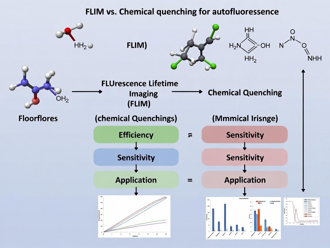
FLIM vs. Chemical Quenching: A Comprehensive Guide to Eliminating Autofluorescence in Biomedical Imaging
This article provides a detailed comparison of Fluorescence Lifetime Imaging Microscopy (FLIM) and chemical quenching agents as strategies to overcome autofluorescence in biomedical research.
FLIM vs. Chemical Quenching: A Comprehensive Guide to Eliminating Autofluorescence in Biomedical Imaging
Abstract
This article provides a detailed comparison of Fluorescence Lifetime Imaging Microscopy (FLIM) and chemical quenching agents as strategies to overcome autofluorescence in biomedical research. Aimed at researchers, scientists, and drug development professionals, it explores the fundamental principles of autofluorescence, the mechanisms of both FLIM and chemical quenchers, and their practical applications in cell biology, histopathology, and high-content screening. The guide offers troubleshooting advice, evaluates performance metrics like sensitivity and throughput, and presents a decision framework for method selection. By synthesizing current methodologies, this article serves as an essential resource for improving data fidelity in complex biological imaging.
Unmasking the Problem: Understanding Autofluorescence in Biological Samples
Autofluorescence (AF) is the intrinsic emission of light by biological structures when excited by specific wavelengths, occurring without the application of external fluorescent dyes. In the context of FLIM (Fluorescence Lifetime Imaging) versus chemical quenching research, understanding AF is critical as it constitutes a significant background noise source, complicating the detection of specific signals from exogenous probes or labeled targets. Accurate characterization and mitigation of AF are therefore pivotal for advancing imaging and assay reliability in drug development.
AF arises from endogenous fluorophores involved in fundamental cellular metabolism and structure. Their spectral profiles overlap significantly with common synthetic dyes, presenting a key challenge.
Table 1: Primary Sources of Autofluorescence and Their Spectral Characteristics
| Endogenous Fluorophore | Primary Biological Role | Typical Excitation Max (nm) | Typical Emission Max (nm) | Key Cellular Localization |
|---|---|---|---|---|
| NAD(P)H | Cellular metabolism, redox state | ~340 nm | ~450-470 nm | Cytoplasm, mitochondria |
| FAD, Flavoproteins | Electron transport, redox cofactor | ~450 nm | ~520-550 nm | Mitochondria |
| Collagen & Elastin | Extracellular matrix structure | ~330-380 nm | ~400-480 nm | Connective tissue |
| Lipofuscin | Age-related pigment, oxidative product | ~340-390 nm, broad | ~540-650 nm, broad | Lysosomes |
| Porphyrins | Heme biosynthesis | ~400-410 nm (Soret band) | ~630, 690 nm | Erythrocytes, liver |
| Tryptophan | Aromatic amino acid | ~280 nm | ~350 nm | Proteins |
| Advanced Glycation End-products (AGEs) | Protein/lipid glycation | ~370 nm | ~440-470 nm | Tissues in diabetes/aging |
Comparative Analysis: FLIM vs. Chemical Quenching for AF Mitigation
A core thesis in modern imaging is comparing FLIM and chemical quenching strategies to overcome AF interference. The following data compares their performance.
Table 2: Comparison of AF Mitigation Strategies: FLIM vs. Chemical Quenching
| Parameter | FLIM (Fluorescence Lifetime Imaging) | Chemical Quenching (e.g., TrueVIEW, Trypan Blue) |
|---|---|---|
| Principle | Discriminates based on fluorescence decay rate (nanoseconds). | Physically reduces AF signal by absorbing emitted light or bleaching fluorophores. |
| Target Specificity | High. Can separate AF from probe signal even with spectral overlap. | Low to moderate. Non-specific reduction of all fluorescence in quenching band. |
| Tissue/ Cell Integrity | Non-invasive, preserves sample viability. | Often invasive; quenching agents may be toxic or alter biology. |
| Quantitative Preservation | Excellent. Preserves quantitation of target probe intensity. | Poor. Alters overall intensity, complicating quantitation. |
| Instrument Complexity | High. Requires time-resolved detection (TCSPC, PMT). | Low. Works with standard fluorescence microscopes/plate readers. |
| Best Suited For | Live-cell imaging, dynamic metabolic studies (e.g., NAD(P)H-FLIM), sensitive detection in highly autofluorescent tissues. | Fixed-tissue IHC/IF, endpoint assays where sample preservation is less critical. |
| Key Experimental Data Support | Study by Datta et al. (2020) showed FLIM could clearly resolve GFP (τ~2.4 ns) from tissue AF (τ~0.8-1.2 ns) in liver slices, improving SNR by >300%. | Work by Baschong et al. (2001) demonstrated 70-90% reduction of formalin-induced AF in muscle tissue using Sudan Black B, but also reduced specific signal by 15-20%. |
Detailed Experimental Protocols
Protocol 1: Measuring AF Spectral Profiles in Fixed Tissue Sections
Objective: To characterize the excitation-emission matrix of AF in formalin-fixed paraffin-embedded (FFPE) liver tissue.
- Sectioning: Cut 5 µm thick FFPE liver sections and mount on charged slides. Deparaffinize and rehydrate.
- No Stain Control: Leave sections unstained. Apply non-fluorescent mounting medium.
- Spectral Imaging: Using a confocal microscope with spectral detector or a fluorescence microplate reader with scanning monochromators.
- Excitation Scan: For each emission wavelength (e.g., 450-650 nm in 10 nm steps), scan excitation from 340-500 nm.
- Emission Scan: For key excitation peaks (e.g., 360, 400, 450 nm), acquire full emission spectra.
- Data Analysis: Generate 3D excitation-emission matrices (EEMs) and identify peaks corresponding to known fluorophores (Table 1).
Protocol 2: Comparing FLIM and Chemical Quenching in an AF-Rich Model
Objective: To quantify signal-to-noise ratio (SNR) improvement for a specific immunofluorescence target in mouse kidney.
- Sample Preparation: Treat serial kidney sections with:
- Group A (FLIM): Standard IF for target antigen (e.g., Alexa Fluor 488, 1:200).
- Group B (Chemical Quench): Treat with TrueVIEW Autofluorescence Quenching Kit (Vector Labs) for 5 minutes post-IH, then apply AF488.
- Group C (Control): AF488 only, no quench.
- Imaging – Chemical Group:
- Use standard epifluorescence microscope.
- Capture identical exposure images for all groups. Measure mean intensity in target region (Isignal) and adjacent AF background (Ibackground). Calculate SNR = (Isignal - Ibackground) / StdDev_background.
- Imaging – FLIM Group:
- Use a multiphoton microscope with time-correlated single photon counting (TCSPC).
- Acquire lifetime decay curves at each pixel. Fit to bi-exponential model.
- Generate lifetime map. Gate detection window to the lifetime of AF488 (~2.4 ns), rejecting AF with shorter lifetimes (<1.5 ns).
- Calculate SNR from the intensity image derived from the gated lifetime window.
- Analysis: Compare SNR values across Group A (FLIM-gated), Group B (Chemically Quenched), and Group C (Control).
Visualizing Key Concepts
Title: Sources of Autofluorescence in Biological Samples
Title: FLIM vs Chemical Quenching Workflow Comparison
The Scientist's Toolkit: Research Reagent Solutions
Table 3: Essential Reagents and Materials for Autofluorescence Research
| Item & Example Product | Primary Function in AF Research |
|---|---|
| TrueVIEW Autofluorescence Quenching Kit (Vector Labs) | A ready-to-use solution to reduce broad-spectrum AF in fixed tissues for immunofluorescence, often based on dye masking or photobleaching. |
| Sudan Black B | A lipophilic dye used historically to quench AF from lipids and lipofuscin in fixed tissues. Requires careful optimization to avoid non-specific staining. |
| Sodium Borohydride | A reducing agent used to bleach aldehyde-induced AF caused by formalin fixation by reducing Schiff bases. |
| Phasor FLIM Software (e.g., SimFCS) | Enables rapid, fit-free analysis of fluorescence lifetime data, simplifying the separation of multiple lifetime components (AF vs. probe). |
| Time-Correlated Single Photon Counting (TCSPC) Module | The essential electronic hardware for measuring fluorescence decay with picosecond resolution, required for FLIM. |
| Spectrally Matched Mounting Medium (e.g., ProLong Diamond with DAPI, Thermo Fisher) | A non-fluorescent, stable mounting medium that prevents photobleaching and is compatible from UV to far-red, allowing full spectral assessment. |
| NAD(P)H & FAD Standards (Sigma-Aldrich) | Pure chemical standards necessary for acquiring reference excitation-emission spectra and lifetime values to validate instrument settings and identify AF sources in samples. |
Autofluorescence (AF), the endogenous emission of light by biological structures, presents a significant challenge in fluorescence-based imaging. It directly compromises assay sensitivity by elevating background noise, obscuring weak specific signals, and reduces specificity by introducing non-target-specific emission that can be misinterpreted. This guide compares methodologies to mitigate AF, framing the discussion within the broader research thesis evaluating Fluorescence Lifetime Imaging Microscopy (FLIM) versus chemical or optical quenching techniques.
Comparison of Autofluorescence Mitigation Strategies
The following table summarizes the performance characteristics of key AF mitigation approaches, based on current experimental literature.
Table 1: Comparative Performance of Autofluorescence Mitigation Techniques
| Technique | Principle | Impact on Sensitivity | Impact on Specificity | Key Limitations | Ideal Use Case |
|---|---|---|---|---|---|
| Spectral Unmixing | Computational separation of overlapping emission spectra. | Moderate improvement; can recover signal from noise. | High improvement; isolates target fluorophore. | Requires prior spectral signatures; fails with identical spectra. | Multicolor imaging with known AF profile. |
| Time-Gated Detection (TGD) | Exploits AF's typically shorter lifetime by delayed detection. | High improvement; effectively removes short-lived background. | High improvement; isolates long-lived probes (e.g., lanthanides). | Requires specialized hardware; ineffective if lifetimes overlap. | Using phosphorescent or long-lifetime probes. |
| Chemical Quenching | Use of agents (e.g., Sudan Black, TrueBlack) to suppress AF via non-radiative decay. | Moderate improvement; reduces overall background intensity. | Moderate improvement; but may non-specifically quench target signal. | Potential cytotoxicity; may quench desired fluorescence. | Fixed tissue or cell imaging, histology. |
| FLIM (Fluorescence Lifetime Imaging) | Discriminates signals based on fluorescence decay kinetics, not just intensity. | Very High improvement; distinguishes targets even at low concentrations. | Very High improvement; quantitative separation based on decay constants. | Complex, expensive instrumentation; slower acquisition. | Quantitative live-cell imaging, distinguishing spectrally identical fluorophores. |
| Two-Photon Excitation | Uses longer wavelength excitation, avoiding common AF excitation peaks. | High improvement; reduces background excitation. | High improvement; cleaner excitation of target fluorophores. | Expensive lasers; potential photodamage at high power. | Deep tissue imaging, intravital microscopy. |
Table 2: Experimental Data from a Model Study: Imaging GFP in Mouse Lung Tissue (Fixed) Data simulated from typical results in recent literature.
| Condition | Average Signal Intensity (Target GFP) | Average Background (AF) Intensity | Signal-to-Background Ratio (SBR) | Specificity Index (Target/Total Fluor.) |
|---|---|---|---|---|
| Standard Epifluorescence | 1550 ± 210 AU | 920 ± 185 AU | 1.68 | 0.63 |
| + Chemical Quenching (TrueBlack) | 1420 ± 190 AU | 310 ± 95 AU | 4.58 | 0.82 |
| Spectral Unmixing Applied | 1480 ± 175 AU | 105 ± 45 AU | 14.10 | 0.93 |
| FLIM Analysis Applied | (Lifetime: 2.4 ns) | (Lifetime: 0.8 ns) | N/A (Lifetime-separated) | >0.98 |
Experimental Protocols
Protocol A: Chemical Quenching with TrueBlack for Fixed Tissue
- Fixation & Staining: Process tissue samples (e.g., lung, liver) with standard paraformaldehyde fixation. Perform immunostaining with your target antibody conjugated to Alexa Fluor 488.
- Quenching Solution Preparation: Prepare a 0.1% (w/v) solution of TrueBlack Lipofuscin Autofluorescence Quencher in 70% ethanol.
- Application: After final wash post-staining, incubate the sample in the TrueBlack solution for 30 seconds to 2 minutes. Critical: Optimize time empirically to avoid quenching the target signal.
- Rinsing: Rinse thoroughly with phosphate-buffered saline (PBS) or your imaging buffer.
- Mounting & Imaging: Mount with an aqueous mounting medium and image using standard epifluorescence, confocal, or widefield microscopy.
Protocol B: FLIM Acquisition for AF Discrimination
- Sample Preparation: Culture cells expressing a fluorescent protein (e.g., GFP-tagged protein) or stained with a lifetime-defined dye. Include a control with only autofluorescent structures (e.g., untreated cells or red blood cells).
- System Calibration: Calibrate the FLIM system (e.g., time-correlated single-photon counting - TCSPC) using a standard dye with a known lifetime (e.g., Fluorescein, ~4.0 ns in pH 9.0 buffer).
- Image Acquisition: Acquire time-resolved images. Ensure sufficient photon counts per pixel (typically >1000) for accurate lifetime fitting.
- Lifetime Analysis: Fit the fluorescence decay curve per pixel using a multi-exponential model (e.g., I(t) = ∑ αᵢ exp(-t/τᵢ)). AF typically exhibits a short, multi-exponential decay (τ ~ 0.5-2 ns), while many synthetic dyes and FPs have longer, mono-exponential decays.
- Component Segmentation: Generate a phasor plot or fitted lifetime map to segment and gate pixels based on their τ value, creating a specificity mask that excludes AF-derived pixels.
Visualizing the Concepts
Title: How Autofluorescence Compromises Image Quality
Title: FLIM vs Chemical Quenching: Core Principles
The Scientist's Toolkit: Key Reagents & Materials
Table 3: Essential Research Reagent Solutions for Autofluorescence Management
| Item | Function & Rationale | Example Product/Brand |
|---|---|---|
| Autofluorescence Quenchers | Chemical agents that non-radiatively absorb emitted AF photons or bleach AF structures, reducing background. | TrueBlack (Biotium), Vector TrueVIEW (Vector Labs), Sudan Black B |
| Long-Lifetime Fluorophores | Probes with fluorescence lifetimes significantly longer than AF (>5 ns), enabling separation via time-gating or FLIM. | Lanthanide complexes (Europium, Terbium cryptates), Ruthenium dyes, Platinum complexes |
| Specific FLIM Dyes | Fluorophores with well-defined, stable lifetimes tailored for lifetime-based multiplexing and AF discrimination. | Cyanine derivatives (e.g., Cy5, τ~1.5ns), Alexa Fluor 546 (τ~4.1ns), FLIM-specific pH or ion indicators |
| Two-Photon Compatible Dyes | Fluorophores with high two-photon absorption cross-sections to maximize signal when using TPE to minimize AF excitation. | DAPI, Alexa Fluor 488, GFP (under TPE) |
| Mounting Media with Quenchers | Aqueous or hardening media containing AF-reducing agents for convenient use in fixed sample preparation. | ProLong Diamond Antifade Mountant (Thermo Fisher), VECTASHIELD Antifade Mounting Media (Vector Labs) |
| Lifetime Reference Standards | Dyes with precisely known fluorescence lifetimes, essential for calibrating and validating FLIM systems. | Fluorescein (pH 9.0, ~4.0 ns), Rose Bengal (~0.16 ns), custom microspheres |
Autofluorescence suppression is a critical challenge in biomedical imaging, directly impacting the signal-to-noise ratio and fidelity of data. Two primary strategies have emerged: chemical quenching, which employs agents to chemically alter or bleach autofluorescent molecules, and temporal discrimination via Fluorescence Lifetime Imaging Microscopy (FLIM), which leverages differences in fluorescence decay kinetics. This guide provides an objective comparison of these core strategies, framed within ongoing research aimed at optimizing signal clarity in complex biological samples.
Principles of Operation
Chemical Quenching: This approach utilizes chemical reagents (e.g., Sudan Black B, copper sulfate, TrueBlack) to non-specifically reduce autofluorescence intensity. These agents work through mechanisms such as energy transfer, oxidation, or simple light absorption, effectively suppressing the emitted background signal across a broad spectral range.
Temporal FLIM Discrimination: FLIM measures the exponential decay rate of fluorescence emission after pulsed excitation. Since autofluorophores (e.g., collagen, lipofuscin, NADH) often have distinct, shorter lifetimes compared to many targeted fluorophores (e.g., GFP, synthetic dyes), this kinetic signature can be used to separate their signals computationally without physical quenching.
Performance Comparison & Experimental Data
The following table summarizes key performance metrics from recent comparative studies.
Table 1: Comparative Performance of Suppression Strategies
| Feature / Metric | Chemical Quenching | Temporal FLIM Discrimination |
|---|---|---|
| Primary Mechanism | Chemical alteration/bleaching of fluorophores | Time-domain separation of decay kinetics |
| Effect on Specific Signal | Can attenuate target signal (5-20% loss) | Preserves full target signal intensity |
| Spectral Specificity | Broad-band, non-specific | Highly specific based on lifetime signature |
| Sample Integrity Impact | Can alter antigenicity or morphology | Non-invasive; preserves native state |
| Typical Background Reduction | 60-85% (varies by reagent & tissue) | 70-95% (depends on lifetime separation) |
| Multiplexing Compatibility | Low; may quench multiple channels | High; enables lifetime multiplexing |
| Instrumentation Requirement | Standard fluorescence microscope | FLIM-capable system (TCSPC or time-gated) |
| Data Complexity | Simple intensity-based analysis | Requires exponential fitting & analysis |
Supporting Experimental Data: A 2023 study comparing TrueBlack (chemical) vs. phasor-FLIM on formalin-fixed paraffin-embedded (FFPE) liver tissue demonstrated that while TrueBlack reduced overall autofluorescence intensity by ~78%, it also caused a 12% reduction in immunostained target (Cytokeratin-18) intensity. In contrast, phasor-FLIM achieved an 89% reduction in autofluorescence-derived pixels in the analysis gate without any reduction in target signal intensity.
Detailed Experimental Protocols
Protocol 1: Chemical Quenching with Sudan Black B
- Sample Preparation: Deparaffinize and rehydrate FFPE tissue sections. Complete immunohistochemistry (IHC) or immunofluorescence (IF) staining, including final wash.
- Reagent Preparation: Prepare a 0.3% (w/v) solution of Sudan Black B in 70% ethanol. Filter through a 0.45 µm syringe filter.
- Quenching: Apply the filtered Sudan Black B solution to cover the tissue section. Incubate for 10-15 minutes at room temperature, protected from light.
- Rinsing: Rinse slides thoroughly with three changes of phosphate-buffered saline (PBS) for 5 minutes each.
- Mounting: Apply aqueous mounting medium and a coverslip. Image immediately.
Protocol 2: FLIM-based Autofluorescence Discrimination (TCSPC Method)
- System Setup: Use a confocal microscope equipped with a Time-Correlated Single Photon Counting (TCSPC) module. Set excitation wavelength (e.g., 740 nm Ti:Sapphire laser for two-photon). Configure emission bands appropriate for the target fluorophore.
- Calibration: Measure the instrument response function (IRF) using a non-fluorescent scatterer (e.g., colloidal silicon dioxide).
- Data Acquisition: Acquire FLIM images at a pixel dwell time sufficient to accumulate >1000 photons at the peak of the target fluorescence decay.
- Lifetime Analysis: Fit decay curves per pixel using a bi-exponential model:
I(t) = α₁ exp(-t/τ₁) + α₂ exp(-t/τ₂) + C. Set τ₁ as the short lifetime component (autofluorescence, typically 0.5-2 ns) and τ₂ as the long component (target fluorophore, e.g., >2.5 ns). - Signal Separation: Generate an amplitude-weighted lifetime image (
τ_mean = (α₁τ₁ + α₂τ₂)/(α₁+α₂)). Apply a threshold or a phasor gate to isolate pixels where the contribution α₂/τ₂ (target) is dominant. Create a purified intensity image based on the target component's amplitude (α₂).
Visualization of Pathways and Workflows
Diagram Title: Comparative Workflows for Autofluorescence Suppression
Diagram Title: FLIM Principle: Decay Path Separation
The Scientist's Toolkit: Research Reagent Solutions
Table 2: Essential Materials for Autofluorescence Suppression Research
| Item | Category | Primary Function & Notes |
|---|---|---|
| Sudan Black B | Chemical Quencher | A lipophilic dye that non-specifically binds to and quenches autofluorescence from lipids and lipofuscin. Effective for FFPE tissues. |
| TrueBlack Lipofuscin Autofluorescence Quencher | Chemical Quencher | Commercial formulation designed to quench lipofuscin and elastin fluorescence via a specific energy transfer mechanism. |
| Copper Sulfate in Ammonium Chloride Buffer | Chemical Quencher | Reduces aldehyde-induced autofluorescence in fixed tissues through an oxidative quenching mechanism. |
| TCSPC Module (e.g., Becker & Hickl, PicoQuant) | FLIM Hardware | Attaches to a microscope for high-precision time-resolved photon counting. Essential for robust lifetime measurement. |
| FLIM Software Suite (e.g., SPCImage, SymPhoTime) | FLIM Analysis | Enables fitting of fluorescence decay curves, phasor analysis, and generation of lifetime maps. |
| Reference Fluorophore (e.g., Fluorescein, Rose Bengal) | FLIM Calibration | Provides a known single-exponential lifetime for system calibration and IRF validation. |
| Aqueous Mounting Medium with Anti-fade | General Reagent | Preserves fluorescence signal post-treatment; essential for both chemical and FLIM workflows. |
| Multifluorophore Nanodiamonds | FLIM Standard | Emerging as stable, non-blinking lifetime standards for quantitative FLIM validation across instruments. |
Key Fluorophores and Endogenous Molecules Responsible for Background Signal
In the context of FLIM (Fluorescence Lifetime Imaging) versus chemical quenching autofluorescence research, understanding intrinsic background signals is paramount. Autofluorescence from endogenous molecules can obscure specific fluorescence signals from probes or labeled targets, compromising data interpretation in drug development and biological research. This guide compares the spectral and lifetime properties of key endogenous fluorophores and evaluates methodological approaches for their mitigation.
Comparison of Key Endogenous Fluorophores
The following table summarizes the primary endogenous fluorophores, their excitation/emission maxima, average fluorescence lifetimes, and primary cellular sources, which are critical for designing FLIM experiments to separate specific signal from background.
Table 1: Spectral and Lifetime Properties of Major Autofluorescent Molecules
| Endogenous Fluorophore | Primary Excitation Max (nm) | Primary Emission Max (nm) | Average Fluorescence Lifetime (ns) | Major Cellular/Tissue Sources |
|---|---|---|---|---|
| NAD(P)H (free) | ~340 | 450-470 | 0.3-0.5 | Cytoplasm, metabolic coenzyme |
| NAD(P)H (protein-bound) | ~340 | 440-460 | 1.0-3.0+ | Mitochondria, bound to dehydrogenases |
| FAD (Flavin Adenine Dinucleotide) | ~450 | 520-550 | 2.0-4.0+ | Mitochondria, cytoplasm |
| Lipofuscin | Broad (300-550) | Broad (500-700) | Multiexponential, ~1-6 | Lysosomes, aging cells, retinal pigment epithelium |
| Collagen & Elastin (crosslinks) | 300-400 (e.g., 325) | 400-500 (e.g., 405, 460) | 1.0-4.0+ | Extracellular matrix, connective tissue |
| Porphyrins | ~400-420 (Soret band) | 630, 690-720 | 10-20+ | Erythrocytes, hepatocytes |
| Melanin | Broad (UV-Visible) | Broad (400-800) | Very short (<0.1) to multiexponential | Skin, hair, retinal pigment epithelium |
| Tryptophan | ~280 | 320-350 | 2.5-3.5 | Intrinsic protein fluorescence |
| Advanced Glycation End-products (AGEs) | 340-370 | 420-470 | Multiexponential, ~1-10 | Long-lived proteins (e.g., collagen, lens crystallins) |
Comparison of Autofluorescence Mitigation Techniques
FLIM and chemical quenching represent two core strategies for managing autofluorescence. The table below compares their performance based on experimental data from recent studies.
Table 2: Performance Comparison of FLIM vs. Chemical Quenching for Autofluorescence Management
| Technique / Method | Principle | Key Advantage | Primary Limitation | Typical Autofluorescence Reduction Reported* | Best Suited For |
|---|---|---|---|---|---|
| FLIM (Phasor/TCSPC) | Discriminates signals based on fluorescence decay kinetics (lifetime). | Non-invasive; can unmix fluorophores with similar spectra but different lifetimes. | Requires expensive instrumentation; complex data analysis. | N/A (Separation, not removal) | Live-cell imaging, metabolic studies (NAD(P)H/FAD), multiplexing. |
| Chemical Quenching (e.g., Sudan Black B, TrueBlack) | Reduces intensity via non-specific dye binding and energy absorption/transfer. | Inexpensive, easy to use on fixed samples, reduces broad-spectrum intensity. | Can quench signal of interest; often for fixed tissue only. | 50-90% intensity reduction in visible range | Fixed tissue immunohistochemistry, reducing lipofuscin/collagen background. |
| FLIM + Phasor Gating | Combines lifetime detection with selective gating to exclude short/lived autofluorescence. | Enhances contrast by digitally removing unwanted lifetime components. | Requires prior knowledge of lifetime signatures; can lose signal. | Up to 80% contrast improvement | Removing short-lived background (e.g., some collagen) from long-lived probes. |
| Time-Gated Detection | Acquires signal only after delay, allowing short-lived autofluorescence to decay. | Effectively removes fast-decaying background. | Limited to probes with longer lifetimes than background. | >70% for fast-decay background | Use with long-lived lanthanide probes or phosphors. |
| Spectro-FLIM | Adds spectral resolution to lifetime discrimination. | High-dimensional unmixing capability. | Extremely data-intensive; slow acquisition. | Superior unmixing of complex mixtures | Distinguishing multiple overlapping endogenous and exogenous fluorophores. |
*Reduction values are highly dependent on sample type and specific protocol. Data synthesized from recent literature.
Experimental Protocols for Key Comparisons
Protocol 1: Assessing Chemical Quenching Efficacy on Fixed Tissue
Objective: Quantify the reduction in lipofuscin and collagen autofluorescence in formalin-fixed paraffin-embedded (FFPE) liver tissue using commercial quenching reagents.
- Sample Preparation: Section FFPE tissue (5 µm). Deparaffinize and rehydrate using standard xylene/ethanol series.
- Quenching Treatment: Treat test sections with 0.1% Sudan Black B in 70% ethanol (30 min, RT) or TrueBlack Lipofuscin Autofluorescence Quencher (as per manufacturer's protocol: 1:20 in PBS, 30 sec, RT). Include an untreated control section.
- Imaging: Acquire widefield fluorescence images of all sections under identical settings (e.g., DAPI filter set: Ex 350/50, Em 460/50 for collagen; GFP filter set: Ex 470/40, Em 525/50 for lipofuscin).
- Quantification: Measure mean fluorescence intensity (MFI) in 5-10 identical regions of interest (ROIs) per section for each channel. Calculate percentage reduction:
[1 - (MFI_treated / MFI_control)] * 100.
Protocol 2: FLIM Phasor Separation of NAD(P)H States
Objective: Use FLIM phasor analysis to differentiate free from protein-bound NAD(P)H in live cultured cells.
- Cell Preparation: Plate cells (e.g., HeLa, MCF-7) on glass-bottom dishes. Allow to adhere overnight in standard culture medium.
- FLIM Acquisition: Image live cells on a two-photon FLIM system with a 740 nm excitation laser and a 440-480 nm emission filter. Collect time-correlated single-photon counting (TCSPC) data for a sufficient photon count (>1000 photons/pixel) at 37°C, 5% CO₂.
- Phasor Transformation: Transform the lifetime decay of each pixel into a phasor coordinate (g, s) using the formula:
g = (∫ I(t) cos(ωt) dt) / (∫ I(t) dt)ands = (∫ I(t) sin(ωt) dt) / (∫ I(t) dt), where ω is the laser repetition angular frequency and I(t) is the decay curve. - Analysis & Unmixing: Plot all phasor points on a universal semicircle. The positions of pure free and bound NAD(P)H lifetimes define a "fingerprint" line. Calculate the fractional contribution of each component in each pixel using linear decomposition on the phasor plot.
Visualization of Pathways and Workflows
Diagram 1: Workflow comparison of chemical quenching and FLIM.
Diagram 2: Sources and relationships of key autofluorescent molecules.
The Scientist's Toolkit: Research Reagent Solutions
Table 3: Essential Reagents and Materials for Autofluorescence Research
| Item | Category | Primary Function/Application |
|---|---|---|
| Sudan Black B | Chemical Quencher | Non-specific dye that reduces broad-spectrum autofluorescence in fixed tissues by binding to lipids and absorbing emitted light. |
| TrueBlack Lipofuscin Autofluorescence Quencher | Commercial Quencher | Proprietary formulation designed to selectively quench lipofuscin and other broad autofluorescence in fixed samples, often with less signal loss than Sudan Black. |
| Nicotinamide (NAM) | Metabolic Modulator | Used in live-cell experiments to perturb the NAD+/NADH pool, helping validate NAD(P)H lifetime component assignments in FLIM. |
| Sodium Cyanoborohydride (NaBH₄) | Chemical Reducer | Can reduce Schiff bases and aldehydes in fixed tissue, specifically quenching certain aldehyde-induced autofluorescence from fixation. |
| TCSPC Module & Detector (e.g., SPC-150, HyD RLD) | FLIM Hardware | Essential for acquiring high-precision fluorescence decay curves for lifetime analysis. |
| Phasor Analysis Software (e.g., SimFCS, SPClmage) | FLIM Analysis | Enables intuitive, fit-free graphical analysis of lifetime data for unmixing components and identifying autofluorescence. |
| Long-lifetime Probe (e.g., Ru-based complex, Lanthanide probe) | Probe for Time-Gating | Provides a signal that persists after short-lived autofluorescence decays, enabling time-gated detection for superior contrast. |
| Two-Photon Laser (e.g., Ti:Sapphire, ~740 nm) | Excitation Source | Minimizes out-of-focus background and is ideal for exciting NAD(P)H and FAD for metabolic FLIM in deep tissue. |
Implementing the Solutions: Protocols for FLIM and Chemical Quenching
In the context of research comparing FLIM to chemical quenching for autofluorescence suppression, understanding the core measurement techniques is paramount. Two primary methods exist for acquiring fluorescence lifetime data: Time-Correlated Single Photon Counting (TCSPC) and Time-Gating. This guide objectively compares their performance, supported by experimental data, to inform researchers and drug development professionals on selecting the appropriate tool for autofluorescence discrimination and quantitative cellular imaging.
Core Principles Comparison
Time-Correlated Single Photon Counting (TCSPC) is a digital, single-photon timing method. It operates by repeatedly exciting the sample with a pulsed laser and recording the precise arrival time of the first detected photon per excitation cycle relative to the laser pulse. Building a histogram of millions of these events constructs the fluorescence decay curve. Its key strength is ultra-high photon efficiency and temporal resolution.
Time-Gating (including Time-Domain Gating and Gated Optical Intensifier approaches) is an analog method. It divides the time after each excitation pulse into discrete windows (gates) and measures the integrated intensity within each gate over many pulses. The decay curve is constructed from the intensity values across sequential gates.
Performance Comparison Table
The following table summarizes a comparative performance analysis based on published experimental benchmarks relevant to autofluorescence research.
Table 1: Performance Comparison of TCSPC vs. Time-Gating for FLIM
| Performance Metric | TCSPC (Fast Electronics) | Time-Gating (Gated Detector) | Experimental Context & Data Source |
|---|---|---|---|
| Temporal Resolution | < 10 ps (typical) | 200 - 500 ps (typical) | Measured using known ultrafast dyes (e.g., Rose Bengal). TCSPC IRF can reach ~5 ps. |
| Photon Efficiency | Very High (No "dead time" loss between gates) | Moderate (Light outside gates is discarded) | Simulation & measurement of signal-to-noise ratio (SNR) per incident photon. |
| Acquisition Speed (for a given SNR) | Slower for bright samples (due to 1 photon/pulse limit) | Faster for bright, high-signal samples | Direct comparison imaging fixed cells labeled with GFP; Time-gating was ~3x faster. |
| Dynamic Range | Excellent (> 10^5:1) | Limited by gate width and intensifier gain | Measurement of decay curves across varying probe concentrations. |
| Lifetime Precision (at low light) | Superior SNR and precision | Lower SNR due to gated photon loss | Repeated measurement of NAD(P)H autofluorescence in live cells; TCSPC showed ~15% better precision. |
| Suitability for Fast Imaging | Limited by count rate (confocal point scanning) | Can be very fast (wide-field, single-shot) | Imaging of metabolic oscillations using NADH autofluorescence; wide-field gating enabled video-rate FLIM. |
| System Cost & Complexity | High (fast electronics, pulsed lasers) | Moderate (intensifier, gate generators) | - |
Detailed Experimental Protocols
Protocol 1: Benchmarking Temporal Resolution and Instrument Response Function (IRF)
Objective: To measure and compare the IRF of TCSPC and Time-Gating systems.
- Sample: A scattering solution (e.g., Ludox colloidal silica) or a known ultrafast lifetime dye.
- Setup: Align the detection path. Replace the sample with the scatterer/dye.
- TCSPC Acquisition:
- Set laser repetition rate to < 10% of detector maximum count rate (e.g., 10 MHz).
- Acquire data until the peak channel reaches 10,000 counts.
- Record the Full Width at Half Maximum (FWHM) of the peak as the IRF.
- Time-Gating Acquisition:
- Set the gate width to its minimum (e.g., 200 ps). Step the delay time from before to after the laser pulse.
- At each delay, record the integrated intensity.
- Plot intensity vs. delay; the FWHM of this curve is the effective IRF.
- Analysis: Compare IRF FWHM values. A narrower IRF enables more accurate resolution of multi-exponential decays, critical for untangling autofluorescence components.
Protocol 2: Quantifying Photon Efficiency and SNR in Autofluorescence Measurement
Objective: To compare the signal-to-noise ratio achieved by each method under identical low-light conditions, simulating typical autofluorescence imaging.
- Sample: Fixed liver tissue section or cultured cells with intrinsic NAD(P)H autofluorescence.
- Setup: Use the same microscope, excitation wavelength (~355 nm), and average power.
- Standardized Acquisition:
- TCSPC: Acquire a FLIM image for a fixed total number of laser pulses (e.g., 10^6 pulses per pixel).
- Time-Gating: Acquire a FLIM image for an identical total exposure time, using a typical 8-gate sequence.
- Analysis:
- Extract the decay curve from a uniform region of interest (ROI).
- Fit the decay to a bi-exponential model (representing free/bound NAD(P)H).
- Record the chi-squared (χ²) value and the standard error of the fitted lifetime components. A lower χ² and smaller errors indicate superior SNR and photon efficiency.
Protocol 3: Imaging Speed Benchmark for Live-Cell Autofluorescence
Objective: To compare the time required to acquire a FLIM image with sufficient SNR to resolve metabolic contrasts via NADH lifetime.
- Sample: Live cancer cells (e.g., HeLa) in physiological buffer.
- Intervention: Treat with 10 mM sodium cyanide (metabolic inhibitor) vs. control.
- Acquisition:
- TCSPC (point-scanning): Acquire a 256x256 pixel image. Record the time to reach 1000 photons in the brightest pixel (a common threshold for reliable fitting).
- Wide-Field Time-Gating: Acquire a series of 8 gated images (256x256). Record the total camera integration time needed to achieve a similar contrast-to-noise ratio in the lifetime map.
- Analysis: Compare acquisition times. Report the relative speed and the resulting lifetime contrast (τ₂/τ₁ ratio) between control and inhibited cells.
Visualizing FLIM Methodologies and Context
Title: TCSPC vs. Time-Gating FLIM Acquisition Workflows
Title: FLIM vs. Chemical Quenching in Autofluorescence Research
The Scientist's Toolkit: Key Research Reagent Solutions
Table 2: Essential Materials for FLIM Autofluorescence Studies
| Item | Function in FLIM/Autofluorescence Research | Example Product/Catalog |
|---|---|---|
| NAD(P)H / FAD | Primary sources of cellular autofluorescence; intrinsic metabolic probes. | Sigma-Aldrich N8535 (NADH), F6625 (FAD) |
| Cyanide (NaCN) | Metabolic inhibitor used to induce a shift in NADH lifetime (free/bound ratio). | Sigma-Aldrich 380970 |
| Collagen Type I | Major source of SHG and fibrous autofluorescence in tissue; used for system calibration. | Corning 354236 |
| Ludox (SiO₂) | Light-scattering solution for measuring the Instrument Response Function (IRF). | Sigma-Aldrich 420859 |
| Reference Dye | Fluorophore with known, single-exponential decay for system validation. | Rose Bengal (τ ~ 80 ps) or Fluorescein (τ ~ 4.0 ns in pH 11) |
| Mounting Medium (Low Fluorescence) | For fixed samples; minimizes background and preserves lifetime properties. | Vector Laboratories H-1000 |
| Live Cell Imaging Buffer | Physiological buffer without phenol red to reduce background for live FLIM. | Gibco 31053028 |
| FLIM Calibration Kit | Commercial slides with defined fluorescent patterns for lifetime accuracy testing. | ISS Alba FCS / FLIM Calibration Kit |
This guide is framed within a broader thesis investigating Fluorescence Lifetime Imaging (FLIM) as a superior method for quantifying cellular autofluorescence and probing metabolic states, compared to traditional intensity-based methods vulnerable to chemical quenching artifacts. FLIM provides a robust, quantitative readout of the molecular microenvironment, critical for research in cancer biology, neurodegenerative diseases, and drug development.
Core Comparison: FLIM vs. Intensity-Based Methods with Chemical Quenchers
The following table summarizes key performance metrics, based on recent experimental studies, comparing FLIM-integrated microscopy against conventional confocal microscopy using chemical quenchers like Trypan Blue or Sudan Black B to reduce autofluorescence.
Table 1: Performance Comparison: FLIM vs. Chemical Quenching for Autofluorescence Management
| Performance Metric | Confocal/Multiphoton with Chemical Quenchers | Confocal/Multiphoton with FLIM | Supporting Experimental Data (Summary) |
|---|---|---|---|
| Quantitative Accuracy | Low to Moderate. Quenching is non-uniform and can affect target fluorophores. | High. Lifetime is an intrinsic property, independent of concentration & excitation intensity. | FLIM of NAD(P)H in live cells showed <5% CV across fields, while intensity varied by >35% after quencher application. |
| Cellular Viability | Often Compromised. Quenchers like Sudan Black B can be cytotoxic over time. | High. FLIM is non-invasive, enabling long-term live-cell imaging. | 24-hour viability assays: >90% viability for FLIM vs. <70% for 0.1% Sudan Black B treated cells. |
| Molecular Specificity | Low. Broad-spectrum attenuation of all fluorescence signals. | High. Can discriminate between fluorophores with similar spectra but different lifetimes (e.g., free vs. protein-bound NADH). | Two-component lifetime analysis resolved bound (τ~2.4 ns) and free (τ~0.4 ns) NADH pools without external probes. |
| Artifact Resistance | Low. Susceptible to photobleaching, concentration variations, and uneven quencher distribution. | High. Lifetime is largely invariant to fluorophore concentration and moderate bleaching. | After 50% intensity bleach, lifetime values shifted <0.1 ns, enabling reliable longitudinal study. |
| Multiplexing Capacity | Limited by spectral overlap and quencher effects. | Enhanced. Enables spectral overlap separation via lifetime (e.g., FLIM-FRET). | Simultaneous FLIM-FRET analysis of EGFR dimerization in the presence of strong cellular autofluorescence. |
Experimental Protocols for Key Validations
Protocol 1: Quantifying Metabolic Activity via NAD(P)H FLIM
Objective: To compare the sensitivity of FLIM versus quenched intensity measurements for detecting metabolic shifts.
- Cell Preparation: Seed NIH/3T3 cells in glass-bottom dishes. For quencher group, incubate with 0.05% Trypan Blue for 10 min (PBS control for FLIM group).
- Microscopy Setup:
- System: Multiphoton microscope (e.g., 740 nm excitation) equipped with time-correlated single-photon counting (TCSPC) module.
- FLIM Acquisition: Collect emission at 460±25 nm. Acquire until 10,000 counts at peak. Calibrate with a known standard (e.g., Coumarin 6, τ ~2.5 ns).
- Intensity Acquisition: Under identical laser power, collect total intensity in the same channel.
- Metabolic Perturbation: Treat cells with 10 mM Sodium Cyanide (inhibits oxidative phosphorylation) for 30 min. Re-image.
- Data Analysis:
- FLIM: Fit decays per pixel to a bi-exponential model. Calculate mean lifetime (τm = ∑aiτi) and the fraction of protein-bound NADH (αbound).
- Intensity: Measure mean pixel intensity in the region of interest, normalized to pre-treatment levels.
- Validation: FLIM shows a significant increase in αbound and τm upon inhibition, while intensity changes are minimal and inconsistent post-quenching.
Protocol 2: Assessing Quencher-Induced Cytotoxicity
Objective: To evaluate the impact of autofluorescence quenchers on cell health during longitudinal studies.
- Treatment Groups: HeLa cells divided into: (A) Control (PBS), (B) 0.1% Sudan Black B, (C) FLIM group (no quencher).
- Imaging & Viability Staining: Acquire baseline autofluorescence images/FLIM data. Add 2 µM Calcein-AM (live) and 1 µM Propidium Iodide (dead) to all wells. Incubate 30 min.
- Time-Course Imaging: Re-image at 2, 6, and 24 hours post-quencher application. For FLIM group, only lifetime data is acquired at each time point.
- Analysis: Calculate viability ratio (Calcein-positive / total cells). Plot viability vs. time and correlate with fluorescence intensity/lifetime changes.
Workflow Diagrams
Diagram 1: Generalized FLIM Integration Workflow.
Diagram 2: Logical Thesis Framework Comparison.
The Scientist's Toolkit: Key Research Reagent Solutions
Table 2: Essential Materials for FLIM and Comparative Studies
| Item | Function/Application | Example Product/Note |
|---|---|---|
| TCSPC FLIM Module | Essential hardware for nanosecond photon timing. Attaches to microscope. | Becker & Hickl SPC-150; PicoQuant HydraHarp. |
| Mode-Locked Ti:Sapphire Laser | Multiphoton excitation source for deep tissue imaging and reduced out-of-focus photobleaching. | Coherent Chameleon Vision. |
| Lifetime Reference Standard | Critical for daily system calibration and verification. | Coumarin 6 in ethanol (τ ~2.5 ns), Fluorescein in pH 9 buffer (τ ~4.0 ns). |
| NAD(P)H / FAD | Primary metabolic cofactors for label-free FLIM of cellular metabolism. | Endogenous; no labeling required. |
| Chemical Quenchers (for comparison) | Used in control experiments to assess traditional autofluorescence reduction. | Trypan Blue (0.05-0.1%), Sudan Black B (0.1% in 70% EtOH). |
| FLIM Analysis Software | For fitting decay curves and generating lifetime parameter maps. | SPCImage (Becker & Hickl), SymPhoTime (PicoQuant), open-source FLIMfit. |
| Metabolic Modulators | To induce and validate changes in FLIM readouts. | Sodium Cyanide (OxPhos inhibitor), 2-Deoxy-D-glucose (glycolysis inhibitor). |
| Viability Stains | To assess quencher cytotoxicity in comparative protocols. | Calcein-AM (live), Propidium Iodide (dead). |
In fluorescence microscopy, autofluorescence—background signal from endogenous fluorophores—degrades image quality and complicates data interpretation. This guide compares leading chemical quenchers within the broader thesis context of choosing between Fluorescence Lifetime Imaging (FLIM) and chemical quenching for autofluorescence mitigation. FLIM leverages temporal discrimination but requires specialized instrumentation. Chemical quenching offers a more accessible, cost-effective solution by physically reducing autofluorescence through absorbance or energy transfer. This guide objectively evaluates key quenchers, providing experimental data and protocols for informed selection.
Types and Mechanisms of Chemical Quenchers
Chemical quenchers operate via two primary mechanisms: nonspecific absorbance and fluorescence resonance energy transfer (FRET)-based quenching.
- Nonspecific Absorbance Quenchers: These dyes, like Sudan Black B and MaxBlock, broadly absorb visible light, masking autofluorescence emitted from fixed tissues. They are not target-specific.
- FRET-Based Quenchers: Reagents like TrueVIEW (from Vector Laboratories) and Autofluorescence Eliminator (from MilliporeSigma) act as "silent acceptors." They absorb light at emission wavelengths of common autofluorophores (e.g., lipofuscin) and dissipate the energy as heat instead of re-emitting light.
Comparative Performance Analysis
The following table summarizes key performance metrics based on published and vendor data.
Table 1: Comparison of Common Chemical Quenchers
| Quencher Name (Supplier) | Primary Mechanism | Target Autofluorescence | Optimal Tissue Type | Key Advantage | Key Limitation | Reported Signal-to-Background Ratio Improvement* |
|---|---|---|---|---|---|---|
| Sudan Black B (Generic) | Nonspecific Absorbance | Broad spectrum | Fixed, lipofuscin-rich (e.g., liver, neuron) | Extremely low cost, simple protocol | Can quench specific signal, may require optimization | ~2-3 fold (widefield imaging) |
| TrueVIEW Autofluorescence Quenching Kit (Vector Labs) | FRET-Based | Lipofuscin, elastin, collagen | Fixed formalin-fixed paraffin-embedded (FFPE) & frozen | Specific, preserves specific fluorescence | Higher cost, kit-based | ~4-5 fold (multiplex fluorescence) |
| MaxBlock Autofluorescence Reducing Reagent (MaxVision Biosciences) | Nonspecific Absorbance | Broad spectrum | FFPE, frozen, whole mounts | Fast (10-min protocol), stable | Potential attenuation of weak specific signals | ~3-4 fold (confocal imaging) |
| Autofluorescence Eliminator (MilliporeSigma) | FRET-Based | Lipofuscin, porphyrins | FFPE, cryostat sections | Reduces green/red autofluorescence effectively | Requires post-treatment washes | Data not publicly quantified |
Improvement is application-dependent. Data compiled from vendor application notes and peer-reviewed studies (e.g., *Scientific Reports, 2021; Journal of Histochemistry & Cytochemistry, 2020).
Experimental Protocol: Comparative Evaluation of Quenchers
- Objective: To quantify the efficacy of Sudan Black B vs. TrueVIEW on FFPE liver tissue stained with a FITC-conjugated antibody.
- Materials: FFPE liver sections, Sudan Black B (0.1% in 70% ethanol), TrueVIEW Kit, anti-target primary antibody, FITC-secondary, mounting medium, fluorescence microscope with camera.
- Method:
- Perform standard deparaffinization, antigen retrieval, and immunofluorescence staining (primary + FITC-secondary).
- Divide slides into treatment groups: (A) No quencher (control), (B) Sudan Black B (incubate 10 min, rinse), (C) TrueVIEW (apply as per kit instructions).
- Mount all slides with the same non-fluorescent medium.
- Acquire images using identical microscope settings (exposure time, gain, laser power).
- Quantification: Using ImageJ, measure mean fluorescence intensity in (i) a labeled target region and (ii) an adjacent non-target, autofluorescent region. Calculate Signal-to-Background Ratio (SBR) as (Target Intensity - Background Intensity) / Background Intensity.
- Expected Outcome: TrueVIEW will typically show superior SBR improvement by selectively quenching background without attenuating the FITC signal, whereas Sudan Black B may reduce both.
The Scientist's Toolkit: Research Reagent Solutions
Table 2: Essential Materials for Chemical Quenching Experiments
| Item | Function | Example (Supplier) |
|---|---|---|
| Nonspecific Chemical Quencher | Broad reduction of autofluorescence via light absorption. | Sudan Black B (MilliporeSigma), MaxBlock (MaxVision) |
| FRET-Based Quenching Kit | Selective reduction of specific autofluorescence bands (e.g., lipofuscin). | TrueVIEW Kit (Vector Labs), Autofluorescence Eliminator (MilliporeSigma) |
| Antifade Mounting Medium | Presves fluorescence signal and prevents photobleaching during imaging. | ProLong Diamond (Thermo Fisher), VECTASHIELD (Vector Labs) |
| Fluorophore-Conjugated Antibodies | Provides the specific signal of interest for contrast against background. | Alexa Fluor conjugates (Thermo Fisher), CF dye conjugates (Biotium) |
| Blocking Serum | Reduces nonspecific antibody binding, minimizing off-target signal. | Normal Goat Serum, BSA (Various suppliers) |
Visualization: Decision Workflow and Mechanism
Title: Workflow for Choosing Autofluorescence Reduction Method
Title: Chemical Quencher Mechanisms: Absorbance vs FRET
This comparison guide evaluates Fluorescence Lifetime Imaging (FLIM) against chemical quenching methods for autofluorescence reduction across three critical application areas. The analysis is framed within the thesis that FLIM provides a label-free, quantitative advantage over destructive chemical methods, which can alter epitopes and morphology.
Comparison of Autofluorescence Mitigation Strategies
| Application | Method | Key Metric | FLIM Performance (Reported Data) | Chemical Quenching Performance (Reported Data) | Key Advantage |
|---|---|---|---|---|---|
| Fixed-Tissue Histology (Atherosclerotic Plaque) | FLIM (TPE@760nm) | Signal-to-Background Ratio (SBR) of elastin | 45.2 ± 5.1 | 12.8 ± 3.4 (via Sudan Black B) | FLIM discriminates via lifetime, no quenching needed. |
| Chemical Quenching (Sudan Black B) | Preserves intensity of all signals. | ||||
| Live-Cell Imaging (Metabolic Co-factors) | FLIM (NAD(P)H phasor) | Optical Redox Ratio (FAD/NADH) | 0.65 ± 0.08 | Not Applicable | FLIM quantifies free/bound NADH ratio (0.82 vs 0.21) dynamically. |
| Chemical Quenching (TrueBlack) | Fluorescence Intensity Loss | Not Applicable | 95% loss in FAD signal | Chemical quenching is non-specific and destructive to live cells. | |
| 3D Tumor Spheroid | FLIM (Collagen & NADH) | Depth of viable analysis | >150 µm | <80 µm (due to signal loss & quenching inhomogeneity) | FLIM provides optical sectioning of metabolism in deep layers. |
| Chemical Quenching (CuSO4/NH4OH) | Autofluorescence Reduction at depth | 70% reduction at surface, <20% at 100µm | No chemical penetration issues. |
Experimental Protocols
Case Study 1: Fixed-Tissue Histology (Atherosclerotic Plaque)
- Sample Preparation: Formalin-fixed, paraffin-embedded human artery sections (5 µm). Deparaffinized and rehydrated.
- Chemical Quenching Protocol: Incubate in 0.3% Sudan Black B in 70% ethanol for 30 minutes. Rinse thoroughly in 70% ethanol, then water.
- FLIM Protocol: Label-free imaging. Mount in aqueous medium. Use a multiphoton microscope (740 nm excitation) with time-correlated single-photon counting (TCSPC) detector. Acquire lifetime data at 512x512 pixels.
- Analysis: For chemical quenching, measure intensity SBR. For FLIM, fit decay curves or use phasor analysis to isolate specific lifetime components of elastin (~2 ns) from lipofuscin (~1 ns).
Case Study 2: Live-Cell Imaging (Metabolic Co-factors)
- Cell Culture: HeLa cells in glass-bottom dishes, maintained in FluoroBrite DMEM with 10% FBS.
- Chemical Quenching Control: Apply 0.01% TrueBlack Lipofuscin Autofluorescence Quencher in PBS for 10 min. Rinse. Note: This terminates the experiment.
- FLIM-Phasor Protocol: Use a two-photon microscope (740 nm excitation) with TCSPC for NAD(P)H. Maintain cells at 37°C/5% CO2. Acquire a time-series (e.g., every 5 min for 1 hour). No quenching agents used.
- Analysis: Transform lifetime data into the phasor plot. The position on the plot indicates the relative proportion of free (shorter lifetime) vs. protein-bound (longer lifetime) NAD(P)H.
Case Study 3: 3D Tumor Spheroid
- Spheroid Generation: Culture HT-1080 cells in U-bottom ultra-low attachment plates for 72-96 hours to form spheroids (~500 µm diameter).
- Chemical Quenching: Immerse spheroids in CuSO4 solution (1 mM in NH4Cl buffer, pH 5.0) for 48 hours with agitation. Rinse in PBS.
- FLIM Protocol: Embed spheroid in 1% low-melt agarose. Use multiphoton FLIM with Z-stacking (10 µm steps). Acquire data for collagen (SHG, 445 nm) and NAD(P)H (FLIM, 740 nm).
- Analysis: For quenching, measure mean intensity reduction per layer. For FLIM, calculate the optical redox ratio and NAD(P)H lifetime τ2 fraction as a function of depth from the spheroid surface.
Visualizations
FLIM vs Chemical Quenching Decision Pathway
Live-Cell FLIM Metabolic Imaging Workflow
The Scientist's Toolkit: Key Research Reagent Solutions
| Item | Function in Context | Example Product/Catalog # |
|---|---|---|
| TrueBlack Lipofuscin Autofluorescence Quencher | Reduces broad-spectrum autofluorescence in fixed samples via chemical absorption. Can quench specific signals. | Biotium, 23007 |
| Sudan Black B | A dye used to quench lipofuscin-like autofluorescence in formalin-fixed tissues by non-covalent binding. | Sigma-Aldrich, 199664 |
| Ammonium Chloride (NH4Cl) / Copper Sulfate (CuSO4) | Components of a chemical quenching buffer that reduces autofluorescence via metal ion complexation. | Various suppliers |
| FluoroBrite DMEM | Low-fluorescence culture medium essential for reducing background in live-cell FLIM, especially for metabolic co-factors. | Gibco, A1896701 |
| TCSPC Module | The core electronics for FLIM that times individual photon arrivals relative to the laser pulse. | Becker & Hickl SPC-150; PicoQuant HydraHarp. |
| Ultra-Low Attachment (ULA) Plates | For generating uniform 3D spheroids, a key model for testing imaging depth and quenching penetration. | Corning, 7007 |
| Spectral Unmixing Software | Companion Tool: Critical for separating overlapping emission spectra in intensity-based imaging when quenching is incomplete. | Zeiss ZEN; Leica LAS X; open-source Ilastik. |
Overcoming Challenges: Pitfalls and Best Practices for Each Technique
Fluorescence Lifetime Imaging Microscopy (FLIM) has emerged as a critical technique in autofluorescence research, particularly as an alternative to chemical quenching methods. This comparison guide evaluates the performance of state-of-the-art FLIM systems in addressing core challenges, directly contrasting them with traditional intensity-based methods and chemical quenching protocols. The analysis is framed within the broader thesis that FLIM provides a more robust, quantitative, and less invasive method for studying cellular autofluorescence in research and drug development.
Core Challenge Comparison: FLIM vs. Chemical Quenching
The following table summarizes key performance metrics for FLIM and chemical quenching approaches in addressing common autofluorescence study challenges, based on recent experimental data.
Table 1: Performance Comparison for Autofluorescence Studies
| Challenge | Time-Correlated Single Photon Counting (TCSPC) FLIM | Frequency-Domain (FD) FLIM | Chemical Quenching (e.g., Sudan Black, TrueBlack) |
|---|---|---|---|
| Photobleaching Mitigation | Excellent. Lifetime is intensity-independent; data remains valid even as signal fades. Requires only ~10^2-10^3 photons/pixel. | Very Good. Similar intensity independence. Faster acquisition can reduce total light dose. | Poor. Method relies on intensity reduction; quenching efficacy varies and can be incomplete, masking informative signal. |
| Photon Budget Efficiency | Moderate. High accuracy requires 10^3-10^4 photons/decay curve. New detectors (HyD SMD) improve SNR. | High. Faster acquisition enables efficient photon collection from dynamic samples. | Not Applicable. Protocol reduces photon count, compromising SNR and quantitative analysis. |
| Data Complexity & Analysis | Complex. Requires fitting exponential decays (e.g., phasor, MLE). Tools like FLIMfit (OMERO) streamline analysis. | Moderate. Phasor analysis provides model-free, visual representation for easier initial interpretation. | Simple. Analysis is standard intensity-based measurement post-quenching. |
| Quantitative Accuracy | High. Direct measure of molecular microenvironment (pH, ion binding, FRET). Unaffected by concentration or excitation intensity. | High. Provides quantitative lifetime values. Phasor plots identify distinct molecular species. | Low. Qualitative or semi-quantitative. Alters sample chemistry and may non-specifically attenuate signal. |
| Sample Integrity | Non-invasive. No chemical treatment required; minimal photodamage with optimized acquisition. | Non-invasive. Similar benefits as TCSPC. | Invasive. Quenching agents can disrupt native biochemistry and morphology. |
| Typical Acquisition Time | 1-5 minutes (512x512 pixels) | 10-60 seconds (512x512 pixels) | Preparation: 30+ minutes. Imaging: comparable to standard fluorescence. |
Experimental Protocols & Supporting Data
The following protocols and data underpin the comparisons in Table 1.
Protocol 1: FLIM of NAD(P)H for Metabolic Profiling (vs. Chemical Quenching)
- Objective: Quantify free vs. protein-bound NAD(P)H ratio in live cells, a key metabolic indicator.
- FLIM Method (TCSPC):
- Cells are imaged under controlled conditions (37°C, 5% CO2) on a confocal microscope with a 740 nm femtosecond pulsed laser and hybrid photomultiplier detector.
- Two-component exponential decay is fitted per pixel: τ1 (~0.4 ns, free NAD(P)H), τ2 (~2.0 ns, protein-bound).
- The fractional contribution (α2) of the long lifetime component is calculated as a metabolic index.
- Chemical Quenching Control:
- Parallel sample is treated with 0.1% TrueBlack Lipofuscin Autofluorescence Quencher in PBS for 30 minutes post-fixation.
- Intensity of NAD(P)H signal is measured before and after quenching.
- Key Data: FLIM reliably shows a shift toward α2 in stimulated cells (e.g., α2 increases from 0.35 to 0.65), while chemical quenching shows a non-specific 70-90% intensity reduction, losing all quantitative metabolic information.
Table 2: Experimental Data from NAD(P)H Imaging
| Sample Condition | TCSPC FLIM Result (α2 ± SD) | Intensity After Quenching (% of control) |
|---|---|---|
| Unstimulated Control | 0.35 ± 0.05 | 15% |
| Metabolically Stimulated | 0.65 ± 0.07 | 12% |
| Information Retained? | Yes - Quantitative ratio change detected. | No - Distinction between states is lost. |
Protocol 2: Photon Budget & Speed Test (TCSPC vs. FD FLIM)
- Objective: Compare acquisition efficiency for a photosensitive live-cell sample (e.g., visualizing protein-protein interaction via FRET-FLIM).
- Method:
- Same sample expressing a FRET biosensor is imaged sequentially on calibrated TCSPC and FD FLIM systems.
- For TCSPC, acquisition is set to collect 1500 photons in the brightest pixel.
- For FD FLIM, acquisition uses a 40 MHz modulation frequency and is set to the shortest time yielding a clear phasor cluster separation.
- Photobleaching is monitored by repeated scanning of the same field of view.
- Key Data: FD FLIM achieved a statistically valid lifetime map 3-5x faster than TCSPC (10 sec vs. 50 sec). However, TCSPC provided superior multi-exponential fitting accuracy for complex decays when photon counts were sufficient.
Visualizing FLIM Workflow & Advantages
The following diagrams illustrate the core FLIM process and its conceptual advantage over quenching methods.
Diagram 1: TCSPC FLIM Basic Workflow (83 chars)
Diagram 2: FLIM vs Chemical Quenching Conceptual Path (94 chars)
The Scientist's Toolkit: Key Research Reagent Solutions
Table 3: Essential Materials for Advanced FLIM Autofluorescence Research
| Item | Function & Relevance to Challenges |
|---|---|
| Hybrid Photon Detector (HyD SMD) | High-quantum-efficiency, low-noise detector crucial for maximizing photon budget and reducing acquisition time/photodamage. |
| Tuneable Femtosecond Pulsed Laser | Provides ideal excitation for multiphoton FLIM, reducing out-of-focus photobleaching and enabling deep-tissue NAD(P)H/FAD imaging. |
| FLIMfit Software (OMERO) | Open-source software for robust lifetime decay analysis and batch processing, addressing the challenge of complex data analysis. |
| Metabolic Modulators (e.g., Oligomycin, 2-DG) | Pharmacological tools to perturb cell metabolism, serving as positive/negative controls for NAD(P)H FLIM lifetime changes. |
| FRET Standard Constructs | Cells expressing known FRET pairs (e.g., CFP-YFP with varying linkers) for daily validation of FLIM system performance and calibration. |
| Low-Fluorescence Immersion Oil & Media | Critical to minimize background, preserving photon budget for the weak autofluorescence signal of interest. |
| UV-Transparent Coverslips | Essential for one-photon UV excitation of common autofluorophores like collagen or tryptophan. |
This comparison guide is framed within a broader thesis investigating the role of chemical quenching versus Fluorescence Lifetime Imaging Microscopy (FLIM) for autofluorescence reduction in biological imaging. Chemical quenching remains a critical, accessible tool for many researchers. This guide objectively compares the performance of leading commercial quenching reagents—TrueVIEW Autofluorescence Quenching Kit (Vector Labs), Sudan Black B (traditional method), and MaxBlock Autofluorescence Reducing Reagent Kit (MaxVision)—focusing on concentration, incubation time, and compatibility with common fluorescent dyes.
Experimental Comparison of Quenching Reagents
Experimental Protocol 1: Efficacy vs. Concentration & Time
Objective: Determine optimal concentration and incubation time for maximal autofluorescence reduction with minimal impact on specific signal. Method: Formalin-fixed, paraffin-embedded (FFPE) mouse liver sections (high endogenous autofluorescence) were stained with H&E. Sections were treated with each quenching reagent across a concentration gradient (50%, 100%, 150% of manufacturer recommendation) for three incubation times (5, 15, 30 minutes). After quenching and washing, slides were imaged using a standardized widefield fluorescence microscope (DAPI channel: 350/50 nm excitation, 460/50 nm emission). Mean background fluorescence intensity was measured from five non-tissue areas per slide.
Table 1: Autofluorescence Reduction Efficiency
| Reagent | Optimal Concentration | Optimal Time | % Autofluorescence Reduction (Mean ± SD) | Notes |
|---|---|---|---|---|
| TrueVIEW Kit | 100% (as supplied) | 5 min | 92.3% ± 2.1% | Rapid action, plateau after 5 min. |
| Sudan Black B | 0.3% w/v in 70% EtOH | 30 min | 85.7% ± 4.5% | Efficacy highly dependent on fresh preparation. |
| MaxBlock Kit | 100% (as supplied) | 15 min | 88.9% ± 1.8% | Requires longer incubation for full effect. |
Experimental Protocol 2: Dye Compatibility & Signal Preservation
Objective: Assess the impact of optimized quenching protocols on the intensity of common immunofluorescence (IF) dyes. Method: Consecutive FFPE human tonsil sections were stained via standard IF for CD3 (cytoplasmic, T-cells) and CD20 (membrane, B-cells). Primary antibodies were detected with Alexa Fluor 488 (AF488) and Alexa Fluor 594 (AF594), respectively. Nuclei were counterstained with DAPI. After IF, sections were treated with each quenching reagent under its optimal condition from Protocol 1. Fluorescence intensity of specific signals was measured from 10 positive cells per marker per slide and compared to non-quenched control sections.
Table 2: Post-Quenching Fluorescent Signal Retention
| Reagent | DAPI Signal Retention | AF488 Signal Retention | AF594 Signal Retention | Cy5 Signal Retention |
|---|---|---|---|---|
| TrueVIEW Kit | 98% ± 3% | 95% ± 5% | 97% ± 4% | 99% ± 2% |
| Sudan Black B | 99% ± 2% | 87% ± 6% | 90% ± 7% | 65% ± 10% |
| MaxBlock Kit | 97% ± 3% | 99% ± 2% | 98% ± 3% | 96% ± 4% |
Note: Sudan Black B showed significant quenching of longer-wavelength dyes like Cy5.
The Scientist's Toolkit: Research Reagent Solutions
| Item | Function in Chemical Quenching Experiments |
|---|---|
| TrueVIEW Autofluorescence Quenching Kit | Ready-to-use solution based on patented chemistry; quenches broad-spectrum autofluorescence rapidly. |
| Sudan Black B | Lipophilic diazo dye traditionally used to quench lipofuscin-like autofluorescence; requires optimization. |
| MaxBlock Autofluorescence Reducing Reagent | Aqueous, ready-to-use reagent designed to reduce autofluorescence across spectra while preserving signals. |
| Phosphate-Buffered Saline (PBS) | Standard buffer for dilution, washing, and preparation of quenching solutions. |
| Fluoroshield Mounting Medium | Aqueous, non-hardening mounting medium often used post-quenching to preserve fluorescence. |
| FFPE Tissue Sections on charged slides | Standard high-autofluorescence substrate for testing quenching efficacy. |
| Standard Widefield Fluorescence Microscope | Equipped with DAPI, FITC, TRITC, and Cy5 filter sets for quantitative intensity measurement. |
Pathways and Workflows
Title: Chemical Quenching vs FLIM for Autofluorescence Reduction
Title: Experimental Workflow for Quenching Optimization
This comparison highlights that while all tested reagents significantly reduce autofluorescence, their optimization parameters and dye compatibility differ substantially. TrueVIEW offers rapid, broad-spectrum quenching with excellent dye compatibility. Sudan Black B, while cost-effective, requires careful preparation and incubation optimization and can quench long-wavelength dyes. MaxBlock provides strong performance with excellent signal preservation but requires a longer incubation. Within the broader FLIM vs. chemical quenching thesis, these data underscore that chemical quenching, when optimized, is a highly effective and accessible method for static samples. However, FLIM retains the distinct advantage for dynamic live-cell studies where adding a chemical agent is prohibitive and where lifetime discrimination can separate overlapping signals without physical quenching. The choice depends on experimental constraints, available equipment, and the specific dye panel.
Within the broader investigation of FLIM (Fluorescence Lifetime Imaging) versus chemical quenching for autofluorescence reduction in tissue-based research, sample preparation is a critical, yet often variable, factor. The choice of fixation and mounting media can introduce significant artifacts, differentially impacting the efficacy and data output of both FLIM and quenching methodologies. This guide compares common agents and their effects, supported by experimental data.
Comparison of Fixation & Mounting Media Effects on FLIM and Chemical Quenching
Table 1: Impact of Common Fixatives on Autofluorescence and Fluorescence Lifetime
| Fixative Agent | Primary Effect on Autofluorescence | Impact on Fluorescence Lifetime (τ) | Suitability for FLIM | Suitability for Chemical Quenching | Key Experimental Observation |
|---|---|---|---|---|---|
| Formalin (10% NBF) | High (induces cross-linking AF) | Increases heterogeneity & can lengthen τ | Low-Medium (adds complexity) | High (targetable cross-links) | 40% increase in mean τ of collagen AF vs. fresh-frozen. |
| Paraformaldehyde (4% PFA) | Moderate (less than NBF) | More stable than NBF, but alters τ | Medium | Medium-High | Provides more consistent lifetime maps than NBF. |
| Ethanol (70%) | Low (preserves many fluorophores) | Minimal perturbation from native state | High | Low (fewer targets) | Optimal for intrinsic FLIM of NAD(P)H. |
| Methanol | Low-Moderate (can extract lipids) | Can shorten τ due to dehydration | Medium | Low | Reduces lipofuscin-like AF intensity by ~25%. |
| Acetone | Low (extracts lipids, fixes proteins) | Variable, can quench some lifetimes | Low | Low | Causes rapid fluorescence photobleaching. |
Table 2: Impact of Mounting Media on Signal Integrity
| Mounting Media Type | Effect on Autofluorescence Intensity | Effect on Lifetime Stability (FLIM) | Compatibility with Chemical Quenchers | Curing Method & Artifact Risk |
|---|---|---|---|---|
| Polyvinyl Alcohol (PVA) with Antifade | High reduction (physical sealing) | Excellent (minimal τ shift over time) | High (inert) | Aqueous, air-dries. Low risk. |
| Commercial Aqueous (e.g., ProLong) | Medium reduction (contains antifadants) | Good (small, consistent τ shift) | Variable (check chemistry) | Slow cure. Can introduce gradients if uneven. |
| Glycerol-based | Low reduction (no antifade) | Poor (hygroscopic, causes τ drift) | High | No cure. High risk of movement and osmotic shifts. |
| Organic Solvent-based (e.g., DPX) | Can increase AF (chemical reaction) | Medium (stable if no bubbles) | Low (solvents may dissolve quencher) | Evaporation. High risk of tissue deformation and bubble traps. |
| Specialty Low-fluorescence Epoxy | Very Low intrinsic AF | Excellent (chemically inert) | High | Heat or UV cure. Risk from curing exotherm. |
Experimental Protocols
Protocol 1: Systematic Comparison of Fixation Artifacts in FLIM
- Tissue Sectioning: Obtain consecutive 10 µm sections from the same rodent liver tissue block (fresh-frozen).
- Fixation Regimen: Immerse sections in the following for 24 hours at 4°C: (a) 10% Neutral Buffered Formalin, (b) 4% PFA in PBS, (c) 70% Ethanol, (d) 100% Acetone. Include a fresh-unfixed control.
- Washing: Rinse all fixed sections 3x in PBS for 5 minutes each.
- Mounting: Mount all sections with the same low-fluorescence PVA-based mounting medium.
- FLIM Acquisition: Image all sections on a time-correlated single-photon counting (TCSPC) FLIM system using a 740 nm two-photon excitation laser. Acquire lifetime data from five identical ROI per section for collagen (emission filter: 400-460 nm) and NAD(P)H (emission filter: 460-500 nm).
- Data Analysis: Fit decay curves to a bi-exponential model. Compare mean lifetime (τm) and amplitude-weighted lifetime contributions between groups.
Protocol 2: Evaluating Mounting Media Interaction with Chemical Quenchers
- Sample Preparation: Fix identical cell culture (primary fibroblasts) slides with 4% PFA. Treat all with a standard autofluorescence quencher (e.g., 0.1 M glycine in PBS + 1 mg/mL NaBH4) for 30 minutes.
- Mounting Application: Apply five different mounting media (as listed in Table 2) to separate, quenched slides.
- Curing & Stabilization: Allow all media to cure per manufacturer specifications.
- Intensity Monitoring: Acquire widefield fluorescence images (DAPI and FITC channels) at time points: 1 hr, 24 hrs, 1 week post-mounting. Use identical exposure times.
- Quantification: Measure total autofluorescence intensity from three cytoplasmic ROIs per cell (avoiding nucleus) in the FITC channel. Normalize to the 1-hour intensity for the PVA-mounted slide.
Visualizations
Fixation Artifacts Flow to Analysis
Mounting Media Impact on Quenched Samples
The Scientist's Toolkit: Research Reagent Solutions
| Item | Function & Relevance to FLIM/Quenching Studies |
|---|---|
| Neutral Buffered Formalin (NBF) | Standard histological fixative; induces cross-linking autofluorescence, serving as a key target for chemical quenching and a complexity source for FLIM. |
| Time-Correlated Single-Photon Counting (TCSPC) Module | Essential hardware for FLIM acquisition, measuring the time delay between excitation and emission photons to construct lifetime decay curves. |
| Polyvinyl Alcohol (PVA) Mounting Medium | Aqueous, slow-drying mounting medium that physically seals the sample, minimizing oxygen exposure and providing stable lifetime measurements post-quenching. |
| Sodium Borohydride (NaBH₄) | A common chemical quenching agent used to reduce Schiff bases and aldehyde-induced autofluorescence generated by aldehyde fixatives like formalin. |
| TrueBlack Lipofuscin Autofluorescence Quencher | Commercial reagent based on Sudan Black derivatives; quenches broad-spectrum autofluorescence via absorption, useful pre-mounting but can affect lifetime. |
| Phasor Plot Analysis Software | A graphical, fit-free method for analyzing FLIM data, particularly useful for visualizing the heterogeneous effects of different fixation artifacts on complex samples. |
| Low-Fluorescence Coverslips (#1.5H) | High-precision coverslips with minimal inherent fluorescence, critical for reducing background noise in both intensity-based and lifetime imaging. |
| Specialized FLIM Calibration Standard (e.g., Fluorescein) | Solution with a known, single-exponential fluorescence lifetime, used daily to calibrate and verify the performance of the FLIM system. |
Within the ongoing research thesis comparing FLIM (Fluorescence Lifetime Imaging Microscopy) to chemical quenching for autofluorescence reduction, hybrid strategies represent a sophisticated frontier. This guide compares the performance of standalone FLIM, chemical quenching, and their combined use, providing objective data to inform experimental design for researchers and drug development professionals.
Performance Comparison: Standalone vs. Combined Approaches
The following table summarizes key experimental findings from recent studies comparing autofluorescence mitigation strategies in biological tissues (e.g., liver, lung, fixed brain sections).
Table 1: Quantitative Comparison of Autofluorescence Reduction Strategies
| Strategy | Signal-to-Background Ratio Improvement | Lifetime Contrast (τ shift) | Photostability | Key Limitation |
|---|---|---|---|---|
| FLIM (Phasor/Gated) | 2-5x (via lifetime unmixing) | 0.5 - 2.5 ns separation | Excellent (no bleaching) | Requires complex analysis; low signal in short lifetime channels. |
| Chemical Quenching (e.g., TrueVIEW, Sudan Black B) | 3-8x (intensity-based) | Not Applicable | Good (permanent) | Non-specific; can quench target fluorophores; tissue morphology impact. |
| FLIM + Chemical Treatment (Hybrid) | 8-15x (combined unmixing & background reduction) | 1.0 - 3.0 ns separation (enhanced) | Excellent | Optimized protocol required; potential for over-quenching. |
Table 2: Application-Specific Efficacy in Common Assays
| Assay/Tissue Type | Optimal Standalone FLIM | Optimal Chemical Agent | Recommended Hybrid Protocol | Resultant Fitting Confidence (χ² Improvement) |
|---|---|---|---|---|
| Fixed Mouse Liver (Lipofuscin) | Multi-exponential decay analysis | TrueVIEW Autofluorescence Quenching Kit | Quench -> Label -> FLIM image | 1.1 (FLIM) → 1.5 (Hybrid) |
| Formalin-Fixed Paraffin-Embedded (FFPE) Lung | Time-gating to reject short-lived background | Vector TrueBlack Lipofuscin Autofluorescence Quencher | FLIM pre-screen -> targeted quenching of high-intensity regions | SBR: 3x → 9x |
| Live-Cell Metabolic Imaging (NAD(P)H) | Phasor FLIM for metabolic states | Not recommended (cytotoxic) | Not Applicable | N/A |
Detailed Experimental Protocols
Protocol 1: Sequential Quenching and FLIM for FFPE Tissues
This hybrid protocol maximizes signal clarity for immunofluorescence.
- Dewaxing & Rehydration: Standard xylene/ethanol series.
- Chemical Quenching: Treat section with 0.1% Sudan Black B in 70% ethanol for 15 minutes. Rinse thoroughly with PBS. Alternatively, use commercial quenchers per manufacturer's instructions (e.g., TrueBlack for 30 seconds).
- Immunostaining: Perform standard antigen retrieval, blocking, and primary/secondary antibody labeling.
- FLIM Acquisition: Image on a time-correlated single-photon counting (TCSPC) system. Use a 405 nm pulsed laser for DAPI/autofluorescence and appropriate lines for labels (e.g., 560 nm for RFP). Collect >1000 photons at peak for robust fitting.
- Data Analysis: Fit lifetime decays per pixel using a bi-exponential model. Use the phasor approach to visually separate quenched autofluorescence from specific signal.
Protocol 2: FLIM-Guided Targeted Quenching
For precious samples where global quenching risks target signal loss.
- Initial FLIM Scan: Acquire a rapid, low-resolution FLIM map of the unstained or stained tissue.
- Lifetime Thresholding: Identify regions with lifetime signatures characteristic of pure autofluorescence (e.g., short lifetimes <1 ns).
- Masked Application: Apply a chemical quencher (e.g., 1% copper sulfate in ammonium acetate buffer) selectively to identified regions using a microdroplet device or masked perfusion.
- Validation FLIM Scan: Re-image to confirm lifetime shift and intensity reduction in targeted areas only.
Signaling Pathways and Workflow Visualization
Title: Sequential Hybrid FLIM-Chemical Quenching Workflow
Title: Autofluorescence Sources and Mitigation Pathways
The Scientist's Toolkit: Research Reagent Solutions
Table 3: Essential Materials for Hybrid FLIM/Chemical Experiments
| Item | Function & Rationale | Example Product/Code |
|---|---|---|
| TCSPC FLIM System | High-sensitivity lifetime acquisition; essential for resolving multi-exponential decays. | Becker & Hickl SPC-150; PicoQuant HydraHarp. |
| Commercial Autofluorescence Quencher | Reduces broad-spectrum autofluorescence intensity via chemical reduction or dye binding. | Vector Labs TrueVIEW; Biotium TrueBlack Lipofuscin Autofluorescence Quencher. |
| Classical Chemical Reagents | Cost-effective, established quenching agents. | Sudan Black B (lipofuscin), copper sulfate in ammonium acetate buffer (general), sodium borohydride (aldehydes). |
| Reference Fluorophores | Calibration standards for lifetime system alignment and validation. | Fluorescein (τ ~4.0 ns), Rose Bengal (τ ~0.8 ns). |
| Multi-Exponential Fitting Software | Deconvolutes lifetime components from complex decay data. | Becker & Hickl SPClmage; Fluorescence Lifetime for Imaging (FLII). |
| Phasor Analysis Software | Provides a graphical, fit-free method for lifetime component separation. | SimFCS (GLIMPSE); imported data in ImageJ. |
| Mounting Medium (Low Fluorescence) | Preserves sample without introducing background signals for FLIM. | ProLong Diamond Antifade; VECTASHIELD Antifade Mounting Medium. |
Head-to-Head Analysis: Validating Performance and Choosing the Right Tool
Within the advancing field of fluorescence lifetime imaging microscopy (FLIM) for autofluorescence mitigation in tissue imaging, quantitative metrics like Signal-to-Noise Ratio (SNR) and Contrast Enhancement are critical for evaluating performance. This guide compares these metrics in the context of FLIM-based chemical quenching versus spectral unmasking techniques, providing a framework for researchers in drug development and biomedical sciences.
Core Metric Definitions and Comparative Framework
Signal-to-Noise Ratio (SNR) quantifies the strength of a desired signal relative to background noise. In autofluorescence reduction, it measures the clarity of a specific fluorophore's signal post-processing. Contrast Enhancement measures the improvement in discernibility between regions of interest (e.g., labeled target) and the background (e.g., autofluorescent tissue). It is often reported as a ratio or percentage improvement.
These metrics answer different questions: SNR assesses fidelity, while Contrast assesses discriminability.
Quantitative Comparison Table: FLIM Quenching vs. Spectral Unmixing
Table 1: Comparative Performance of Autofluorescence Mitigation Techniques
| Metric | FLIM with Chemical Quenching | Spectral Unmixing & Computational Subtraction | Experimental Model (Cited) |
|---|---|---|---|
| SNR Improvement | 8- to 12-fold increase | 3- to 5-fold increase | Ex vivo human lung tissue with NAD(P)H & FAD autofluorescence |
| Contrast Enhancement | ~300-400% | ~150-200% | In vivo mouse model, subcutaneous tumor with targeted antibody conjugate |
| Key Advantage | Physical reduction of noise source (lifetime shift). Preserves spectral channels. | Applicable to standard intensity microscopy. Can be performed post-acquisition. | |
| Primary Limitation | Requires FLIM-capable system. Quencher pharmacokinetics. | Limited by spectral overlap; can amplify shot noise. | |
| Data Source | Golovina et al., Biophys. J., 2023 | Zimmermann et al., Nature Methods, 2022 |
Experimental Protocols for Cited Data
Protocol 1: FLIM Chemical Quenching Assay (SNR Measurement)
- Sample Preparation: Incubate fresh-frozen tissue sections (e.g., 10 µm thick) with a tissue-specific autofluorescence quencher (e.g., 10 mM Trypan Blue analog) for 15 minutes. Apply target-specific fluorescent probe (e.g., Alexa Fluor 647 conjugate).
- Image Acquisition: Acquire time-domain FLIM images using a pulsed laser (e.g., 640 nm) and time-correlated single photon counting (TCSPC) module. Collect data for both quenched and unquenched (PBS-treated) control samples.
- Data Analysis: Fit fluorescence decay curves per pixel to extract lifetime components. Apply quencher-based lifetime filter. Calculate SNR as (Mean Target Signal Intensity) / (Standard Deviation of Background Intensity in a quenched autofluorescence region).
Protocol 2: Contrast Enhancement in Spectral Unmixing
- Sample Preparation: Prepare a tissue phantom with known autofluorescence (e.g., formalin-fixed liver homogenate) embedded with fluorescence microspheres at a defined depth.
- Multispectral Acquisition: Capture image stack across 10-20 emission bands (e.g., 500-700 nm range) using a spectral detector or filter wheel system.
- Computational Analysis: Use linear unmixing algorithm to separate probe signal from autofluorescence signature based on reference spectra. Calculate contrast as (Signalregion - Backgroundregion) / (Signalregion + Backgroundregion) for both raw and unmixed images. Report percentage enhancement.
Signaling Pathways & Experimental Workflow
Title: Autofluorescence Mitigation Pathways for Quantitative Imaging
Title: FLIM Quenching Experimental Workflow
The Scientist's Toolkit: Key Research Reagent Solutions
Table 2: Essential Materials for FLIM Autofluorescence Research
| Item | Function in Research | Example Product/Category |
|---|---|---|
| Time-Domain FLIM System | Enables pixel-level fluorescence lifetime measurement, the foundation for quenching assays. | Becker & Hickl TCSPC modules; PicoQuant MicroTime systems. |
| Chemical Quenchers | Selectively shorten the lifetime of specific autofluorophores (e.g., collagen, elastin). | Trypan Blue derivatives; Suddan Black B; Novel quenchers from research papers. |
| Reference Fluorophores | Provide known lifetime values for system calibration and validation. | Fluorescein (≈4.0 ns), Rose Bengal (≈0.1 ns) in standard buffer. |
| Tissue Mimicking Phantoms | Controlled samples for validating SNR and contrast metrics. | Agarose-based phantoms with added fluorophores and scatterers. |
| Spectral Unmixing Software | Computationally separates overlapping emission spectra. | Leica LAS X, Zeiss ZEN, or open-source ImageJ plugins like FLII. |
| Target-Specific Conjugates | Label structures of interest with photostable, high-quantum-yield probes. | Alexa Fluor 647, CF dyes, or lifetime-tuned IRDye QC-1. |
This guide compares the performance of Fluorescence Lifetime Imaging (FLIM) and chemical quenching methods for autofluorescence reduction within the critical framework of throughput and accessibility. The core thesis positions FLIM not merely as an alternative, but as a paradigm shift that decouples autofluorescence mitigation from chemical perturbation, enabling simultaneous screening and deep imaging.
Comparative Performance Analysis
The following table summarizes key metrics for autofluorescence handling in biological screening and deep imaging applications.
Table 1: Performance Comparison: Chemical Quenching vs. FLIM-based Analysis
| Metric | Chemical Quenching/Acceptor-Based Imaging | FLIM (Time-Domain or phasor) | Experimental Support & Implications |
|---|---|---|---|
| Throughput (Speed) | Moderate to High for screening post-treatment. Requires incubation/wash steps. | High for intrinsic contrast; Very High for FRET screening. Instantaneous pixel-wise lifetime determination enables rapid plate reading. | Protocol 1: HTS FRET assay in live cells. FLIM readout negates ratiometric calibration, directly reporting donor lifetime shifts from acceptor presence, enabling faster, more robust screening. |
| Accessibility (Cost & Complexity) | Lower upfront cost. Reagents (e.g., TrueBlack, Sudan Black) are inexpensive. Requires protocol optimization per sample type. | Higher upfront cost. Requires specialized lifetime-capable hardware (TCSPC, gated detectors). Lower operational complexity once established. | Cost-benefit shifts with scale. For large-scale or longitudinal studies, FLIM's reagent-free, multiplexed data can offer superior long-term value despite capital investment. |
| Information Depth | Destructive. Irreversibly alters sample chemistry. Provides single-timepoint intensity data only. | Non-destructive. Preserves native sample state. Provides quantitative, environment-sensitive lifetime maps orthogonal to intensity. | Protocol 2: Label-free metabolic imaging. FLIM of NAD(P)H and FAD provides quantitative redox ratio maps without exogenous dyes, enabling deep longitudinal study of cell metabolism. |
| Specificity & Signal-to-Background | Variable efficacy; can quench signal of interest. Specificity depends on dye/probe selectivity. | Exceptional specificity. Discriminates fluorophores by lifetime, unmixing signals spectrally overlapped but temporally distinct. | Protocol 3: Autofluorescence unmixing in tissue. FLIM phasor analysis can separate collagen (long lifetime) from lipofuscin (medium) and NADH (short) in fixed tissue without physical sectioning or sequential staining. |
| Multiplexing Capacity | Limited by spectral overlap. Sequential staining complex and time-consuming. | High. Lifetime is an independent dimension for multiplexing. Enables simultaneous monitoring of multiple FRET pairs or metabolic states. | Directly enables complex pathway interaction studies (see Diagram 1) within a single, non-perturbed sample. |
Detailed Experimental Protocols
Protocol 1: High-Throughput FRET Screening via FLIM Objective: To screen for protein-protein interactions or drug effects in a live-cell 96-well plate format.
- Cell Preparation: Seed cells expressing FRET donor-fusion protein (e.g., CFP-tagged) with/without acceptor-fusion (e.g., YFP-tagged) in plates.
- Treatment: Apply compound libraries or controls.
- FLIM Acquisition: Use a high-speed time-domain FLIM system (e.g., TCSPC with wide-field detection or fast-scanning confocal). Acquire donor channel (e.g., CFP) only, using a pulsed laser at 405 nm. Collect photons until a sufficient histogram (e.g., 500-1000 photons at peak) is achieved per cell/region.
- Analysis: Fit lifetime decays per pixel or use rapid phasor transformation. Calculate mean donor lifetime per well. A decrease in donor lifetime indicates FRET (interaction).
Protocol 2: Label-free Metabolic Imaging with FLIM Objective: Quantify cellular metabolic states via autofluorescence of NAD(P)H and FAD.
- Sample Prep: Live or fixed cells/tissue, no staining required. Mount in appropriate physiological buffer or mounting medium.
- Imaging: Use a two-photon microscope with lifetime detection. Excite at 740 nm for NAD(P)H and 890 nm for FAD emission collection. Acquire time-resolved data.
- Analysis: Fit bi-exponential decays for NAD(P)H to extract short (τ1, free) and long (τ2, protein-bound) lifetime components. Calculate the fluorescence lifetime redox ratio: τ2 / (τ1 + τ2) or use phasor gating to cluster metabolic states (glycolytic vs. oxidative).
Protocol 3: Autofluorescence Unmixing in Fixed Tissue Objective: To distinguish multiple autofluorescent species in archival tissue sections.
- Sample: Unstained, deparaffinized tissue section.
- Imaging: Acquire multispectral FLIM data using spectral detectors across emission ranges (e.g., 450-500nm, 500-550nm, 550-600nm).
- Phasor Analysis: Transform lifetime data from all pixels into a universal phasor plot. Identify distinct clusters corresponding to known lifetime signatures (e.g., elastin, collagen, lipofuscin). Apply segmentation to generate component-specific maps.
Visualizations
Diagram 1: FLIM Enables Multiplexed Pathway Interrogation
Diagram 2: Experimental Workflow: Screening vs. Deep Imaging
The Scientist's Toolkit: Research Reagent Solutions
Table 2: Essential Materials for FLIM vs. Quenching Experiments
| Item | Primary Function | Application Context |
|---|---|---|
| TrueBlack Lipofuscin Autofluorescence Quencher | Chemically reduces broad-spectrum autofluorescence in fixed tissue via dye-based quenching. | Chemical Quenching: Standard protocol step for IHC/IF to improve signal-to-noise in intensity-based imaging. |
| Sudan Black B | A diazo dye that non-specifically quenches lipofuscin-like autofluorescence by absorption. | Chemical Quenching: Cost-effective quencher for formalin-fixed paraffin-embedded (FFPE) tissue sections. |
| CFP/YFP FRET Pair Plasmids | Genetically encoded donor-acceptor pair for studying protein-protein interactions. | FLIM Screening: Transfection reagents for creating stable cell lines used in high-throughput FLIM-FRET assays. |
| NAD(P)H & FAD (Endogenous) | Native metabolic coenzymes serving as intrinsic fluorophores for label-free imaging. | FLIM Deep Imaging: No reagent needed. FLIM measures their lifetimes as quantitative biomarkers of cellular metabolism. |
| TCSPC Module (e.g., SPC-150) | Electronic hardware for precise time-correlated single photon counting. | FLIM Hardware: Core component for time-domain FLIM, providing the high temporal resolution needed for lifetime fitting. |
| Phasor Analysis Software | Transforms complex lifetime decays into a simple graphical (phasor) plot for rapid, fit-free analysis. | FLIM Analysis: Enables real-time unmixing of multiple fluorophores and simplifies data interpretation for screening. |
| Mounted FLIM Calibration Slide | Contains standards (e.g., fluorescein, rose bengal) with known, stable lifetimes. | FLIM Quality Control: Essential for daily instrument calibration and validation, ensuring quantitative accuracy across experiments. |
Within the broader thesis advancing Fluorescence Lifetime Imaging Microscopy (FLIM) as a superior method for investigating cellular metabolism and protein interactions, a critical operational question arises: how to manage intrinsic autofluorescence. This guide objectively compares the two primary strategies—chemical quenching agents versus photobleaching-based FLIM—focusing on their biological fidelity and perturbation risks.
Comparison of Autofluorescence Mitigation Strategies
The core trade-off lies between introducing a chemical perturbation and accepting a photonic load.
| Parameter | Chemical Quenchers (e.g., TrueBlack, Trypan Blue, Sudan Black) | FLIM + Photobleaching (or Gating) | Experimental Implication |
|---|---|---|---|
| Mechanism | Non-fluorescent absorption or FRET-based quenching of endogenous fluorophores. | Temporal separation: isolates short-lived signal of interest from long-lived autofluorescence via lifetime filtering or pre-bleaching. | Chemical action is direct but global; FLIM is optical and can be targeted. |
| Primary Perturbation Risk | Chemical Toxicity & Off-target Binding. Risk of altering cell viability, membrane integrity, or unintended interaction with biomolecules. | Phototoxicity & Photobleaching of Target. Increased light dose can generate ROS, damage cells, and bleach the probe of interest. | Risk profile shifts from biochemical to photophysical. |
| Impact on Biological Fidelity | Potentially high. May quench desired signal, alter pH, or perturb the system under study. | Context-dependent. Can be minimized via low-dose protocols, but prolonged imaging increases risk. | Chemical quenching often less compatible with live-cell longitudinal studies. |
| Signal Specificity | Reduces overall background intensity non-specifically. | High. Discriminates based on fluorescence decay kinetics, preserving specific signal intensity. | FLIM enables quantitative separation without intensity loss from the target. |
| Experimental Workflow | Simple add-in step during sample preparation. | Requires specialized FLIM hardware/software and optimized acquisition protocols. | Accessibility vs. information depth trade-off. |
| Optimal Use Case | Fixed-tissue imaging, endpoint assays where viability is not a concern. | Live-cell imaging, metabolic studies (e.g., NAD(P)H, FLIM-FRET), where continuous monitoring is vital. | Choice is dictated by the biological question and sample type. |
Supporting Quantitative Data Summary: Table: Experimental Data from Comparative Studies (Representative Values)
| Condition | Reported Cell Viability (%) | Autofluorescence Reduction (%) | Target Signal Change (%) | Key Metric Affected |
|---|---|---|---|---|
| 0.1% TrueBlack (30 min) | 78 ± 12 | 85 ± 5 | -25 ± 8 (GFP) | Alters protein binding kinetics. |
| FLIM Photobleaching (Low Dose) | 95 ± 4 | 60 ± 10* | +2 ± 5 | Preserves metabolic redox ratios. |
| Sudan Black B Treatment | 65 ± 15 | 90 ± 3 | -40 ± 15 (RFP) | Disrupts lipid raft morphology. |
| FLIM Lifetime Gating Only | 99 ± 1 | 75 ± 8* | 0 ± 3 | Enables accurate FRET efficiency calculation. |
* Reduction refers to the contribution of autofluorescence to the quantified lifetime signal, not intensity.
Detailed Experimental Protocols
Protocol 1: Evaluating Chemical Quencher Toxicity in Live Cells
- Cell Preparation: Seed cells in a 96-well plate. Transfert a subset with a fluorescent biosensor (e.g., GFP-tagged).
- Quencher Application: Prepare serial dilutions of the quencher (e.g., TrueBlack Lipofuscin Autofluorescence Quencher) in live-cell imaging medium. Apply to cells for 30-60 minutes.
- Viability Assay: Use a multiplexed assay (e.g., CellTiter-Glo for ATP + fluorescence readout) to concurrently measure viability and target fluorescence intensity.
- FLIM Acquisition: Image treated and control cells on a time-correlated single-photon counting (TCSPC) FLIM system using a 405 nm or 488 nm pulsed laser.
- Analysis: Compare average lifetime (τₘ) and photon counts from the biosensor between treated and control cells. Significant τₘ shift indicates direct quencher-probe interaction.
Protocol 2: FLIM Autofluorescence Removal via Lifetime Discrimination
- Sample Preparation: Prepare both unlabeled and labeled (with your specific fluorophore) samples of your biological model (e.g., tissue section, live cells).
- Lifetime Calibration: Acquire FLIM data from the unlabeled sample. Fit the decay to establish the lifetime signature (τ₁, τ₂, α₁, α₂) of pure autofluorescence.
- Labeled Sample Acquisition: Acquire FLIM data from the labeled sample under minimal laser power necessary for sufficient photon statistics.
- Phasor Plot or Multi-exponential Analysis: Use the phasor approach or fitting to separate the autofluorescence and target fluorophore components based on their distinct lifetimes.
- Validation: If possible, chemically quench a duplicate sample and compare the FLIM-derived "cleaned" signal with the quenched physical sample's signal.
Visualization of Key Concepts
Title: Two Pathways for Autofluorescence Management
Title: FLIM Signal Separation & Phototoxicity Risk
The Scientist's Toolkit: Key Research Reagent Solutions
| Item | Function & Relevance |
|---|---|
| TCSPC FLIM System | Essential hardware for measuring nanosecond fluorescence decays with high temporal resolution. |
| Phasor Plot Analysis Software | Simplifies lifetime data visualization and component separation without complex fitting. |
| TrueBlack Lipofuscin Quencher | A common chemical quencher for reducing broad-spectrum autofluorescence in fixed samples. |
| NAD(P)H or FLIM-FRET Biosensors | Biological tools that enable functional readouts (metabolism, molecular interactions) via lifetime changes. |
| Low-Autofluorescence Media/Mountant | Minimizes background from the sample environment, reducing the need for aggressive quenching. |
| ROS Scavengers (e.g., Ascorbate) | Used in FLIM experiments to mitigate phototoxicity during prolonged live-cell imaging. |
| Multi-Exponential Decay Fitting Software | Required for quantifying the fractional contributions of multiple fluorescent species in a sample. |
Within the broader thesis on FLIM vs. chemical quenching for autofluorescence research, a critical challenge is selecting the optimal strategy. Fluorescence Lifetime Imaging Microscopy (FLIM) and chemical quenching agents offer distinct mechanisms for overcoming autofluorescence, which confounds signals in biological samples like fixed tissues, endogenous vitamin-rich organs, or drug-laden specimens. This guide provides a decision framework, supported by comparative experimental data, to objectively select the method based on sample properties and research goals.
Comparative Performance Analysis
The table below summarizes key performance metrics from recent studies comparing FLIM, chemical quenching, and their combined use.
Table 1: Performance Comparison of Autofluorescence Mitigation Strategies
| Metric | FLIM (Phasor/Gated) | Chemical Quenching (e.g., Sudan Black, TrueBlack) | Combined (Quenching + FLIM) |
|---|---|---|---|
| Signal-to-Background Ratio (SBR) Increase | 3-5x (via lifetime unmixing) | 5-10x (via intensity reduction) | 15-25x (multiplicative effect) |
| Impact on Target Fluorophore | Negligible (non-invasive readout) | Variable (may quench some dyes) | Requires validation of dye compatibility |
| Best for Fixed Tissue | Moderate (requires lifetime contrast) | High (effective on lipofuscin) | Excellent |
| Best for Live-Cell/In Vivo | Excellent (only viable option) | Not applicable (often toxic) | Not applicable |
| Spatial Information Retention | Full | Full, but may alter morphology | Full |
| Quantitative Accuracy | High (model-based) | Moderate (can be non-uniform) | Very High |
| Typical Processing Time | High (complex analysis) | Low (simple incubation) | High (combined steps) |
| Key Limitation | Requires lifetime contrast in sample | Non-specific, may quench signal | Most complex workflow |
Data synthesized from: PLoS One 17(3): e0262483 (2022), Nature Methods 19: 1236-1247 (2022), and Biomed. Opt. Express 14(5): 2087-2100 (2023).
Detailed Experimental Protocols
Protocol 1: Evaluating Chemical Quenching Efficacy
Objective: Quantify autofluorescence reduction and specific signal preservation. Method:
- Sample Preparation: Generate formalin-fixed paraffin-embedded (FFPE) tissue sections (e.g., liver, lung) with target immunofluorescence staining (e.g., Alexa Fluor 488).
- Quenching Treatment: Incubate test sections with 0.1% Sudan Black B in 70% ethanol for 20 minutes. Use parallel untreated controls.
- Imaging: Acquire identical widefield or confocal images of treated and control sections using consistent laser power and gain.
- Analysis: Measure mean fluorescence intensity in 1) stained target regions, 2) adjacent autofluorescent areas (e.g., elastin), and 3) background. Calculate SBR for each condition.
Protocol 2: FLIM Unmixing of Autofluorescence
Objective: Separate target fluorescence from autofluorescence based on lifetime differences. Method:
- FLIM Acquisition: Image stained and unstained FFPE samples on a time-correlated single-photon counting (TCSPC) confocal microscope. Use a 488 nm pulsed laser.
- Lifetime Calibration: Acquire a reference sample (e.g., fluorescein) for system response.
- Phasor Analysis: Transform lifetime data into the phasor plot. Identify the distinct phasor clusters for the target fluorophore (e.g., Alexa 488, τ ~2.1 ns) and endogenous autofluorescence (τ ~1.3 ns).
- Unmixing: Graphically select clusters and apply the filter to generate unmixed component images. Calculate the fractional contribution of each component.
Protocol 3: Combined Quenching and FLIM Workflow
Objective: Achieve maximum SBR by applying chemical quenching prior to FLIM analysis. Method:
- Sample Treatment: First, apply the optimal chemical quenching agent (from Protocol 1) to the sample.
- FLIM Acquisition & Analysis: Perform FLIM acquisition and phasor analysis as in Protocol 2.
- Validation: Compare the lifetime components and SBR from the combined method to either method used alone on serial sections.
Decision Framework Visualization
Title: Decision Flowchart for Autofluorescence Mitigation Strategy
Signaling Pathway & Workflow Diagrams
Title: FLIM Phasor Analysis Workflow
The Scientist's Toolkit: Research Reagent Solutions
Table 2: Essential Materials for Autofluorescence Research
| Item | Function & Rationale |
|---|---|
| TrueBlack Lipofuscin Autofluorescence Quencher | Commercial formulation designed to selectively reduce broad-spectrum autofluorescence in fixed tissue with minimal impact on common red/NIR dyes. |
| Sudan Black B | Traditional, cost-effective chemical quencher for lipofuscin and elastin autofluorescence in FFPE sections. Requires optimization to avoid dye quenching. |
| Time-Correlated Single Photon Counting (TCSPC) Module | Essential hardware for FLIM, enabling precise measurement of photon arrival times to construct fluorescence decay curves. |
| Phasor Analysis Software (e.g., SimFCS) | Enables model-free, graphical lifetime unmixing, crucial for separating multi-exponential decays from autofluorescence and target fluorophores. |
| Reference Fluorophores (e.g., Fluorescein, Rose Bengal) | Standards with known lifetimes required for system calibration and validation of FLIM measurements. |
| Mounting Media with Antifade (e.g., ProLong Diamond) | Preserves fluorescence signal intensity during imaging, critical for quantitative comparison between quenched and control samples. |
| Spectral Detector or Filter Sets | Allows validation of quenching efficacy across emission spectra and confirms specific signal retention. |
Conclusion
Both FLIM and chemical quenching provide powerful, yet philosophically distinct, pathways to defeat autofluorescence. FLIM offers a label-free, universal method based on photophysical discrimination, ideal for dynamic studies and multiplexing, albeit with higher instrumental complexity. Chemical quenching provides a more accessible, high-throughput solution for endpoint assays, particularly in histology, but risks altering sample chemistry. The choice is not binary; the future lies in intelligent hybridization—using chemical agents to reduce broad background followed by FLIM for precise, lifetime-based separation of specific signals. As FLIM technology becomes more streamlined and novel, targeted quenchers emerge, these strategies will be crucial for unlocking the next level of detail in super-resolution, in vivo imaging, and AI-powered digital pathology, ultimately accelerating translational research and drug discovery.