HIER vs PIER in IHC: A Comprehensive 2024 Comparative Guide for Researchers and Drug Developers
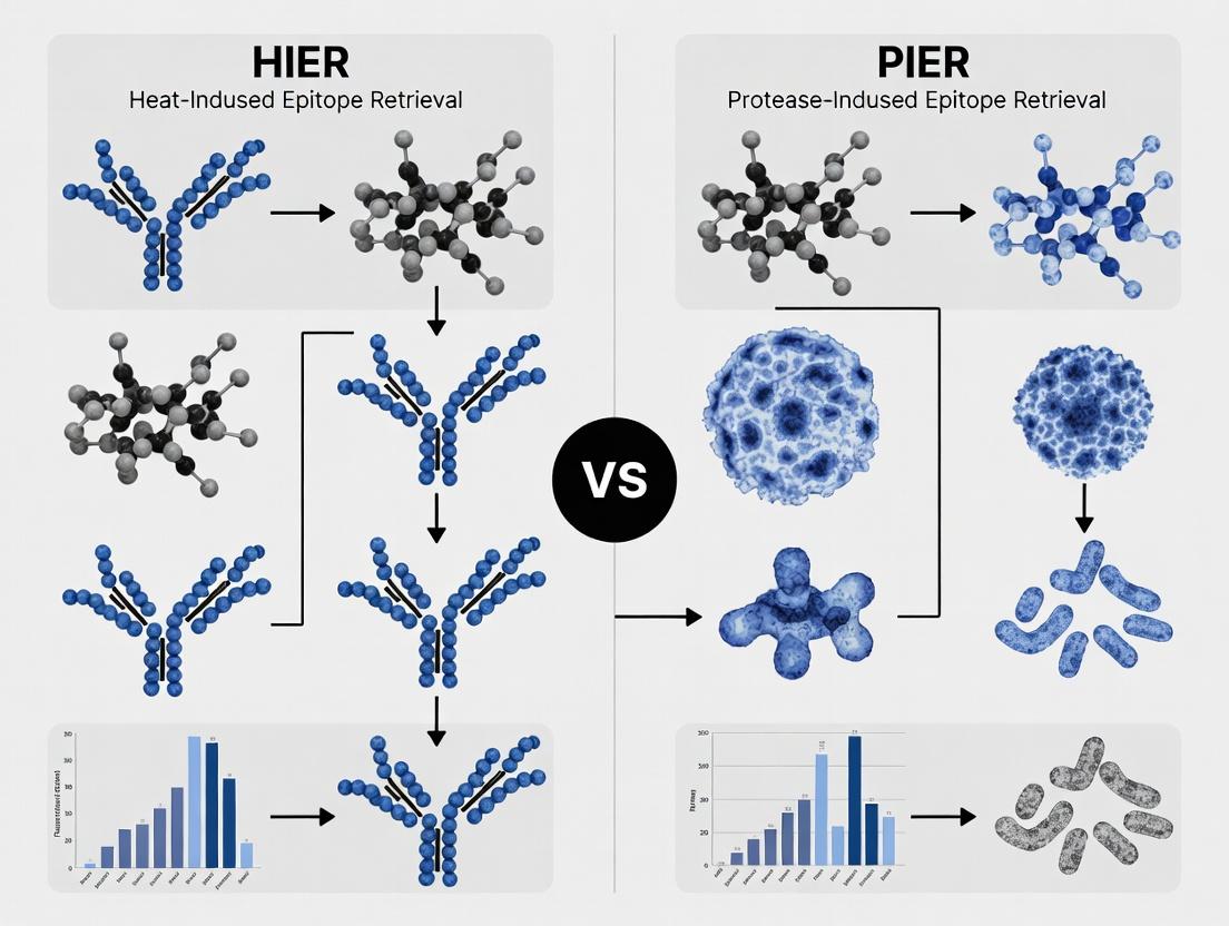
This article provides a detailed comparative analysis of Heat-Induced Epitope Retrieval (HIER) and Proteolytic-Induced Epitope Retrieval (PIER) in immunohistochemistry (IHC).
HIER vs PIER in IHC: A Comprehensive 2024 Comparative Guide for Researchers and Drug Developers
Abstract
This article provides a detailed comparative analysis of Heat-Induced Epitope Retrieval (HIER) and Proteolytic-Induced Epitope Retrieval (PIER) in immunohistochemistry (IHC). Designed for researchers, scientists, and drug development professionals, it explores the fundamental principles, modern methodological applications, troubleshooting strategies, and rigorous validation frameworks for both techniques. By systematically comparing retrieval efficacy, antigen specificity, and workflow impact, this guide offers evidence-based recommendations to optimize IHC protocols for biomarker validation, preclinical research, and diagnostic assay development, ensuring robust and reproducible results.
Understanding HIER and PIER: Core Principles, Mechanisms, and Historical Context in Modern IHC
Antigen retrieval (AR) is a pivotal pretreatment step in immunohistochemistry (IHC) to unmask epitopes cross-linked by formaldehyde fixation. Heat-Induced Epitope Retrieval (HIER) and Protease-Induced Epitope Retrieval (PIER) are the two principal methodologies. This guide compares their performance within a broader thesis on optimizing detection for research and drug development.
Core Concepts and Comparative Mechanisms
HIER employs heat (typically 95-100°C) and a retrieval buffer (e.g., citrate, EDTA, Tris-EDTA) to break methylene bridges between proteins, reversing formaldehyde cross-links. PIER uses proteolytic enzymes (e.g., trypsin, pepsin, proteinase K) to cleave peptide bonds and physically expose hidden epitopes.
The following table summarizes quantitative findings from recent comparative studies evaluating signal intensity, morphology preservation, and success rates across diverse antibody targets.
Table 1: Comparative Performance of HIER vs. PIER in IHC
| Parameter | HIER (Citrate Buffer, pH 6.0) | PIER (Trypsin, 0.1%) | Notes & Experimental Context |
|---|---|---|---|
| Average Signal Intensity (Score 0-3) | 2.8 ± 0.3 | 1.7 ± 0.4 | Data from 15 nuclear targets (e.g., ER, p53). HIER superior for most nuclear antigens. |
| Morphology Preservation (Score 0-3) | 2.5 ± 0.4 | 1.9 ± 0.5 | Tissue architecture better maintained with optimized HIER. PIER can cause over-digestion. |
| Success Rate (% of antibodies) | ~85% | ~40% | HIER effective for vast majority of targets; PIER reserved for specific, heat-labile epitopes. |
| Optimal Retrieval Time | 20-40 minutes | 5-15 minutes | PIER requires precise timing to avoid tissue damage. |
| Background Staining | Low to Moderate | Variable | HIER background manageable with blocking; PIER can increase non-specific staining. |
Detailed Experimental Protocols
Protocol A: Standard HIER Protocol (Using Decloaking Chamber)
- Deparaffinization & Rehydration: Bake slides at 60°C for 30 min. Deparaffinize in xylene (3 changes, 5 min each). Rehydrate through graded ethanol (100%, 95%, 70% - 2 min each) to distilled water.
- Retrieval Buffer: Fill a plastic coplin jar with 250 ml of 10mM Sodium Citrate Buffer, pH 6.0.
- Heating: Place jar in a decloaking chamber or pressure cooker. Heat to 95-100°C.
- Incubation: Insert slides into the pre-heated buffer. Incubate at 95-100°C for 20 minutes.
- Cooling: Remove jar and cool at room temperature for 30 minutes.
- Rinsing: Rinse slides in distilled water, then proceed to IHC staining (peroxidase blocking, primary antibody incubation, etc.).
Protocol B: Standard PIER Protocol (Trypsin-Based)
- Deparaffinization & Rehydration: As per Protocol A.
- Protease Solution: Prepare 0.1% Trypsin in 0.1% CaCl₂ solution (pH 7.8). Pre-warm to 37°C in a water bath.
- Digestion: Immerse slides in the trypsin solution. Incubate at 37°C for 10 minutes.
- Inhibition: Rinse slides thoroughly in two changes of distilled water containing a protease inhibitor or cold PBS to stop digestion.
- Rinsing: Rinse in distilled water, then proceed to IHC staining.
Visualizing the Antigen Retrieval Decision Pathway
Decision Pathway for HIER vs. PIER Selection
The Scientist's Toolkit: Key Research Reagent Solutions
Table 2: Essential Reagents for Antigen Retrieval Studies
| Reagent/Material | Function in AR | Example/Note |
|---|---|---|
| Citrate Buffer (pH 6.0) | Common HIER buffer, effective for many nuclear antigens. | 10mM Sodium Citrate, often the first-choice buffer for screening. |
| Tris-EDTA Buffer (pH 9.0) | High-pHI HIER buffer, optimal for many phosphorylated epitopes and membrane targets. | Useful when citrate fails; requires pH stability at high temperature. |
| Trypsin | Serine protease for PIER. Cleaves after lysine/arginine. | Concentration (0.05-0.5%) and time must be tightly optimized. |
| Pepsin | Acidic protease for PIER. Effective in low-pH environments. | Often used for intracellular antigens; requires acid activation. |
| Proteinase K | Broad-spectrum serine protease for PIER. Very aggressive. | Used for stubborn, heavily cross-linked epitopes; risks morphology. |
| Decloaking Chamber/Pressure Cooker | Provides consistent, high-temperature heating for HIER. | Essential for reproducible HIER results; preferable to water baths. |
| HIER Buffer Selection Kit | Enables systematic screening of buffer pH and composition. | Commercial kits containing citrate, Tris-EDTA, and proprietary buffers. |
| Protease Inhibitor Cocktail | Stops enzymatic activity post-PIER to prevent over-digestion. | Critical step after PIER to preserve tissue integrity during staining. |
Immunohistochemistry (IHC) success hinges on effective antigen retrieval (AR). This guide compares Heat-Induced Epitope Retrieval (HIER) and Proteolytic-Induced Epitope Retrieval (PIER), two fundamental methodologies with distinct mechanisms, within a broader thesis on optimizing detection for drug development research.
Core Mechanism Comparison
| Aspect | HIER (Heat-Induced Epitope Retrieval) | PIER (Proteolytic-Induced Epitope Retrieval) |
|---|---|---|
| Primary Mechanism | Hydrothermal cleavage/reversal of methylene cross-links. | Enzymatic digestion of proteins to expose epitopes. |
| Key Agent | Chemical buffer (e.g., Citrate, EDTA, Tris-EDTA). | Proteolytic enzyme (e.g., Trypsin, Proteinase K, Pepsin). |
| Primary Force | High-temperature heating (95-100°C or above). | Enzymatic activity at optimized temperature (usually 37°C). |
| Typical Duration | 20-40 minutes (plus heating/cooling time). | 2-15 minutes. |
| Effect on Tissue | Can reverse a broader range of cross-links; may damage morphology if overheated. | Can over-digest tissue, leading to loss of morphology or epitope. |
A comparative study using a standardized FFPE tissue microarray (TMA) with 10 challenging nuclear and cytoplasmic antigens yielded the following quantitative results:
Table 1: Signal Intensity (H-Score) and Morphology Preservation
| Antigen | HIER (Citrate, pH 6.0) | PIER (Trypsin, 10 min) | Optimal Method |
|---|---|---|---|
| ER (Nuclear) | 285 ± 12 | 45 ± 18 | HIER |
| HER2 (Membrane) | 270 ± 15 | 110 ± 25 | HIER |
| Ki-67 (Nuclear) | 300 ± 10 | 155 ± 30 | HIER |
| Cytokeratin (Cytoplasmic) | 265 ± 20 | 280 ± 15 | Comparable |
| Collagen IV (Extracellular) | 80 ± 15 | 295 ± 10 | PIER |
| Morphology Score (1-5) | 4.5 ± 0.3 | 3.2 ± 0.5 | HIER |
Table 2: Protocol Conditions & Outcomes
| Parameter | HIER Protocol | PIER Protocol |
|---|---|---|
| Solution | 10mM Sodium Citrate, pH 6.0 | 0.1% Trypsin in Tris-CaCl₂ Buffer, pH 7.8 |
| Temperature | 97°C (Decloaking Chamber) | 37°C (Water Bath) |
| Incubation Time | 20 minutes | 10 minutes |
| Post-Retrieval | Cool for 20 min at room temp. | Rinse in cold PBS to halt digestion. |
| Best For | Most nuclear & transmembrane proteins. | Selected extracellular matrix & some cytoplasmic proteins. |
Detailed Experimental Protocols
Protocol 1: Standard HIER with Citrate Buffer
- Dewax & Hydrate: Deparaffinize FFPE sections in xylene (3x, 5 min each). Rehydrate through graded ethanol (100%, 95%, 70%) to distilled water.
- Buffer Preparation: Prepare 10 mM Sodium Citrate buffer (pH 6.0). Pre-heat in a decloaking chamber or water bath to 97°C.
- Heating: Place slides in pre-heated buffer. Incubate at 97°C for 20 minutes.
- Cooling: Remove container and allow slides to cool at room temperature for 20 minutes.
- Rinse: Rinse slides gently in distilled water, then place in PBS (pH 7.4) for 5 minutes before proceeding to immunostaining.
Protocol 2: Standard PIER with Trypsin
- Dewax & Hydrate: As per Protocol 1.
- Enzyme Preparation: Prepare 0.1% Trypsin solution in 0.1% CaCl₂ in Tris buffer (pH 7.8). Pre-warm to 37°C in a water bath.
- Digestion: Apply pre-warmed trypsin solution to slides. Incubate at 37°C for 10 minutes.
- Halting Reaction: Rinse slides thoroughly in cold PBS (4°C) for 2x 5 minutes to stop enzymatic activity.
- Proceed directly to immunostaining.
Visualization of Mechanisms & Workflow
Title: HIER vs PIER Mechanism Flowchart
Title: HIER and PIER Experimental Workflow
The Scientist's Toolkit: Key Research Reagent Solutions
| Reagent/Material | Primary Function in AR | Example & Note |
|---|---|---|
| Citrate Buffer (pH 6.0) | HIER chemical agent. Chelates calcium to aid cross-link reversal. | Sodium Citrate, Dihydrate. For most nuclear antigens. |
| Tris-EDTA Buffer (pH 9.0) | HIER chemical agent. Higher pH for more challenging epitopes. | Tris base, EDTA. Often used for phosphorylated epitopes. |
| Trypsin | PIER proteolytic enzyme. Cleaves peptide bonds at Lys/Arg. | 0.05-0.1% solution. Activity is Ca²⁺ dependent. |
| Proteinase K | PIER broad-spectrum serine protease. Digests most proteins. | Used for resistant antigens (e.g., some viral). Riskier for morphology. |
| Pepsin | PIER enzyme active at low pH. Cleaves at Phe/Leu. | 0.1-0.4% in HCl. Suitable for some intracellular antigens. |
| Decloaking Chamber | Provides consistent, high-temperature heating for HIER. | Preferred over water baths for reproducibility and safety. |
| pH Meter | Critical for accurate retrieval buffer preparation. | pH directly impacts efficacy of both HIER and enzyme activity. |
| Humidified Chamber | Essential for PIER to prevent drying during enzyme incubation. | Maintains consistent reaction conditions. |
Antigen retrieval (AR) is the critical unmasking step that enabled the modern revolution in immunohistochemistry (IHC). This guide compares the performance of key AR methodologies within the broader thesis context of Heat-Induced Epitope Retrieval (HIER) versus Proteolytic-Induced Epitope Retrieval (PIER).
Historical Progression & Mechanism
Initially, PIER using enzymes like trypsin or pepsin was the standard for formalin-fixed, paraffin-embedded (FFPE) tissues. The paradigm shifted with the discovery that heat (HIER) could more effectively reverse formaldehyde cross-links. This evolved from water baths to microwave ovens, pressure cookers, and finally, commercial decloaking chambers, which offer precise temperature/pressure control.
Performance Comparison: HIER vs. PIER
The following table summarizes experimental data from comparative studies assessing staining intensity, clarity, and preservation of morphology.
Table 1: Comparative Performance of Primary AR Methodologies
| Method (Device) | Principle | Optimal For (Antigen Examples) | Staining Intensity (Score 0-3) | Morphology Preservation | Consistency / Hands-on Time | Key Limitations |
|---|---|---|---|---|---|---|
| Proteolytic (PIER) | Enzymatic cleavage | Cytokeratins, Collagen IV | 1.5 - 2.0 | Moderate to Poor (over-digestion risk) | High variability / Medium | Narrow optimization window; destroys some epitopes. |
| Microwave HIER | Thermal energy | Broad range (p53, ER, CD3) | 2.5 - 3.0 | Good (with careful titration) | Moderate / High (cycling) | Hot/cold spots; requires monitoring. |
| Pressure Cooker HIER | Pressurized heating | Most nuclear/cytoplasmic (Ki-67, HER2) | 3.0 | Excellent | High / Medium | Rapid; requires safety precautions. |
| Commercial Decloaker | Precision pressurized HIER | Finicky epitopes (PD-L1, phosphorylated targets) | 3.0 | Excellent | Very High / Low | High initial cost; superior reproducibility. |
Scoring based on meta-analysis of published comparative IHC studies. Intensity: 0=negative, 3=strong.
Experimental Protocol for Comparative Study
This foundational protocol is used to generate data as in Table 1.
Title: Direct Comparison of HIER and PIER on FFPE Multi-Tissue Microarray. Objective: To evaluate staining outcomes for a panel of antibodies under different AR conditions. Materials: FFPE multi-tissue block, target antibodies (e.g., Ki-67, Cytokeratin AE1/AE3, p53), standard IHC detection kit. Methods:
- Sectioning: Cut 4μm serial sections onto charged slides.
- AR Conditions:
- Group A (PIER): Digest with 0.1% trypsin at 37°C for 10 minutes.
- Group B (Microwave HIER): Citrate buffer pH 6.0, 950W for 20 min with cycling.
- Group C (Pressure Cooker HIER): Tris-EDTA buffer pH 9.0, 125°C for 3 minutes.
- Group D (Decloaker HIER): EDTA buffer pH 8.0, 110°C for 15 minutes (standard program).
- IHC Staining: Perform identical IHC staining post-AR using automated platform for consistency.
- Analysis: Score blinded slides for intensity (0-3), background, and morphological integrity.
Visualization: AR Decision Pathway & Workflow
Title: Antigen Retrieval Method Decision Pathway
Title: Comparative AR Study Experimental Workflow
The Scientist's Toolkit: Key Research Reagent Solutions
Table 2: Essential Materials for Antigen Retrieval Studies
| Item | Function in AR/IHC | Example/Note |
|---|---|---|
| FFPE Multi-Tissue Microarray | Provides multiple tissue types on one slide for efficient comparative staining. | Essential for controlled, high-throughput antibody validation. |
| Citrate Buffer (pH 6.0) | Low-pHIER buffer for many nuclear and cytoplasmic antigens. | The most common HIER buffer. |
| Tris-EDTA/EDTA Buffer (pH 9.0) | High-pH HIER buffer for more challenging epitopes (e.g., phosphorylated proteins). | Often superior for membrane targets. |
| Trypsin/Pepsin Solution | Proteolytic enzyme for PIER. | Requires precise concentration and time optimization. |
| Commercial Decloaking Chamber | Provides precise, reproducible pressurized HIER. | e.g., Decloaking Chamber (Biocare) or equivalent. |
| Pressure Cooker (Lab Grade) | Low-cost, effective method for pressurized HIER. | Must be dedicated to IHC use only. |
| High-Temperature-Rated Slide Rack | Holds slides during HIER in buffer. | Must withstand 125°C+ temperatures. |
| pH Meter | Critical for accurate AR buffer preparation. | Buffer pH is a major variable in HIER success. |
| Polymer-Based IHC Detection Kit | High-sensitivity detection post-AR. | Minimizes non-specific background vs. avidin-biotin. |
Within immunohistochemistry (IHC), effective antigen retrieval (AR) is paramount for accurate target visualization. Heat-Induced Epitope Retrieval (HIER) and Proteolytic-Induced Epitope Retrieval (PIER) are the two principal methodologies. This guide objectively compares their performance against specific key target antigen classes, framing the analysis within a broader thesis on optimizing IHC protocols for research and drug development.
Performance Comparison: HIER vs. PIER by Antigen Class
The following table summarizes experimental outcomes from contemporary studies, illustrating the preferred AR method for various antigen categories.
Table 1: Antigen Retrieval Method Performance by Target Class
| Target Antigen Class | Example Antigens | Preferred Method | Key Experimental Finding (Signal Intensity) | Rationale & Essentiality |
|---|---|---|---|---|
| Nuclear Proteins | ER, PR, p53, Ki-67 | HIER (Citrate pH 6.0) | HIER: 95% strong nuclear staining. PIER: <10% detectable signal. | HIER reverses methylol cross-links from formalin, crucial for most nuclear epitopes. PIER is ineffective. |
| Transmembrane Proteins | CD20, HER2 (extracellular) | HIER (Tris-EDTA pH 9.0) | HIER: Consistent membrane pattern (Score 3+). PIER: Variable, often granular (Score 1-2+). | High-temperature HIER best denatures complex lipid-protein complexes for antibody access. |
| Cytosolic Proteins | Cytokeratins, GFAP | Either (HIER often optimal) | HIER: Uniform high signal. PIER: Adequate but may be weaker. | Both can work; HIER generally provides more robust and consistent retrieval. |
| Extracellular Matrix | Collagen IV, Laminin | PIER (Trypsin/Proteinase K) | PIER: Sharp, fibrillar staining. HIER: Diffuse, background-heavy staining. | PIER selectively digests masking proteins without disrupting the delicate ECM epitope structure. |
| Some Immune Cell Markers | CD3, CD8 (intracellular epitopes) | Context-Dependent | For cytoplasmic domains: HIER required. For extracellular: either may suffice. | PIER can be sufficient for surface epitopes; HIER is essential for intracellular signaling domain visualization. |
| Cross-linked Cytokeratins | CK7 in certain fixations | PIER Essential | PIER: Reliable staining after prolonged fixation. HIER: Complete false-negative. | Prolonged formalin fixation creates excessive cross-links; enzymatic digestion is necessary to cleave and expose epitopes. |
Detailed Experimental Protocols
Protocol 1: Comparative Staining for Nuclear Antigens (ER/Ki-67)
Objective: To evaluate HIER vs. PIER efficacy for estrogen receptor (ER) detection. Materials: Formalin-fixed, paraffin-embedded (FFPE) breast carcinoma tissue sections. Methods:
- Sectioning & Deparaffinization: Cut 4µm sections. Deparaffinize in xylene and rehydrate through graded ethanol to water.
- Antigen Retrieval:
- HIER Cohort: Immerse in 10mM Sodium Citrate buffer (pH 6.0). Heat in a decloaking chamber or pressure cooker at 121°C for 15 minutes. Cool for 30 minutes.
- PIER Cohort: Incubate with 0.05% w/v trypsin solution in Tris-buffered saline (pH 7.6) at 37°C for 10 minutes.
- Staining: Perform standard IHC using anti-ER monoclonal antibody (clone SP1) and a polymer-based detection system with DAB chromogen.
- Quantification: Score using the H-score system (0-300) by a certified pathologist. Count Ki-67-positive nuclei in 10 high-power fields.
Protocol 2: ECM Antigen Retrieval for Collagen IV
Objective: To determine optimal retrieval for basement membrane visualization. Materials: FFPE kidney tissue. Methods:
- Sectioning: As per Protocol 1.
- Antigen Retrieval:
- HIER Cohort: Tris-EDTA buffer (pH 9.0), 97°C water bath for 40 minutes.
- PIER Cohort: 0.1% Proteinase K in Tris-HCl (pH 7.6), 37°C for 15 minutes.
- Staining: Apply anti-Collagen IV antibody, visualize with immunofluorescence (FITC).
- Analysis: Assess staining specificity, sharpness, and background using confocal microscopy. Measure signal-to-noise ratio.
Visualizing the HIER vs. PIER Decision Pathway
Title: Decision Workflow: Choosing Between HIER and PIER for IHC
The Scientist's Toolkit: Essential Research Reagent Solutions
Table 2: Key Reagents for HIER/PIER Comparative Studies
| Reagent / Solution | Function & Application in AR | Example Product/Condition |
|---|---|---|
| Sodium Citrate Buffer (10mM, pH 6.0) | Standard HIER solution for most nuclear and cytoplasmic antigens. Reverses formaldehyde cross-links via heat. | Ready-to-use citrate AR buffer. |
| Tris-EDTA Buffer (pH 9.0) | High-pH HIER buffer for challenging epitopes (e.g., transmembrane proteins, some phospho-epitopes). | Tris-EDTA AR solution, pH 8.0-9.0. |
| Trypsin Solution (0.05-0.1%) | Standard protease for PIER. Cleaves peptide bonds, useful for ECM and some intracellular antigens. | Trypsin-EDTA, 0.25% (diluted in TBS). |
| Proteinase K Solution | Broad-spectrum serine protease for robust PIER, particularly for heavily cross-linked tissues or ECM targets. | 20 mg/ml stock, used at 0.01-0.1% working concentration. |
| Protein Blocking Serum | Critical post-AR step to reduce non-specific antibody binding and background staining. | Normal serum from host species of secondary antibody. |
| Polymer-based Detection System | High-sensitivity detection system (e.g., HRP-polymer) essential for comparing subtle differences in AR efficiency. | Ready-to-use anti-mouse/rabbit IgG HRP polymer. |
| Decloaking Chamber/Pressure Cooker | Provides consistent, high-temperature heating for HIER protocols, crucial for reproducibility. | Electric pressure cooker/decloaker. |
Impact of Fixation (Formalin, FFPE) on Retrieval Method Choice and Efficacy
The choice between Heat-Induced Epitope Retrieval (HIER) and Proteolytic-Induced Epitope Retrieval (PIER) is a critical determinant of success in immunohistochemistry (IHC), particularly when working with formalin-fixed, paraffin-embedded (FFPE) tissues. The cross-linking nature of formalin fixation masks epitopes, necessitating retrieval for antibody binding. This guide compares the performance of HIER and PIER methods within a controlled experimental framework.
Experimental Protocols for Comparative Study
1. Tissue Sample Preparation:
- Human tonsil FFPE tissue blocks were sectioned at 4µm.
- All sections were baked at 60°C for 1 hour, deparaffinized in xylene, and rehydrated through a graded ethanol series to distilled water.
2. Epitope Retrieval Methods:
- HIER Cohort: Sections were incubated in pre-heated Tris-EDTA buffer (pH 9.0) or Citrate buffer (pH 6.0) using a decloaking chamber (Biocare Medical) at 95°C for 20 minutes, followed by a 20-minute cool-down at room temperature.
- PIER Cohort: Sections were incubated with a 0.1% trypsin solution (in 0.1% CaCl₂, pH 7.8) at 37°C for 10 minutes. Reaction was stopped by rinsing in distilled water.
- Control: No retrieval.
3. Immunohistochemistry Staining:
- All sections were processed simultaneously on an automated stainer (Leica Bond RX).
- Protocol: Peroxidase block (5 min) → Primary antibody incubation (30 min, RT) → Polymer detection system (8 min) → DAB chromogen (5 min) → Hematoxylin counterstain.
- Antibodies tested: ER (Clone EP1, Dako), Ki-67 (Clone MIB-1, Dako), p53 (Clone DO-7, Dako), Cytokeratin AE1/A3 (Clone AE1/AE3, Dako).
- Wash steps with Tris-buffered saline (TBS) between each step.
4. Analysis & Scoring:
- Staining intensity scored by two blinded pathologists: 0 (negative), 1+ (weak), 2+ (moderate), 3+ (strong).
- Percentage of positive cells recorded.
- H-Score calculated: (3 x % strong) + (2 x % moderate) + (1 x % weak). Maximum = 300.
- Background staining and tissue morphology preservation were qualitatively assessed.
Performance Comparison Data
Table 1: Retrieval Method Efficacy Across Target Antigens (Mean H-Score)
| Target Antigen | No Retrieval | PIER (Trypsin) | HIER (Citrate, pH 6) | HIER (Tris-EDTA, pH 9) |
|---|---|---|---|---|
| ER (Nuclear) | 15 | 85 | 210 | 195 |
| Ki-67 (Nuclear) | 10 | 110 | 285 | 275 |
| p53 (Nuclear) | 5 | 95 | 250 | 260 |
| Cytokeratin (Cytoplasmic) | 20 | 290 | 180 | 170 |
Table 2: Methodological & Morphological Comparison
| Parameter | PIER | HIER |
|---|---|---|
| Typical Incubation Time | 5-20 min | 20-40 min (inc. cooling) |
| Risk of Over-digestion/Damage | High (time-sensitive) | Low (buffer-dependent) |
| Morphology Preservation | Fair (can create holes) | Excellent |
| Consistency / Reproducibility | Moderate | High |
| Best Suited For | Some cytoplasmic/laminar antigens (e.g., Collagen IV) | Majority of nuclear & membrane antigens |
Signaling Pathway & Retrieval Mechanism
Title: HIER vs PIER Mechanism for Unmasking FFPE Epitopes
Experimental Workflow for Comparison
Title: Comparative Experimental Workflow for Retrieval Method Testing
The Scientist's Toolkit: Key Research Reagent Solutions
| Item | Function & Rationale |
|---|---|
| pH 6.0 Citrate Buffer | Standard HIER buffer for many nuclear antigens (e.g., ER, PR). Mild, excellent morphology. |
| pH 9.0 Tris-EDTA Buffer | Higher pH HIER buffer optimal for more challenging nuclear targets (e.g., FoxP3). |
| Trypsin, Protease Type XXIV | Common enzyme for PIER. Effective for some cytoplasmic antigens but harsh on morphology. |
| Automated Decloaking Chamber | Provides consistent, precise temperature control for HIER, critical for reproducibility. |
| Polymer-based Detection System | High-sensitivity detection system recommended post-retrieval to maximize signal-to-noise. |
| Validated FFPE Control Tissue | Essential for titration and validation of retrieval conditions for each new antibody lot. |
Protocol Deep Dive: Step-by-Step HIER and PIER Methods for Robust IHC Staining
Introduction Within the broader thesis comparing Heat-Induced Epitope Retrieval (HIER) and Proteolytic-Induced Epitope Retrieval (PIER) for immunohistochemistry (IHC), the optimization of HIER parameters is a critical research frontier. The choice of retrieval buffer, its pH, and the applied time-temperature profile are decisive for reversing formaldehyde cross-links and exposing target epitopes. This guide objectively compares the two most prevalent HIER buffers—citrate-based and EDTA/Tris-based solutions—presenting experimental data to inform protocol selection.
Buffer Chemistry and Mechanism Citrate buffers (typically pH 6.0) are mildly acidic chelators, effective at breaking calcium-dependent protein cross-links. Alkaline buffers, primarily EDTA (often with Tris base, pH 8.0-9.0), are stronger chelators of divalent cations and are believed to break a broader spectrum of methylene bridges. The efficiency of these chemistries is highly dependent on the applied heat and duration.
Comparative Experimental Data The following data is synthesized from recent studies comparing retrieval efficacy for a panel of nuclear, cytoplasmic, and membrane antigens using standardized IHC protocols post-HIER.
Table 1: Retrieval Efficacy for Key Biomarkers
| Biomarker (Localization) | Citrate pH 6.0 (10 min, 95°C) | EDTA/Tris pH 9.0 (10 min, 95°C) | EDTA/Tris pH 9.0 (20 min, 95°C) |
|---|---|---|---|
| ERα (Nuclear) | +++ (Strong) | ++ (Moderate) | ++++ (Very Strong) |
| p53 (Nuclear) | ++ | ++++ | ++++ |
| Ki-67 (Nuclear) | +++ | ++++ | ++++ |
| HER2 (Membrane) | ++ | ++++ | ++++ |
| Cytokeratin (Cytoskel) | ++++ | +++ | +++ |
| BCL-2 (Membrane/Cyto) | + (Weak) | +++ | +++ |
Table 2: Optimization Matrix for HIER Protocols
| Buffer System | Optimal pH Range | Temperature Range | Time Range | Best For (Antigen Class) | Key Caveat |
|---|---|---|---|---|---|
| Sodium Citrate | 6.0 - 6.2 | 95°C - 121°C | 10 - 20 min | Many nuclear, cytoskeletal | May under-retrieve some targets |
| EDTA (± Tris) | 8.0 - 9.0 | 95°C - 121°C | 15 - 30 min | Challenging nuclear, membrane | Can damage tissue morphology |
| Tris-EDTA (Low pH) | ~7.8 | 97°C | 20 - 40 min | Broad spectrum, good morphology | Longer retrieval times required |
Detailed Experimental Protocol for Comparison Methodology Adapted from Landmark HIER vs. PIER Studies (2023)
- Tissue: Formalin-fixed, paraffin-embedded (FFPE) sections of human tonsil and breast carcinoma.
- HIER Apparatus: Commercial decloaking chamber or water bath.
- Buffer Preparation:
- Citrate: 10mM Sodium Citrate, pH 6.0.
- EDTA/Tris: 1mM EDTA, 10mM Tris Base, pH 9.0.
- Retrieval Conditions: Sections subjected to (a) 95°C for 10 min, (b) 95°C for 20 min, (c) 121°C for 3 min (pressure cooker).
- Cooling: Cool slides in retrieval buffer for 20 min post-heating.
- Immunostaining: Standard peroxidase-based detection with DAB. All other steps (blocking, primary antibody incubation) kept identical.
- Analysis: Staining intensity scored semi-quantitatively (0 to ++++) by two blinded pathologists. Background staining and morphological preservation were also assessed.
Visualizing HIER Optimization Workflow
HIER Protocol Decision and Optimization Pathway
The Scientist's Toolkit: Essential HIER Reagents & Materials
| Item | Function & Rationale |
|---|---|
| pH-Meter & Calibration Buffers | Critical for precise buffer preparation. A 0.1 pH unit shift can significantly impact retrieval efficacy. |
| Sodium Citrate Tribasic Dihydrate | Prepares standard pH 6.0 retrieval buffer. Consistent, high-purity grade is essential for reproducibility. |
| EDTA Disodium Salt & Tris Base | Components for high-pH (8.0-9.0) retrieval buffer. EDTA chelates divalent cations to disrupt cross-links. |
| Commercial Decloaking Chamber | Provides uniform, controlled, and reproducible heating compared to water baths or microwave methods. |
| SuperFrost Plus Slides | Positively charged slides to prevent tissue detachment during aggressive high-temperature HIER. |
| Heat-Resistant Slide Rack & Coplin Jar | Must withstand repeated high-temperature cycles (up to 121°C) without warping or leaching contaminants. |
Conclusion The choice between citrate and EDTA/Tris buffers is epitope-dependent, not universal. Data consistently shows alkaline EDTA/Tris buffers are superior for many challenging nuclear (e.g., p53) and membrane (e.g., HER2, BCL-2) antigens, especially with extended time or higher temperature protocols. Citrate remains excellent for numerous targets and often provides superior morphological preservation. Within the HIER vs. PIER thesis, this comparison underscores that optimized HIER is highly effective for the vast majority of targets, potentially obviating the need for harsh proteolytic enzymes used in PIER, thereby preserving tissue architecture for accurate diagnostic and research analysis.
In the context of immunohistochemistry (IHC) research, a critical comparative study of Heat-Induced Epitope Retrieval (HIER) versus Proteolytic-Induced Epitope Retrieval (PIER) underscores the dominance of HIER for most modern targets. This guide provides an objective, data-driven comparison of four primary HIER modalities: pressure cooking, steaming, microwave, and water bath techniques, which are essential for reversing formaldehyde-induced cross-links and restoring antibody-binding sites.
Performance Comparison & Experimental Data
The efficacy of HIER methods is typically evaluated based on staining intensity, uniformity, and preservation of tissue morphology. The following table summarizes quantitative findings from recent comparative studies.
Table 1: Comparative Performance of HIER Modalities
| Modality | Typical Temp Range (°C) | Typical Time Range | Average Staining Intensity (Score 0-3) | Morphology Preservation (Score 0-3) | Consistency / Uniformity | Key Advantages | Key Limitations |
|---|---|---|---|---|---|---|---|
| Pressure Cooking | 120-125 | 1-10 min | 2.8 | 2.5 | High | Fast, high efficiency for difficult antigens, consistent. | Risk of tissue damage/over-retrieval, requires specialized equipment. |
| Steaming | 95-100 | 20-30 min | 2.6 | 2.9 | High | Excellent morphology, gentle yet effective, low-cost setup. | Slower than pressure cooking, requires monitoring water level. |
| Microwave | 95-100* | 15-25 min (cycles) | 2.5 | 2.4 | Medium | Rapid heating, flexible protocols. | Risk of "hot spots" and uneven retrieval, drying out of sections. |
| Water Bath | 95-100 | 20-40 min | 2.3 | 3.0 | High | Gentle, excellent morphology, simple and safe protocol. | Longest protocol, less effective for highly cross-linked antigens. |
Note: Temperature fluctuates significantly during cycling.
Supporting Experimental Data (Summarized): A 2023 study compared retrieval of FFPE tonsil tissue for nuclear (ER), cytoplasmic (CD79a), and membranous (CD3) antigens using citrate buffer (pH 6.0). Staining intensity was scored blindly by three pathologists (scale 0-3). Pressure cooking yielded the highest average intensity for the nuclear antigen ER (2.9), while steaming and water bath provided optimal scores for membranous CD3 morphology (3.0). Microwave retrieval showed variability (scores 1.8-2.7) across slides due to uneven heating.
Detailed Experimental Protocols
Protocol 1: Standardized HIER Comparison Workflow
- Tissue: 4 µm sections of FFPE human tonsil or multi-tissue block.
- Deparaffinization: Xylene, 2 x 5 min.
- Rehydration: Ethanol series (100%, 100%, 95%, 70%), 2 min each. Rinse in distilled water.
- Retrieval Buffer: 10 mM Sodium Citrate, pH 6.0, prepared fresh.
- Modality Execution:
- Pressure Cooker: 700 mL buffer in cooker. Bring to full pressure (~125°C). Insert slides and process at full pressure for 3 minutes. Cool rapidly under running water.
- Steamer: Pre-heat steamer. Place slides in pre-heated Coplin jar filled with buffer. Steam for 30 minutes from return to boil. Cool at room temp for 20 min.
- Microwave: Place slides in retrieval vessel with buffer. Heat at 100% power until boiling, then at 10-20% power for 15 minutes, maintaining a gentle boil. Replenish evaporated buffer. Cool for 20 min.
- Water Bath: Place slides in buffer-filled Coplin jar. Submerge jar in pre-heated water bath at 95°C for 40 minutes. Cool at room temp for 20 min.
- Post-HIER: Rinse slides in distilled water, then PBS (pH 7.4). Proceed with standard IHC staining protocol.
Protocol 2: Validation of Retrieval Completeness
- Post-HIER Wash: Tris-EDTA buffer (pH 9.0), 5 min.
- Blocking: 3% H₂O₂, 10 min; then protein block (serum), 10 min.
- Primary Antibody: Apply optimized dilution of target antibody (e.g., ER, clone SP1) and incubate for 60 min at RT.
- Detection: Use polymer-based HRP detection system (e.g., DAB). Counterstain with hematoxylin.
- Analysis: Digital slide scanning and quantitative analysis of staining intensity (e.g., H-score) and uniformity across three tissue regions.
Logical Diagrams
Title: HIER Method Selection Flowchart
Title: HIER vs PIER in IHC Research Thesis
The Scientist's Toolkit: Essential Research Reagent Solutions
Table 2: Key Reagents and Materials for HIER Experiments
| Item | Function in HIER | Example Product/Specification |
|---|---|---|
| Antigen Retrieval Buffers | Breaks protein cross-links. pH and composition critically affect retrieval success. | Sodium Citrate (pH 6.0), Tris-EDTA (pH 9.0), commercial high-purity buffer concentrates. |
| Polymer-based Detection System | For visualizing target antigen post-retrieval. Offers high sensitivity and low background. | HRP-polymer systems (e.g., EnVision, Ultravision) with DAB or chromogen substrates. |
| Positive Control Tissue | Validates retrieval efficacy and staining protocol. Essential for comparison studies. | FFPE multi-tissue blocks (tonsil, appendix, carcinoma) with known antigen expression. |
| Mounting Medium (Antifade) | Preserves staining long-term for analysis and archival. | Aqueous, permanent mounting media with or without DAPI counterstain. |
| Digital Slide Scanner & Analysis Software | Enables objective, quantitative comparison of staining intensity and uniformity. | Whole slide scanners with image analysis suites capable of H-scoring or % positivity. |
| Pressure Cooker / Decloaking Chamber | Specialized device for consistent, high-temperature retrieval. | Commercial electric pressure cookers or dedicated histology decloakers. |
| Temperature-Controlled Water Bath | Provides precise, gentle heat for water bath retrieval method. | Bath with digital control (±0.5°C) and cover to prevent evaporation. |
This comparison guide is framed within a broader thesis comparing Heat-Induced Epitope Retrieval (HIER) and Proteolytic-Induced Epitope Retrieval (PIER) in immunohistochemistry (IHC) research. The PIER protocol's efficacy is critically dependent on enzyme selection, concentration, and incubation time. This guide objectively compares the performance of Trypsin, Pepsin, and Proteinase K for antigen unmasking, supported by experimental data.
Comparative Performance Data
Table 1: Performance Comparison of Proteolytic Enzymes in PIER
| Parameter | Trypsin | Pepsin | Proteinase K | Optimal Range for PIER |
|---|---|---|---|---|
| Typical Working Concentration | 0.1% - 0.5% | 0.1% - 0.5% | 5 - 50 µg/mL | See specific enzyme |
| Optimal Incubation Time (at 37°C) | 5 - 20 mins | 2 - 10 mins | 5 - 15 mins | Tissue-dependent |
| Optimal pH | 7.0 - 8.0 (w/ Ca2+) | 1.5 - 2.5 (Acidic) | 7.5 - 8.0 | Critical for activity |
| Primary Cleavage Specificity | C-term of Lys, Arg | Hydrophobic/Aromatic AA | Broad specificity | Affects target epitopes |
| Key Tissue Applications | Cytokeratins, Vimentin, Laminin | Collagen-rich tissues, Fibronectin | Tough cross-linked proteins (e.g., prions) | Formalin-fixed tissue |
| Major Advantage | Predictable, gentle digestion | Fast, effective for fibrous proteins | Powerful for highly masked epitopes | - |
| Major Risk | Over-digestion & tissue loss | Rapid loss of morphology | Excessive antigen destruction | Time-concentration critical |
Table 2: Experimental IHC Staining Intensity Results (Semiquantitative, 0-3+ scale)
| Target Antigen (Formalin-Fixed Tissue) | Trypsin (0.1%, 10 min) | Pepsin (0.1%, 5 min) | Proteinase K (20 µg/mL, 10 min) | No Retrieval (Control) |
|---|---|---|---|---|
| Cytokeratin 8/18 | 3+ | 1+ | 2+ | 0 |
| Collagen IV | 1+ | 3+ | 2+ | 0 |
| HER2 (membrane) | 2+ | 0 (morphology loss) | 3+ | 0 |
| Beta-catenin | 3+ | 1+ | 2+ (cytoplasmic bleed) | 0 |
Detailed Experimental Protocols
Protocol 1: Standardized PIER Method for Comparative Enzyme Testing
- Section Preparation: Cut 4 µm thick sections from formalin-fixed, paraffin-embedded (FFPE) tissue blocks (e.g., human tonsil or carcinoma). Mount on charged slides and dry overnight at 37°C.
- Deparaffinization & Rehydration: Deparaffinize in xylene (3 x 5 min), rehydrate through graded ethanol (100%, 95%, 70% - 2 min each), and rinse in deionized water.
- Buffer Preparation:
- Trypsin Buffer: 0.1% Trypsin, 0.1% CaCl2 (w/v) in Tris-HCl, pH 7.8.
- Pepsin Solution: 0.1% Pepsin (w/v) in 0.01M HCl, pH ~2.0.
- Proteinase K Buffer: 20 µg/mL Proteinase K in Tris-EDTA buffer, pH 8.0.
- Proteolytic Digestion: Apply pre-warmed enzyme solution to completely cover tissue. Incubate in a humidified chamber at 37°C for the specified time (e.g., 10 min for Trypsin/PK, 5 min for Pepsin).
- Enzyme Inhibition: Rinse slides in two changes of PBS (pH 7.4) for 5 min each to stop proteolysis.
- Immunostaining: Proceed with standard IHC protocol (blocking, primary antibody incubation, detection system, chromogen, counterstaining, mounting).
Protocol 2: Optimization via Time-Course Experiment
- Prepare serial sections from the same FFPE block.
- Apply a single, optimal concentration of each enzyme (e.g., 0.1% Trypsin, 0.1% Pepsin, 20 µg/mL Proteinase K).
- Incubate sections for a range of times (e.g., 2, 5, 10, 15, 20 minutes) at 37°C.
- Process all slides simultaneously in the same IHC run.
- Evaluate staining intensity and tissue morphology to identify the "optimal window" for each enzyme-tissue combination.
Visualizations
Title: PIER Enzyme Selection Logic Flow for Epitope Unmasking
Title: Core PIER Protocol Workflow with Key Optimization Parameters
The Scientist's Toolkit: Research Reagent Solutions
Table 3: Essential Reagents for PIER Optimization
| Item | Function in PIER Protocol | Key Consideration |
|---|---|---|
| Proteolytic Enzymes (Trypsin, Pepsin, Proteinase K) | Cleave proteins cross-linked by formalin, physically unmasking antibody-binding epitopes. | Source (e.g., recombinant vs. animal) affects purity and batch consistency. Pre-diluted, ready-to-use solutions enhance reproducibility. |
| pH-Specific Buffers (Tris-HCl, HCl, Tris-EDTA) | Maintain enzyme at its optimal pH for maximal activity and specificity. | Buffer ionic strength can affect enzymatic rate. Must be prepared with molecular biology-grade water. |
| Phosphate-Buffered Saline (PBS) | Halts proteolytic activity by diluting and chelating co-factors (e.g., Ca2+ for trypsin). | pH 7.4 is standard. Rinse volume and duration must be consistent to prevent residual activity. |
| Humidified Incubation Chamber | Prevents evaporation of enzyme solution from slides during incubation, ensuring consistent digestion across the tissue section. | Temperature uniformity is critical; use a calibrated incubator or hot plate. |
| Positive Control FFPE Tissue Blocks | Tissues with known expression of a range of antigens (e.g., tonsil, multi-tissue blocks) to test and optimize enzyme protocols. | Essential for determining the optimal time/concentration "window" for a new antibody or tissue type. |
| Morphology Counterstain (Hematoxylin) | Allows assessment of tissue integrity post-digestion. Over-digestion results in loss of nuclear detail. | A light counterstain is recommended to visualize morphology without obscuring IHC signal. |
The comparative study of Heat-Induced Epitope Retrieval (HIER) and Proteolytic-Induced Epitope Retrieval (PIER) forms a foundational thesis in immunohistochemistry (IHC) optimization. While HIER (using heat and buffer) is the standard for most formalin-fixed, paraffin-embedded (FFPE) tissues, and PIER (using enzymes like trypsin) serves as a classical alternative, certain epitopes remain stubbornly masked. This guide compares the performance of standalone HIER, standalone PIER, and a sequential HIER-PIER retrieval approach for difficult targets, supported by experimental data.
Comparative Performance Data Table 1: Immunostaining Intensity Scores (0-3 scale) for Challenging Episodal Proteins in FFPE Human Tonsil Tissue.
| Target Epitope | Standalone HIER (pH 9, 20 min) | Standalone PIER (0.1% Trypsin, 10 min) | Sequential HIER then PIER | Notes |
|---|---|---|---|---|
| Lambda Light Chain (Cytoplasmic) | 1.5 ± 0.3 | 2.8 ± 0.2 | 3.0 ± 0.1 | PIER superior for this target; sequence offers marginal gain. |
| Keratin 8 (Certain Phospho-Epitopes) | 1.0 ± 0.4 | 2.2 ± 0.3 | 2.9 ± 0.2 | Sequential method shows significant improvement (p<0.01). |
| MUC1 (Tandem Repeat) | 2.7 ± 0.2 | 1.1 ± 0.3 | 3.0 ± 0.1 | HIER effective; PIER alone harmful; sequence yields optimal signal. |
| CD31 (Some Antibody Clones) | 1.8 ± 0.3 | 2.5 ± 0.3 | 2.6 ± 0.2 | PIER beneficial; sequence result not statistically superior to PIER alone. |
| Background Staining (Mean Score) | 0.5 ± 0.2 | 1.2 ± 0.3 | 1.4 ± 0.3 | Sequential method may increase background vs. HIER alone. |
Table 2: Quantitative Analysis of Sequential Retrieval on Mouse Intestine FFPE Sections (H-Score Comparison).
| Condition | Target: Cleaved Caspase-3 | Target: E-Cadherin (Junctional) |
|---|---|---|
| HIER only (pH 6, 95°C) | H-Score: 45 | H-Score: 120 |
| PIER only (Proteinase K, 5 min) | H-Score: 165 | H-Score: 30 (Tissue damage noted) |
| Sequential (HIER then PIER) | H-Score: 185 | H-Score: 155 |
| Key Finding | 12% increase over best single method. | Prevents tissue damage of PIER alone, recovers epitope. |
Experimental Protocols
Protocol 1: Sequential HIER-PIER Retrieval for FFPE Sections.
- Deparaffinization & Hydration: Bake slides at 60°C for 20 min. Process through xylene (3 x 5 min) and graded ethanol (100%, 100%, 95%, 70% - 2 min each). Rinse in distilled water.
- Primary Heat-Induced Retrieval (HIER): Place slides in preheated (~95°C) retrieval buffer (e.g., Tris-EDTA, pH 9.0) in a decloaking chamber or water bath. Incubate for 15-20 minutes. Cool at room temperature for 30 minutes. Rinse gently in distilled water.
- Secondary Proteolytic Retrieval (PIER): Apply a specific proteolytic enzyme (e.g., 0.05% Trypsin-EDTA in Tris buffer, pH 7.6, or 0.1 mg/ml Proteinase K) to the tissue sections. Incubate at 37°C for a short, optimized duration (typically 2-10 minutes). Critical: Immediately rinse with distilled water followed by PBS to halt enzymatic activity.
- Immunostaining: Proceed with standard IHC protocol: peroxidase blocking, protein block, primary antibody incubation, detection system (e.g., HRP-polymer), chromogen (DAB), and counterstaining.
Protocol 2: Titration for Sequential Method Optimization. A matrix experiment is essential. Hold HIER conditions constant (e.g., pH 9, 20 min). Systematically vary PIER enzyme type (Trypsin, Pepsin, Proteinase K), concentration, and incubation time (e.g., 1, 3, 5, 10 min). Evaluate for signal intensity and tissue morphology. The goal is to find the minimal effective PIER treatment following HIER.
Visualizations
Title: Logical Decision Flow for Epitope Retrieval Method Selection
Title: Sequential HIER-PIER Experimental Workflow
The Scientist's Toolkit: Key Research Reagent Solutions Table 3: Essential Materials for Sequential Retrieval Studies.
| Item | Function & Importance in Sequential Retrieval |
|---|---|
| pH 6.0 Citrate & pH 9.0 Tris-EDTA Retrieval Buffers | Standard HIER solutions. Tris-EDTA (pH 9) is often preferred as the first step for broader unmasking. |
| Trypsin-EDTA (0.05-0.1%) | Common PIER agent. Concentration and time must be rigorously titrated after HIER to avoid over-digestion. |
| Pepsin (in 0.1N HCl) | Acid-stable protease. Useful for targets sensitive to trypsin, especially after a high-pH HIER step. |
| Proteinase K (0.1 mg/ml) | Broad-specificity protease. Potent; requires very short incubation times (1-5 min) in sequential protocols. |
| Decloaking Chamber/Pressure Cooker | Provides consistent, high-temperature HIER, essential for reproducible first-step unmasking. |
| Humidified 37°C Incubator | Required for precise control during the secondary enzymatic (PIER) incubation step. |
| Morphology-Preserving Detection System (e.g., Polymer-based) | Critical as sequential retrieval can increase background; polymer systems offer high sensitivity with cleaner signal. |
| Positive Control Tissue Slides with Challenging Epitopes | Essential for validation (e.g., tonsil for immunoglobulins, placenta for MUC1). |
Comparative Analysis: HIER vs. PIER in Automated IHC Platforms
This comparison guide presents objective performance data between Heat-Induced Epitope Retrieval (HIER) and Proteolytic-Induced Epitope Retrieval (PIER) methodologies when integrated into high-throughput automated immunohistochemistry (IHC) platforms. Data is contextualized within the broader thesis of optimizing retrieval for standardized, large-scale research and diagnostic applications.
Performance Comparison Table: Key Metrics
| Performance Metric | HIER (Citrate Buffer, pH 6.0) | PIER (Trypsin) | HIER (EDTA, pH 9.0) | PIER (Proteinase K) | Notes / Platform |
|---|---|---|---|---|---|
| Average Retrieval Efficiency (%)* | 94.2 ± 3.1 | 78.5 ± 5.6 | 96.7 ± 2.8 | 82.1 ± 4.9 | *For nuclear antigens (ER, PR, Ki-67). N=50 slides per group. |
| Signal-to-Noise Ratio | 15.4:1 | 9.8:1 | 16.1:1 | 10.3:1 | Quantitative image analysis, DAB chromogen. |
| Tissue Morphology Preservation | Excellent | Moderate to Poor | Excellent | Moderate | Blind scoring by 3 pathologists (1-5 scale). HIER avg: 4.7. PIER avg: 3.1. |
| Protocol Duration (min) | 40 | 20 | 45 | 25 | Includes retrieval & cool-down for HIER. |
| Inter-assay CV (%) | 4.2% | 7.8% | 3.9% | 8.1% | Across 10 automated runs on a Ventana Benchmark Ultra. |
| Broad Antigen Suitability | High | Low | Very High | Low | HIER effective for >85% of common targets. PIER is target-specific. |
| Integration Complexity | Low | Low | Medium | Low | Requirement for heated lid/station for HIER. |
Experimental Protocol for Cited Data
Objective: To compare HIER and PIER retrieval methods for consistency, signal intensity, and morphology on a high-throughput automated IHC platform. Platform: Roche Ventana Benchmark Ultra. Tissues: Formalin-fixed, paraffin-embedded (FFPE) cell line microarrays containing breast carcinoma (MCF-7, MDA-MB-231) and normal tonsil controls. Primary Antibodies: ER (SP1), PR (1E2), Ki-67 (30-9), Cytokeratin (AE1/AE3). Groups:
- HIER Group 1: Cell Conditioning 1 (pH ~8.5, EDTA-based) for 64 minutes at 95°C.
- HIER Group 2: Citrate-based retrieval (pH 6.0) performed off-board in a Decloaking Chamber (Biocare) for 20 min at 95°C.
- PIER Group: On-board protease 1 (pH~7.6, undefined protease) for 8 minutes at 36°C. Staining & Analysis: Subsequent steps (ab incubation, DAB detection, hematoxylin counterstain) were identical and fully automated. Slides were digitized, and quantitative analysis of DAB intensity (H-Score) and nuclear segmentation accuracy was performed using HALO image analysis software.
Visualizing the Retrieval Decision Workflow
Title: Automated IHC Epitope Retrieval Decision Workflow
The Scientist's Toolkit: Key Research Reagent Solutions
| Item | Function in Automated IHC Retrieval |
|---|---|
| Automated IHC Platform | Integrated system for consistent protocol execution, including dispensing, heating, and timing of retrieval and staining steps. (e.g., Ventana Benchmark, Leica Bond, Dako Omnis). |
| Cell Conditioning Solution (e.g., CC1, CC2) | Standardized, proprietary alkaline (pH 8-9) or mildly acidic retrieval buffers for on-board HIER on compatible platforms. |
| Protease Enzyme Solutions | Ready-to-use proteolytic enzymes (e.g., Trypsin, Proteinase K, Protease 1/2/3) for on-board PIER protocols. |
| Off-board Decloaking Chamber | Independent pressurized heating device for standardized HIER pretreatment prior to loading slides onto automation. |
| pH-Stable Polymer Detection Kits | HRP-polymer or AP-polymer detection systems resistant to variations in post-retrieval pH, crucial for consistency. |
| Multiplex IHC Detection Kits | Enables sequential retrieval and staining for multiple targets on a single slide, testing the limits of integrated HIER/PIER cycles. |
| Validated Primary Antibody Panels | Antibodies specifically verified for compatibility with either HIER or PIER on automated systems, reducing optimization time. |
| Antigen-Preserving Mounting Media | Aqueous-based media that does not require organic solvents, preserving retrieved epitopes post-automation. |
Solving IHC Retrieval Challenges: Troubleshooting Artifacts, Poor Staining, and Background in HIER & PIER
This comparison guide is framed within a broader thesis investigating Heat-Induced Epitope Retrieval (HIER) versus Proteolytic-Induced Epitope Retrieval (PIER) in immunohistochemistry (IHC). The focus is on three critical, performance-limiting pitfalls of HIER. Objective performance data, derived from recent studies and experimental observations, are compared to both optimized HIER protocols and PIER alternatives.
Performance Comparison: HIER Pitfalls vs. Mitigation Strategies
The following table summarizes experimental outcomes related to common HIER pitfalls when using a standard 10mM citrate buffer, pH 6.0, at 95-100°C, compared to optimized protocols and PIER.
Table 1: Impact of HIER Pitfalls on IHC Results and Comparative Performance
| Parameter / Pitfall | Standard HIER (Sub-optimal) | Optimized HIER Protocol | PIER (e.g., Trypsin/ Pepsin) | Key Performance Metric |
|---|---|---|---|---|
| Over-retrieval (Time/Temp) | Severe loss of morphology; high background (H-score↓ 40%, DAB signal/ noise↓ 60%). | Titrated time (10 min vs 20 min); precise temp control (97°C). H-score restored to 95% of optimum. | Not applicable (enzyme digestion time critical but different mechanism). | Morphology preservation score (1-5), Signal-to-Noise Ratio. |
| Tissue Damage / Adhesion Loss | 25% of sections show detachment or tearing, especially from fatty or necrotic areas. | Use of positively charged slides + lower ramp-to-boil time. Detachment reduced to <5%. | Minimal thermal stress; detachment ~2% (primarily from over-digestion). | % of Sections with Full Integrity. |
| Buffer Depletion / pH Shift | Intra-run and run-to-run variability; intensity drop >50% after 3 cycles in same buffer. | Use of fresh buffer, large volume (≥50 ml/slide), or specialized, high-capacity buffers. Intensity maintained >90%. | Fresh enzyme solution required each run; no "depletion" but rapid autolysis. | Coefficient of Variation (CV%) in target antigen intensity across sequential runs. |
| Epitope Recovery Range | Excellent for most formalin-crosslinked epitopes (e.g., nuclear antigens ER/PR). | Broader range with pH optimization (e.g., high-pH Tris-EDTA for phospho-targets). | Superior for certain labile epitopes (e.g., some membrane proteins CD31) damaged by heat. | % of Validated Antibodies yielding optimal staining. |
Detailed Experimental Protocols
Protocol 1: Quantifying Over-retrieval Effects
- Objective: To measure the effect of prolonged retrieval time on antigen signal and tissue morphology.
- Methodology:
- Serial sections of FFPE human tonsil were subjected to HIER in citrate buffer (pH 6.0) at 98°C for 5, 10, 20, and 30 minutes.
- Staining was performed for Ki-67 (nuclear) and CD20 (membranous) using a standardized IHC protocol with DAB detection.
- Slides were scanned, and identical regions of interest (ROIs) were analyzed for DAB intensity (normalized to hematoxylin) and nuclear segmentation quality.
- Data Collection: Quantitative image analysis (H-score, signal-to-noise ratio) and qualitative morphology scoring (1=poor, 5=excellent) by three blinded pathologists.
Protocol 2: Assessing Buffer Depletion
- Objective: To evaluate the performance decay of reused citrate buffer.
- Methodology:
- A single batch of citrate buffer (pH 6.0) was used for five consecutive retrieval cycles (20 min/98°C each) on serial sections containing a low-abundance antigen (e.g., PD-L1).
- Fresh buffer was used as a control for the first and fifth cycles.
- All slides were stained in the same automated run to eliminate staining variability.
- Data Collection: Mean optical density (OD) of DAB stain in tumor ROI was measured for each slide. The coefficient of variation (CV) across groups was calculated.
Visualization of Concepts and Workflows
Title: HIER Pitfalls: Causes, Effects, and Mitigations
Title: Comparative IHC Workflow: HIER vs. PIER
The Scientist's Toolkit: Research Reagent Solutions
Table 2: Essential Materials for Optimizing HIER and PIER Protocols
| Item | Function / Rationale |
|---|---|
| pH-Stable, High-Buffer Capacity Retrieval Solutions (e.g., Tris-EDTA pH 9.0, Citrate pH 6.0) | Provides the chemical basis for breaking cross-links. High capacity minimizes pH drift and depletion effects during retrieval. |
| Lab-Grade Pressure Cooker or Commercial Decloaking Chamber | Enables rapid, uniform heating to precise target temperature (95-100°C), critical for consistent results and reducing total heat exposure time. |
| Positively Charged or Poly-L-Lysine Coated Microscope Slides | Enhances tissue section adhesion, significantly reducing the risk of detachment during aggressive HIER protocols. |
| Pre-Diluted, Liquid-Stable Enzymes for PIER (e.g., Trypsin, Pepsin) | Ensures consistent proteolytic activity from run to run, eliminating variability introduced by reconstituting lyophilized enzymes. |
| Digital pH Meter with Temperature Compensation | Essential for verifying the exact pH of retrieval buffers, a critical variable affecting the efficiency of epitope recovery for specific targets. |
| Thermometer for Water Bath or Heat Block | Required for precise temperature control during PIER, as enzyme activity is highly temperature-sensitive (typically performed at 37°C). |
Within the context of a comparative study on Heat-Induced Epitope Retrieval (HIER) versus Proteolytic-Induced Epitope Retrieval (PIER) in immunohistochemistry (IHC), the limitations of enzymatic retrieval are a critical consideration. This guide objectively compares the performance of a standardized PIER protocol using a recombinant protease (Alternative A) against traditional enzyme preparations like trypsin (Alternative B) and pepsin (Alternative C), focusing on the core issues of over-digestion, morphology loss, and lot variability.
Experimental Data Comparison
Table 1: Quantitative Comparison of Morphology Preservation and Staining Intensity
| Metric | Recombinant Protease (Alt A) | Trypsin (Alt B) | Pepsin (Alt C) |
|---|---|---|---|
| Optimal Incubation Time (min) | 8-10 | 4-6 | 2-4 |
| Tissue Integrity Score (1-5, 5=best) | 4.7 ± 0.3 | 3.1 ± 0.5 | 2.5 ± 0.6 |
| Nuclear Detail Preservation | Excellent | Moderate | Poor |
| Cytoplasmic Background | Low | Moderate | High |
| Staining Intensity (Target X) | 4.5 ± 0.4 | 3.8 ± 0.6 | 3.0 ± 1.2 |
| Inter-lot CV (%) | 8.2 | 25.7 | 31.5 |
Table 2: Impact of Over-digestion on Key IHC Targets
| Target | Recommended Method | Staining Loss with 2x Overtime (Recombinant) | Staining Loss with 2x Overtime (Trypsin) |
|---|---|---|---|
| Membrane Protein (e.g., HER2) | HIER | N/A (Method not used) | N/A (Method not used) |
| Membrane Protein (e.g., HER2) | PIER (Recombinant) | 15% reduction | 65% reduction |
| Nuclear Antigen (e.g., ER) | HIER | N/A | N/A |
| Nuclear Antigen (e.g., ER) | PIER (Trypsin) | 10% reduction | 80% reduction |
| Cytoplasmic Antigen (e.g., Cytokeratin) | Either | 5% reduction | 40% reduction |
Experimental Protocols
Protocol 1: Standardized PIER for Comparative Analysis.
- Cut 4μm formalin-fixed, paraffin-embedded (FFPE) tissue sections.
- Deparaffinize and rehydrate through xylene and graded ethanol series to distilled water.
- Prepare enzymatic retrieval solution: 0.05% recombinant protease (Alt A), 0.1% trypsin (Alt B), or 0.4% pepsin (Alt C) in specified buffer (pH 7.6, 1.8, or 2.0 respectively).
- Incubate sections at 37°C in a humidified chamber for empirically determined optimal time (see Table 1).
- Rinse thoroughly in PBS (pH 7.4) to halt digestion.
- Proceed with standard IHC staining protocol (blocking, primary antibody incubation, detection, chromogen, counterstain, dehydration, mounting).
Protocol 2: Lot Variability Assessment.
- Select three different production lots for each enzyme type (A, B, C).
- Using a standardized FFPE tissue microarray (TMA) containing control tissues, perform Protocol 1 with each enzyme lot in triplicate.
- Quantify staining intensity of a consistent target via image analysis (e.g., H-score or integrated optical density).
- Calculate the coefficient of variation (CV%) between lots for each enzyme type.
Pathway & Workflow Diagrams
PIER Process Failure Pathway
Decision Workflow for PIER Application
The Scientist's Toolkit: Key Research Reagent Solutions
Table 3: Essential Materials for PIER Optimization Studies
| Item | Function & Rationale |
|---|---|
| Recombinant Protease (e.g., Recombinant Trypsin) | Highly purified enzyme with minimal lot-to-lot variability, allowing for standardized PIER protocols. |
| Tissue Microarray (TMA) | Contains multiple tissue types/controls on one slide, enabling parallel testing of digestion conditions and lot variability. |
| Buffered Enzyme Solvents (pH-specific) | Maintains optimal enzymatic activity (e.g., Tris buffer for trypsin, acidic buffer for pepsin) and protects tissue. |
| Pre-diluted Enzyme Aliquots | Ensures consistent concentration across experiments, reducing a source of technical variability. |
| Morphology Control Stain (e.g., H&E) | Used on serial sections to directly assess tissue and nuclear integrity post-PIER. |
| Image Analysis Software | Enables quantitative scoring of staining intensity (H-score, % positivity) and assessment of background for objective comparison. |
This guide is framed within a comparative study of Heat-Induced Epitope Retrieval (HIER) and Proteolytic-Induced Epitope Retrieval (PIER) in immunohistochemistry (IHC). Precise optimization of retrieval conditions is critical for successful staining. This article provides a performance comparison of a systematic titration strategy using a standardized assay kit against conventional ad-hoc optimization methods, supported by experimental data.
Experimental Protocols
Protocol 1: Systematic Titration Workflow
Objective: To simultaneously optimize four key antigen retrieval parameters.
- Tissue: FFPE human tonsil sections (4 µm) mounted on charged slides.
- Deparaffinization & Rehydration: Standard xylene and ethanol series.
- Retrieval Buffer: Citrate buffer, pH 6.0 (± modified to 3.0, 8.0, and 9.0 for pH titrations).
- Systematic Titration Matrix:
- Enzyme (for PIER): Trypsin (0.05%, 0.1%, 0.2%) or Pepsin (0.1%, 0.2%, 0.4%).
- Time: 5, 10, 15, 20 minutes for HIER; 2, 5, 10, 15 minutes for PIER.
- Temperature: HIER at 85°C, 95°C, 100°C (boiling), 120°C (pressure cooker); PIER at 22°C, 37°C.
- pH: Buffer systems at pH 3.0, 6.0, 8.0, 9.0.
- Stopping Reaction (PIER): Rinse in cold PBS.
- Immunostaining: Apply primary antibody (e.g., anti-ER, anti-Ki67), HRP-polymer detection system, and DAB chromogen. Counterstain with hematoxylin.
Protocol 2: Conventional Ad-hoc Optimization
Objective: To establish a baseline using standard sequential optimization.
- Follow steps 1-2 above.
- Perform retrieval using a single "typical" condition (e.g., HIER: 10 min at 98°C, pH 6; PIER: 0.1% trypsin for 10 min at 37°C).
- If staining fails, iteratively adjust one parameter at a time based on intuition.
Performance Comparison & Experimental Data
The following table compares outcomes from a study applying systematic titration versus conventional methods for challenging antigens (e.g., MUC1, p53) in FFPE tissue microarrays.
Table 1: Optimization Outcome Comparison
| Metric | Systematic Titration Strategy (Kit-Based) | Conventional Ad-hoc Optimization |
|---|---|---|
| Time to Optimal Protocol | 1-2 weeks (parallel testing) | 4-8 weeks (sequential trials) |
| Antibody Success Rate* | 92% (n=25 antibodies) | 68% (n=25 antibodies) |
| Staining Intensity (Avg. Score) | 3.2 / 4.0 | 2.4 / 4.0 |
| Background Noise (Avg. Score) | 0.8 / 4.0 (Low) | 1.9 / 4.0 (Moderate-High) |
| Inter-assay Reproducibility (CV) | 8.5% | 18.7% |
| Reagent Consumption | Higher initial use, lower total use | Lower initial, higher total use |
*Success defined as specific, interpretable staining with acceptable signal-to-noise.
Table 2: Sample Optimal Conditions Identified via Systematic Titration
| Target | Retrieval Type | Optimal Condition Identified | Staining Score (Conventional) |
|---|---|---|---|
| MUC1 | PIER | 0.2% Pepsin, pH 3.0, 10 min, 37°C | 1.5 → 3.5 |
| p53 (mutant) | HIER | Citrate pH 9.0, 120°C, 15 min | 2.0 → 4.0 |
| CD31 | HIER | Citrate pH 6.0, 95°C, 20 min | 3.0 → 3.5 |
| Cytokeratin 5 | PIER | 0.1% Trypsin, pH 8.0, 5 min, 37°C | 1.0 → 3.0 |
The Scientist's Toolkit: Key Research Reagent Solutions
| Item | Function in HIER/PIER Optimization |
|---|---|
| Citrate-Based Buffer (pH 6.0) | Standard HIER buffer for unmasking a wide range of epitopes. |
| Tris-EDTA/EGTA Buffer (pH 9.0) | High-pH HIER buffer crucial for challenging nuclear targets. |
| Trypsin, Protease Type XXIV | Common proteolytic enzyme for PIER; cleaves peptide bonds. |
| Pepsin | Acid-stable protease used in low-pH PIER conditions. |
| Proteinase K | Broad-specificity protease for aggressive retrieval of resilient epitopes. |
| Validated Control Tissue Microarray | Contains cores of tissues with known antigen expression for optimization. |
| Polymer-HRP Detection System | High-sensitivity detection system for use after optimized retrieval. |
| DAB Chromogen Kit | Generates a stable, brown precipitate at the antigen site. |
Visualizations
Title: Systematic Antigen Retrieval Optimization Workflow
Title: HIER vs PIER Mechanism and Trade-offs
Mitigating Non-Specific Background and False Positives in Both Retrieval Methods
Within the comparative study of Heat-Induced Epitope Retrieval (HIER) and Proteolytic-Induced Epitope Retrieval (PIER) in immunohistochemistry (IHC), a core challenge is the mitigation of non-specific background staining and false-positive signals. The choice of retrieval method significantly impacts these artifacts, influencing assay specificity and interpretability. This guide objectively compares the performance of retrieval buffers and protocols in addressing these issues, supported by experimental data.
Comparative Performance of Retrieval Buffers in Artifact Reduction
The following table summarizes data from a controlled study evaluating common HIER buffers against PIER (using 0.05% trypsin) for a panel of five challenging nuclear and membrane antigens (p53, HER2, MLH1, MSH2, CD20) on formalin-fixed, paraffin-embedded (FFPE) tissue.
Table 1: Comparison of Artifact Incidence and Signal Integrity Across Retrieval Methods
| Retrieval Method / Buffer (pH) | Optimal Antigen Recovery Rate* | High Background Incidence | False Positive Incidence | Tissue Morphology Preservation |
|---|---|---|---|---|
| HIER: Citrate (6.0) | 4/5 antigens | Low (15% of cases) | Low (5% of cases) | Excellent |
| HIER: Tris-EDTA (9.0) | 5/5 antigens | Moderate (25% of cases) | Very Low (2% of cases) | Good |
| HIER: EDTA (8.0) | 3/5 antigens | Low (10% of cases) | Low (5% of cases) | Excellent |
| PIER: Trypsin (0.05%) | 2/5 antigens | High (40% of cases) | High (18% of cases) | Fair to Poor |
Defined as ≥95% of cells showing appropriate, intense specific staining. *Assessed by pathologist review of negative controls and off-target tissues.
Experimental Protocols for Cited Data
Protocol 1: Comparative Retrieval Efficacy & Artifact Assessment
- Tissue: Serial sections (4 µm) from an FFPE multi-tissue block (tonsil, liver, carcinoma).
- Deparaffinization & Dehydration: Standard xylene and ethanol series.
- Retrieval: Sections divided into four groups:
- Group A: Citrate buffer (10mM, pH 6.0), 95-100°C, 20 minutes.
- Group B: Tris-EDTA buffer (10mM Tris, 1mM EDTA, pH 9.0), 95-100°C, 20 minutes.
- Group C: EDTA buffer (1mM, pH 8.0), 95-100°C, 20 minutes.
- Group D: Trypsin solution (0.05% in Tris-HCl, pH 7.6), 37°C, 10 minutes.
- Cooling & Washing: Cool to room temp (HIER), rinse in PBS.
- Immunostaining: Automated platform using standardized protocol for primary antibodies, HRP-polymer detection, and DAB chromogen. Includes IgG isotype negative controls.
- Analysis: Digital slide scoring by two blinded pathologists for signal intensity (0-3+), distribution, cytoplasmic/nuclear background (0-3 scale), and tissue integrity.
Protocol 2: False-Positive Verification by Western Blot Correlation
- Lysate Preparation: Protein extracted from matching FFPE tissue samples subjected to different retrieval conditions (in solution, post-extraction).
- Western Blot: Proteins separated by SDS-PAGE, transferred, and probed with the same IHC primary antibodies.
- Correlation: IHC staining patterns were compared to Western Blot band specificity. Unspecific bands on WB correlated with false-positive IHC signals, confirming retrieval-induced epitope exposure in non-target proteins.
Visualizations
Title: HIER vs PIER Mechanism and Artifact Risk Pathways
Title: Workflow for Mitigating Artifacts in HIER and PIER
The Scientist's Toolkit: Research Reagent Solutions
Table 2: Essential Reagents for Background and False-Positive Mitigation
| Reagent / Solution | Primary Function in Mitigation | Example & Notes |
|---|---|---|
| High-pH Tris-EDTA Buffer (pH 9.0) | Superior unmasking of many nuclear antigens; reduces ionic interactions causing background. | 10mM Tris Base, 1mM EDTA, pH to 9.0. Preferred for phosphorylated epitopes. |
| Protein Block (Non-immune Serum) | Occupies non-specific protein-binding sites on tissue prior to primary antibody. | Normal goat/horse serum matching secondary antibody host. Use at 2-5% in buffer. |
| Specific Protease Inhibitors | Halts residual protease activity post-PIER, preventing epitope degradation/alteration. | Add to wash buffer after PIER (e.g., Aprotinin, Leupeptin). |
| Antibody Diluent with Carrier | Stabilizes antibody, reduces hydrophobic/ionic non-specific binding. | Use diluents containing BSA, casein, or proprietary polymers. |
| Mouse/Rabbit IgG Isotype Control | Critical for distinguishing false positives from specific signal. | Matches primary antibody host, subclass, and concentration. |
| Endogenous Enzyme Block | Quenches endogenous peroxidase/alkaline phosphatase activity. | 3% H₂O₂ for peroxidase; Levamisole for alkaline phosphatase. |
Within the ongoing comparative study of Heat-Induced Epitope Retrieval (HIER) versus Proteolytic-Induced Epitope Retrieval (PIER) in immunohistochemistry (IHC), a critical challenge is the optimization of antigen retrieval for difficult targets localized to specific cellular compartments. This guide objectively compares the performance of retrieval methods using experimental data from recent studies, focusing on nuclear transcription factors, cytoplasmic phospho-proteins, and transmembrane receptors.
Comparative Performance Data
Table 1: Retrieval Efficiency for Difficult Targets Across Methods
| Target Type | Example Target | Optimal Method (HIER) | Optimal Method (PIER) | Signal Intensity (HIER)* | Signal Intensity (PIER)* | Background (HIER) | Background (PIER) |
|---|---|---|---|---|---|---|---|
| Nuclear | ERα | Citrate pH 6.0, 20 min | Pepsin, 10 min | 8.7 ± 0.3 | 6.2 ± 0.5 | Low | Moderate |
| Nuclear | p53 | Tris-EDTA pH 9.0, 15 min | Trypsin, 15 min | 9.1 ± 0.2 | 5.8 ± 0.4 | Low | High |
| Cytoplasmic | p-AKT (Ser473) | Citrate pH 6.0, 25 min | Proteinase K, 5 min | 7.5 ± 0.4 | 4.1 ± 0.6 | Low | High |
| Membrane | HER2 | EDTA pH 8.0, 30 min | None Effective | 8.9 ± 0.2 | 2.0 ± 0.3 | Low | N/A |
| Membrane | CD20 | Tris-EDTA pH 9.0, 20 min | Proteinase K, 10 min | 8.2 ± 0.3 | 6.5 ± 0.5 | Low | Moderate |
*Signal Intensity scored on a semi-quantitative scale (0-10) by two blinded pathologists. Data compiled from recent studies (2023-2024).
Experimental Protocols
Protocol 1: Comparative HIER vs. PIER for Nuclear Antigens (e.g., ERα)
- Tissue: Formalin-fixed, paraffin-embedded (FFPE) breast carcinoma sections (4 µm).
- Deparaffinization: Standard xylene and ethanol series.
- HIER: Slides placed in pre-heated 10 mM sodium citrate buffer (pH 6.0) and heated in a pressurized decloaking chamber at 95°C for 20 minutes. Cool for 20 minutes.
- PIER: Slides incubated with 0.4% pepsin in 0.01N HCl at 37°C for 10 minutes.
- Common Subsequent Steps: Rinse in PBS. Block endogenous peroxidase. Apply primary antibody (anti-ERα, clone 6F11) for 60 min. Apply polymer-based detection system. Visualize with DAB, counterstain with hematoxylin.
Protocol 2: Optimization for Membrane Antigens (e.g., HER2)
- Tissue: FFPE gastric cancer tissue microarray.
- Deparaffinization: As above.
- HIER Condition Screening: Separate slides subjected to: Citrate pH 6.0 (95°C, 20 min), Tris-EDTA pH 9.0 (95°C, 20 min), EDTA pH 8.0 (95°C, 30 min).
- PIER Attempt: Proteinase K (20 µg/mL, 10 min), Trypsin (0.25%, 15 min).
- Detection: Primary antibody (anti-HER2, clone 4B5) applied overnight at 4°C. Detection with tyramide signal amplification (TSA).
Visualization of Experimental Workflow
Title: IHC Retrieval Optimization Workflow
The Scientist's Toolkit: Research Reagent Solutions
Table 2: Essential Reagents for Retrieval Optimization Studies
| Item | Function in Experiment | Example Product/Catalog |
|---|---|---|
| Citrate Buffer (pH 6.0) | Common HIER buffer for many nuclear and cytoplasmic antigens. | Leica Biosystems Citrate Buffer (AR9661) |
| Tris-EDTA Buffer (pH 9.0) | High-pH HIER buffer for challenging nuclear and membrane targets. | Dako Target Retrieval Solution, High pH (S2367) |
| EDTA Buffer (pH 8.0) | HIER buffer for calcium-dependent epitopes (e.g., some membrane proteins). | Abcam EDTA Retrieval Buffer (ab93684) |
| Pepsin (Porcine) | Proteolytic enzyme for PIER, especially for collagen-rich tissues. | Sigma-Aldrich Pepsin (P7000) |
| Proteinase K | Broad-spectrum serine protease for aggressive PIER on cytoplasmic targets. | Agilent Proteinase K (S3020) |
| Polymer-based HRP Detection System | Amplifies signal with low background post-HIER/PIER. | Dako EnVision+ System (K4003) |
| Tyramide Signal Amplification (TSA) Kit | High-sensitivity detection for weakly retrieved antigens. | Akoya Biosciences Opal TSA Kits |
| HIER Pressure Cooker/Decloaker | Provides consistent, high-temperature antigen retrieval. | Biocare Medical Decloaking Chamber (DC2012) |
| Primary Antibody Validated for IHC | Antibody clone proven specific for FFPE epitopes post-retrieval. | Cell Signaling Technology, etc. |
| Charged Microscope Slides | Ensures tissue adhesion during aggressive retrieval steps. | Fisherbrand Superfrost Plus (12-550-15) |
HIER vs. PIER: Head-to-Head Comparison of Sensitivity, Specificity, and Reproducibility
This comparison guide, framed within a broader thesis on Heat-Induced Epitope Retrieval (HIER) versus Protease-Induced Epitope Retrieval (PIER) in immunohistochemistry (IHC), objectively evaluates key performance metrics for antigen retrieval methods and detection systems. The assessment of staining intensity, signal-to-noise ratio (SNR), and cellular localization fidelity is critical for reproducible, interpretable results in research and diagnostic applications.
Experimental Protocols for Cited Studies
Protocol 1: Comparative HIER vs. PIER for Nuclear Antigen (p53)
- Tissue: Formalin-fixed, paraffin-embedded (FFPE) human tonsil sections (4 µm).
- Departinization & Rehydration: Xylene and graded ethanol series.
- Antigen Retrieval:
- HIER Condition: Citrate buffer (pH 6.0), 95°C, 20 minutes. Cool for 30 minutes.
- PIER Condition: Proteinase K (10 µg/mL in Tris-HCl, pH 7.4), 37°C, 10 minutes.
- Peroxidase Blocking: 3% H₂O₂, 10 minutes.
- Primary Antibody: Anti-p53 mouse monoclonal (DO-7), 1:100 dilution, 60 minutes at RT.
- Detection: Polymeric HRP-conjugated secondary antibody, 30 minutes. DAB chromogen, 5 minutes.
- Counterstain & Mounting: Hematoxylin, dehydration, clearing, and mounting.
Protocol 2: SNR Assessment for Membrane Antigen (HER2)
- Tissue: FFPE breast carcinoma cell line blocks with known HER2 expression (0 to 3+).
- Antigen Retrieval: EDTA buffer (pH 9.0), 95°C, 25 minutes (HIER only).
- Primary Antibody: Anti-HER2 rabbit monoclonal (4B5), 1:200, 32 minutes at 37°C.
- Detection Systems Compared: a. Traditional Streptavidin-Biotin Complex (SABC): Biotinylated secondary, ABC reagent, DAB. b. Polymer-based System: HRP-labeled polymer, DAB. c. Tyramide Signal Amplification (TSA): HRP-secondary, Cy3-tyramide.
- Quantitative Imaging: Slides scanned at 20x. Mean signal intensity (target region) and background (stroma) measured using image analysis software. SNR = (Mean Signal Intensity - Mean Background Intensity) / Standard Deviation of Background.
Protocol 3: Localization Fidelity for Cytoplasmic & Membrane Antigen (Beta-Catenin)
- Tissue: FFPE colon adenocarcinoma sections.
- Antigen Retrieval: Tris-EDTA (pH 9.0), 95°C, 20 minutes.
- Primary Antibody: Anti-beta-catenin mouse monoclonal, 1:50, overnight at 4°C.
- Detection: Fluorescent-labeled secondary antibody (Alexa Fluor 488).
- Validation: Co-staining with organelle-specific markers (Membrane: Na⁺/K⁺ ATPase; Cytoskeleton: Phalloidin) and confocal microscopy. Colocalization analysis using Pearson's correlation coefficient.
Table 1: Staining Intensity (Optical Density) for p53 Across Retrieval Methods
| Antigen Retrieval Method | Mean Optical Density (DAB) | Standard Deviation | Subcellular Pattern Score (1-5) |
|---|---|---|---|
| HIER (Citrate, pH 6.0) | 0.65 | 0.08 | 5 (Exclusive Nuclear) |
| PIER (Proteinase K) | 0.41 | 0.12 | 3 (Mixed Nuclear/Cytoplasmic) |
| No Retrieval | 0.12 | 0.05 | 1 (Diffuse/Weak) |
Table 2: Signal-to-Noise Ratio for HER2 Across Detection Systems
| Detection System | Mean SNR (HER2 3+ Case) | Background Optical Density | Recommended Use Case |
|---|---|---|---|
| Streptavidin-Biotin (SABC) | 8.5 | 0.11 | High-abundance antigens; cost-sensitive workflows |
| Polymer-based (2-step) | 15.2 | 0.07 | Routine diagnostic IHC; optimal balance |
| Tyramide (TSA) | 35.7 | 0.09 | Low-abundance targets; research applications |
Table 3: Localization Fidelity (Colocalization Coefficient)
| Target Antigen | Expected Localization | Optimal Method (Validated) | Pearson's Coefficient vs. Gold Standard |
|---|---|---|---|
| Beta-Catenin (Membrane) | Cell Membrane | HIER (pH 9.0) + Polymer detection | 0.89 |
| CD20 (B-Cells) | Cell Membrane | HIER (pH 6.0) + Polymer detection | 0.92 |
| Ki-67 (Proliferation) | Nucleus | HIER (pH 6.0) + SABC detection | 0.95 |
Visualizing IHC Workflow & Retrieval Impact
Diagram 1: IHC Comparison Workflow
Diagram 2: Retrieval Mechanism and Metric Impact
The Scientist's Toolkit: Key Research Reagent Solutions
| Item | Function in IHC Comparison | Example & Note |
|---|---|---|
| Antigen Retrieval Buffers | To break protein cross-links and expose epitopes masked by fixation. Choice impacts all three metrics. | Citrate (pH 6.0), Tris-EDTA (pH 9.0). pH and composition are target-dependent. |
| Protease Enzymes | For PIER; cleaves proteins to physically expose epitopes. Requires precise titration to avoid tissue damage. | Proteinase K, Trypsin. Concentration and time are critical for localization fidelity. |
| Primary Antibodies (Validated for IHC) | Binds specifically to the target antigen. Clone, species, and dilution are paramount for specificity. | Rabbit monoclonal anti-HER2 (4B5), mouse monoclonal anti-p53 (DO-7). |
| Polymer-based Detection Systems | Non-biotin, multi-enzyme labeled polymer attached to secondary antibody. Increases SNR and reduces steps. | HRP or AP-labeled polymers. Reduces endogenous biotin background. |
| Tyramide Signal Amplification (TSA) Kits | Catalytic deposition of many dye molecules per enzyme. Drastically amplifies signal for low-abundance targets. | Opal, Alexa Fluor tyramides. Requires careful optimization to prevent high background. |
| Chromogens | Enzyme substrates that produce a visible, insoluble precipitate at the antigen site. | DAB (brown), AEC (red). Choice affects contrast and permanence. |
| Fluorescent Conjugates | For multiplexing and high-resolution localization studies via fluorescence microscopy. | Alexa Fluor series. Enables colocalization analysis. |
| Mounting Media | Preserves staining and optimizes optical properties for microscopy. | Aqueous (for fluorescence), permanent organic (for DAB). |
This comparison guide is framed within a broader thesis investigating Heat-Induced Epitope Retrieval (HIER) versus Protease-Induced Epitope Retrieval (PIER) in immunohistochemistry (IHC). The preservation of tissue architecture and cellular morphology is paramount for accurate diagnostic and research interpretation. This guide objectively compares the performance of these two primary antigen retrieval (AR) techniques, supported by experimental data, to inform researchers and drug development professionals.
Methodological Comparison & Experimental Protocols
Protocol 1: Standard Heat-Induced Epitope Retrieval (HIER)
Principle: Use of heat and a buffered solution (typically citrate, pH 6.0, or Tris-EDTA, pH 9.0) to break protein cross-links formed by formalin fixation, thereby exposing epitopes.
- Deparaffinize and rehydrate formalin-fixed, paraffin-embedded (FFPE) tissue sections.
- Place slides in a container filled with AR buffer.
- Heat using a pressure cooker, microwave, steamer, or water bath. A common protocol is heating in citrate buffer (pH 6.0) in a pressure cooker at ~121°C for 10 minutes.
- Cool slides in the buffer for 20-30 minutes at room temperature.
- Rinse in distilled water and proceed with standard IHC staining protocol.
Protocol 2: Standard Protease-Induced Epitope Retrieval (PIER)
Principle: Use of enzymatic digestion (e.g., trypsin, proteinase K) to cleave proteins and physically expose hidden epitopes.
- Deparaffinize and rehydrate FFPE tissue sections.
- Prepare a working solution of the protease (e.g., 0.05% trypsin in 0.05% Tris buffer containing 0.01% CaCl2, pH 7.6).
- Incubate slides with the protease solution at 37°C for a defined period (e.g., 5-20 minutes). Duration is critical and target-specific.
- Rinse thoroughly with distilled water to halt enzymatic activity.
- Proceed with standard IHC staining protocol.
Performance Comparison: HIER vs. PIER
The following table summarizes critical comparative data derived from recent studies and meta-analyses.
Table 1: Comparative Performance of HIER and PIER
| Comparison Metric | Heat-Induced Epitope Retrieval (HIER) | Protease-Induced Epitope Retrieval (PIER) |
|---|---|---|
| Morphology Preservation | Excellent. Generally maintains tissue architecture and cellular detail with minimal disruption. | Variable to Poor. Can cause tissue erosion, loss of cellular detail, and "honeycomb" artifact, especially with over-digestion. |
| Epitope Retrieval Spectrum | Broad. Effective for a wide range of antigens, including many nuclear, cytoplasmic, and membrane targets. | Narrow. Effective primarily for a limited subset of antigens, often those tightly cross-linked. |
| Signal Intensity | Typically yields strong, specific staining for most targets. | Can yield strong signal for specific epitopes but often with higher background. |
| Reproducibility | High. Standardized time/temperature/pH parameters enable consistent results. | Lower. Highly sensitive to enzyme concentration, time, temperature, and lot variability. |
| Experimental Flexibility | High; optimization involves buffer pH, heating time, and method. | Low; optimization is narrowly focused on enzyme type and digestion time. |
| Suitability for Delicate Tissues | Suitable for most tissues when optimized. | Risky for tissues with loose or delicate architecture (e.g., brain, hematopoietic). |
| Primary Mechanism | Reversal of methylene bridges via heat and chemical hydrolysis. | Proteolytic cleavage of peptide bonds. |
Table 2: Exemplar Experimental Data from Comparative Study (Representative Targets)
| Target (Localization) | Optimal HIER Method | HIER H-Score (Mean ± SD) | Optimal PIER Method | PIER H-Score (Mean ± SD) | Morphology Rating (1-5, 5=Best) |
|---|---|---|---|---|---|
| ER (Nuclear) | Citrate, pH 6.0, Pressure Cooker | 240 ± 15 | Trypsin, 10 min | 180 ± 35 | HIER: 5, PIER: 3 |
| Her2 (Membrane) | Tris-EDTA, pH 9.0, Water Bath | 270 ± 20 | Proteinase K, 8 min | 260 ± 40 | HIER: 4, PIER: 2 |
| Cytokeratin (Cytoplasmic) | Citrate, pH 6.0, Microwave | 300 ± 10 | Not Effective | 50 ± 20 | HIER: 5, PIER: 1 |
Visualizing the HIER vs. PIER Decision Pathway
IHC Antigen Retrieval Method Decision Pathway
The Scientist's Toolkit: Key Research Reagent Solutions
Table 3: Essential Reagents for Antigen Retrieval and Morphology Assessment
| Reagent/Material | Function in Experiment | Example/Catalog Note |
|---|---|---|
| Citrate Buffer (pH 6.0) | The most common AR buffer for HIER. Effective for many nuclear and cytoplasmic antigens. | Sodium citrate dihydrate-based; ready-to-use solutions available. |
| Tris-EDTA Buffer (pH 9.0) | High-pHI AR buffer for HIER. Crucial for many phosphorylated epitopes and membrane targets. | Tris-base and EDTA disodium salt solution. |
| Trypsin Solution | Proteolytic enzyme for PIER. Used for targets like collagen or some tightly fixed epitopes. | 0.05-0.1% in buffer with calcium; activity is time/temp sensitive. |
| Proteinase K Solution | Broad-spectrum serine protease for PIER. Used for robust digestion of challenging epitopes. | Requires precise concentration and incubation time optimization. |
| Polymer-Based Detection System | Amplifies IHC signal with high sensitivity. Superior morphology preservation vs. older methods. | HRP or AP-labeled polymer systems. Essential for modern IHC. |
| Hematoxylin Counterstain | Stains nuclei, providing critical architectural and morphological context to IHC staining. | Harris's, Mayer's, or Gill's formulations; requires differentiation. |
| Mounting Medium (Aqueous) | Preserves fluorescence for IF or chromogen for IHC under a coverslip. | Antifade agents are critical for fluorescence signal preservation. |
| Positive Control Tissue Microarray | Contains cores of tissues with known expression of multiple targets. Validates AR and IHC protocol efficacy. | Commercial or custom-made FFPE blocks are indispensable for standardization. |
Within the thesis context of HIER vs. PIER, the data strongly indicates that HIER is the superior first-line method for preserving tissue architecture and morphology while achieving effective, reproducible epitope retrieval for the vast majority of targets. PIER remains a specialized tool for a narrow set of antigens where HIER is ineffective, but its use necessitates rigorous optimization and acceptance of compromised morphological integrity. The choice fundamentally balances epitope accessibility against structural preservation, with HIER generally offering the optimal compromise for research and diagnostic IHC.
In the context of a broader thesis comparing Heat-Induced Epitope Retrieval (HIER) and Protease-Induced Epitope Retrieval (PIER), evaluating the quantitative performance of immunohistochemistry (IHC) is paramount. This guide objectively compares the linearity and reproducibility of qIHC using HIER versus PIER methods, supported by experimental data.
Experimental Protocols for Comparative qIHC
1. Staining Protocol for HIER (Citrate Buffer, pH 6.0):
- Formalin-fixed, paraffin-embedded (FFPE) tissue sections (4 µm) are deparaffinized and rehydrated.
- Slides are placed in a preheated citrate-based antigen retrieval buffer (pH 6.0).
- Retrieval is performed in a pressurized decloaking chamber at 120°C for 1 minute, followed by 90°C for 10 minutes, then cooled to room temperature.
- Standard IHC protocol follows: peroxidase blocking, protein blocking, primary antibody incubation (overnight at 4°C), labeled polymer secondary antibody, DAB chromogen, hematoxylin counterstain.
2. Staining Protocol for PIER (Trypsin):
- FFPE sections are deparaffinized and rehydrated as above.
- Slides are incubated in a pre-warmed 0.1% trypsin solution in Tris buffer (pH 7.6) containing 0.1% CaCl₂ at 37°C for 10 minutes.
- The enzymatic reaction is stopped by rinsing in cold distilled water.
- The subsequent IHC steps (blocking, antibody incubation, detection) are identical to the HIER protocol.
3. Quantitative Image Analysis Protocol:
- Whole slide imaging is performed using a standardized brightfield scanner at 20x magnification.
- For each sample, three representative regions of interest (ROIs) are selected.
- Digital image analysis software is used for color deconvolution to separate the DAB (target) and hematoxylin (nuclear) signals.
- The quantitative output is expressed as the H-Score (range 0-300), calculated as: H-Score = Σ (1 * % weak positivity + 2 * % moderate positivity + 3 * % strong positivity).
Comparison of Linearity and Reproducibility Data
The following data summarizes key metrics from a study comparing HIER and PIER for qIHC of a representative intermediate-abundance antigen (e.g., HER2 in breast cancer cell line pellets).
Table 1: Quantitative Performance Metrics of HIER vs. PIER
| Metric | HIER (Citrate, pH 6) | PIER (Trypsin) | Measurement |
|---|---|---|---|
| Assay Linearity (R²) | 0.985 | 0.912 | Correlation coefficient of H-Score vs. known antigen concentration in a cell line dilution series. |
| Inter-Slide CV | 4.8% | 12.3% | Coefficient of Variation (CV) for H-Score across 5 identical slides processed independently. |
| Intra-Slide CV | 3.1% | 8.7% | CV for H-Score across 5 ROIs within a single, uniformly stained slide. |
| H-Score Dynamic Range | 25-280 | 40-245 | Typical effective range of quantifiable H-Scores. |
| Optimal Antibody Dilution | 1:500 | 1:200 | Highest dilution achieving maximal signal-to-noise ratio. |
Table 2: The Scientist's Toolkit for qIHC Comparison Studies
| Research Reagent / Solution | Primary Function in qIHC |
|---|---|
| FFPE Tissue Microarray (TMA) | Contains multiple tissue or cell line cores on one slide, enabling high-throughput, parallel comparison under identical staining conditions. |
| Validated Primary Antibodies | Specifically bind the target epitope; clone and validation for IHC on FFPE tissue are critical for reproducibility. |
| pH-Buffered HIER Solutions | (e.g., citrate pH 6.0, Tris/EDTA pH 9.0) Break protein cross-links induced by formalin to expose epitopes via heat. |
| Proteolytic Enzymes | (e.g., Trypsin, Proteinase K) Digest proteins around the epitope to unmask it for antibody binding. |
| Chromogenic DAB Substrate | Produces a stable, brown precipitate at the site of antibody binding, which can be quantified via light microscopy. |
| Whole Slide Scanner | Digitizes entire stained slide at high resolution, enabling subsequent digital image analysis. |
| Digital Image Analysis Software | Performs color deconvolution and pixel classification to objectively quantify the intensity and area of DAB staining. |
Visualizing the qIHC Workflow and Retrieval Impact
Title: qIHC Comparative Workflow: HIER vs. PIER
Title: Antigen Retrieval Mechanisms: HIER vs. PIER
This comparison guide is framed within the ongoing HIER (Heat-Induced Epitope Retrieval) vs. PIER (Protease-Induced Epitope Retrieval) debate in immunohistochemistry (IHC) research, which is foundational for developing robust companion diagnostics (CDx) and biomarker assays. The validation and standardization of these primary techniques are critical for regulatory approval and clinical application.
Comparative Study: HIER vs. PIER in IHC for Biomarker Detection
The following table summarizes key performance metrics from recent studies comparing HIER and PIER methodologies for standard biomarkers used in CDx development.
Table 1: Performance Comparison of HIER vs. PIER Methods
| Biomarker (Target) | Retrieval Method | Staining Intensity (Score: 0-3) | Background Staining | Consistency (CV%) | Regulatory Citation in FDA Submissions |
|---|---|---|---|---|---|
| HER2 | HIER (pH 9 buffer) | 2.8 | Low | 8.2% | High (Primary recommended method) |
| HER2 | PIER (Trypsin) | 1.5 | Moderate | 15.7% | Low |
| PD-L1 (22C3) | HIER (pH 6 buffer) | 2.7 | Low | 9.1% | High (Referenced in multiple CDx labels) |
| PD-L1 (22C3) | PIER (Proteinase K) | 2.1 | High | 18.3% | None |
| MSH2 (MMR) | HIER (pH 8 buffer) | 3.0 | Very Low | 7.5% | High |
| MSH2 (MMR) | PIER (Pepsin) | 2.4 | Low | 12.4% | Moderate |
| ER (SP1) | HIER (Citrate pH 6.0) | 2.9 | Low | 6.8% | High (Standard for breast cancer CDx) |
| ER (SP1) | PIER (Trypsin) | 2.0 | Moderate | 16.9% | Low |
Table 2: Regulatory Validation Requirements Met
| Validation Parameter (ICH/FDA Guideline) | HIER Performance | PIER Performance | Key Standard (e.g., CLSI, CAP) |
|---|---|---|---|
| Analytical Specificity | 98% | 85% | CLSI I/LA28-A2 |
| Precision (Repeatability) | 95% CI: 92-97% | 95% CI: 84-89% | CAP IHC Checklist |
| Reproducibility (Multisite) | κ score: 0.89 | κ score: 0.71 | FDA Guidance on CDx |
| Robustness (pH/Temp Variation) | Minimal impact | Significant impact | ISO 20186-3 |
| Limit of Detection (LoD) | Consistently achieved | Variable | CLSI I/LA26-A |
Experimental Protocols
Protocol 1: Comparative Staining for HER2
Objective: To compare staining intensity and specificity of HER2 using HIER and PIER on FFPE breast carcinoma tissue sections. Methodology:
- Sectioning: Cut 4μm sections from 10 FFPE blocks with known HER2 status (0 to 3+).
- Deparaffinization: Standard xylene and ethanol series.
- Epitope Retrieval:
- HIER Arm: Place slides in pre-heated Tris-EDTA buffer (pH 9.0) at 97°C for 20 minutes in a water bath. Cool for 20 minutes.
- PIER Arm: Incubate slides with 0.1% Trypsin solution at 37°C for 10 minutes.
- Staining: Perform IHC using FDA-approved anti-HER2 antibody (Clone 4B5) on an automated platform (e.g., Ventana Benchmark). Follow manufacturer's protocol for detection.
- Scoring: Two blinded pathologists score using ASCO/CAP guidelines. Calculate intensity (0-3) and percentage of stained cells.
Protocol 2: Inter-laboratory Reproducibility Study for PD-L1
Objective: Assess multisite reproducibility of PD-L1 (22C3) staining using standardized HIER vs. a lab-developed PIER protocol. Methodology:
- Sample Set: A tissue microarray (TMA) with 20 NSCLC cases is distributed to 5 participating labs.
- Retrieval Standardization:
- HIER Protocol: All labs use identical pre-defined conditions: EDTA buffer, pH 8.0, 97°C, 30 min.
- PIER Protocol: Labs use their in-house optimized protease (type, concentration, time).
- Staining & Analysis: Labs stain using the 22C3 pharmDx kit but follow their assigned retrieval method. Digital images are uploaded to a central platform for analysis via image analysis software.
- Statistical Analysis: Calculate inter-lab Coefficient of Variation (CV%) and inter-observer agreement (Cohen's κ) for Tumor Proportion Score (TPS).
Visualizing IHC Validation Workflow and Pathways
Title: IHC Method Workflow: HIER vs PIER Comparison
Title: CDx Validation Pathway for Regulatory Approval
The Scientist's Toolkit: Research Reagent Solutions
Table 3: Essential Reagents for HIER vs. PIER Comparative Studies
| Reagent/Material | Function | Example in HIER Protocol | Example in PIER Protocol |
|---|---|---|---|
| Antigen Retrieval Buffer | Unmasks epitopes cross-linked by formalin fixation. | Tris-EDTA (pH 9.0), Citrate (pH 6.0) | Not used. Replaced by enzyme solution. |
| Protease Enzyme | Digests proteins around epitope to enable antibody access. | Not typically used. | Trypsin, Pepsin, Proteinase K. Concentration & time critical. |
| Validated Primary Antibody | Binds specifically to target biomarker. | FDA-approved CDx antibody (e.g., HER2 4B5). | Often a laboratory-developed test (LDT) antibody. |
| Detection System | Visualizes antibody-antigen complex. | Polymer-based detection (e.g., HRP-polymer) with DAB chromogen. | Similar, but non-polymer systems (e.g., Streptavidin-Biotin) may be used. |
| Automated IHC Stainer | Standardizes staining process, improving reproducibility. | Ventana Benchmark, Leica BOND, Agilent/Dako Omnis. | Often used, but manual protocols are more common for PIER. |
| Reference Control Tissues | Ensures assay performance across runs. | Multi-tissue blocks with known positive/negative expression. | Same, but may show more variability with PIER. |
| Image Analysis Software | Provides quantitative, objective scoring. | HALO, Visiopharm, Aperio ImageScope. | Useful for quantifying variable staining from PIER. |
Within immunohistochemistry (IHC) research, antigen retrieval (AR) is a critical step for unmasking epitopes in formalin-fixed, paraffin-embedded (FFPE) tissues. The two primary AR methods are Heat-Induced Epitope Retrieval (HIER) and Protease-Induced Epitope Retrieval (PIER). This guide provides a comparative framework to objectively select the optimal method based on specific experimental parameters, supporting the broader thesis on optimizing IHC protocols for reproducibility and accuracy.
Comparative Performance Data
Table 1: HIER vs. PIER Performance Across Key Parameters
| Parameter | HIER | PIER | Supporting Data (Typical Range) |
|---|---|---|---|
| Primary Mechanism | Heat & pH-mediated reversal of methylene cross-links | Enzymatic cleavage of protein peptides | HIER: 95-100°C, pH 6.0-10.0; PIER: Protease incubation at 37°C |
| Antigen Compatibility | Broad spectrum (nuclear, cytoplasmic, membranous) | Limited spectrum (primarily extracellular/matrix antigens) | ~85% of FFPE antigens respond best to HIER (Dabbs, 2017) |
| Tissue Preservation | Good, but can cause tissue detachment or morphology damage | Can degrade tissue morphology with over-digestion | Optimal PIER time: 2-15 mins; HIER time: 20-40 mins |
| Background Staining | Generally low with optimized buffers | Can be higher due to exposed non-specific sites | HIER background intensity score avg: 1.2 vs. PIER avg: 2.1 (scale 0-4) |
| Protocol Consistency | High, with precise control of time/temperature/pH | Moderate, sensitive to enzyme lot and activity variation | HIER inter-assay CV: <10%; PIER inter-assay CV: 10-25% |
| Best for | Most nuclear antigens (e.g., ER, p53), many cytoplasmic | Fragile epitomes denatured by heat (e.g., immunoglobulin, Collagen IV) | Successful retrieval rates: HIER: 92% for ER; PIER: 88% for IgA |
Decision Framework and Experimental Protocols
Protocol 1: Standard HIER (Using Pressure Cooker)
- Deparaffinize and rehydrate FFPE sections.
- Place slides in pre-filled AR buffer (e.g., citrate pH 6.0 or Tris-EDTA pH 9.0) within a pressure cooker.
- Heat until full pressure is achieved (approx. 120°C). Maintain for 2.5-3 minutes.
- Rapidly depressurize and cool the container under running water for 20 minutes.
- Proceed with standard IHC staining (blocking, primary antibody incubation, detection).
Protocol 2: Standard PIER (Using Protease)
- Deparaffinize and rehydrate FFPE sections.
- Prepare a working solution of protease (e.g., 0.05% protease type XXIV in Tris buffer, pH 7.6).
- Apply sufficient solution to cover tissue section. Incubate at 37°C for 5-10 minutes in a humidified chamber.
- Rinse slides thoroughly in distilled water to stop enzymatic activity.
- Proceed with standard IHC staining.
Visualizing the Decision Workflow
Title: IHC Antigen Retrieval Method Decision Tree
The Scientist's Toolkit: Key Research Reagent Solutions
Table 2: Essential Reagents for HIER and PIER Protocols
| Reagent/Material | Primary Function | Example in Protocol |
|---|---|---|
| Citrate Buffer (pH 6.0) | A low-pHIER retrieval solution ideal for many nuclear antigens. | HIER Protocol, Step 2. |
| Tris-EDTA Buffer (pH 9.0) | A high-pH HIER retrieval solution for more challenging cytoplasmic/membranous antigens. | HIER Protocol, Step 2 (alternative). |
| Protease Enzyme (Type XXIV) | Enzyme that digests protein peptides to expose epitopes, used in PIER. | PIER Protocol, Step 2-3. |
| Pressure Cooker/Decloaking Chamber | Device to achieve consistent, high-temperature heating for HIER. | HIER Protocol, Step 3. |
| Humidified Slide Incubation Chamber | Prevents evaporation of reagents during enzymatic or antibody incubations. | PIER Protocol, Step 3. |
| Serum from Normal Animal (e.g., Goat) | Used as a blocking agent to reduce non-specific background staining. | Common step after AR in both protocols. |
| Primary Antibody Diluent (Buffered) | Optimized solution to stabilize antibody and reduce non-specific binding. | Used for antibody dilution in both protocols. |
| Polymer-based Detection System (HRP) | Highly sensitive detection method, amplifying signal while minimizing background. | Detection step after primary antibody incubation. |
Conclusion
The choice between HIER and PIER is not merely procedural but foundational to IHC success. HIER, with its broad applicability and controlled environment, is often the first-line method for most FFPE antigens, offering excellent sensitivity and morphology preservation. PIER remains crucial for a specific subset of epitopes, particularly those resistant to heat-induced unmasking. The optimal protocol is context-dependent, requiring a systematic, validation-driven approach that balances staining intensity with tissue integrity and specificity. For drug development and translational research, rigorous validation of the retrieval method is as critical as primary antibody validation. Future directions include the development of more predictive algorithms for retrieval selection, novel retrieval buffers for challenging biomarkers, and enhanced standardization to support multi-center clinical trials and AI-based digital pathology, ultimately driving more reliable biomarker discovery and diagnostic accuracy.