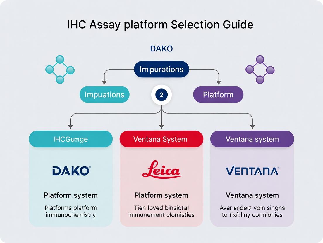
IHC Platform Selection Guide 2025: Dako vs. Leica vs. Ventana for Precision Research & Diagnostics
This comprehensive guide provides researchers, scientists, and drug development professionals with a critical analysis for selecting an immunohistochemistry (IHC) assay platform.
IHC Platform Selection Guide 2025: Dako vs. Leica vs. Ventana for Precision Research & Diagnostics
Abstract
This comprehensive guide provides researchers, scientists, and drug development professionals with a critical analysis for selecting an immunohistochemistry (IHC) assay platform. We explore the foundational technologies of Dako (Agilent), Leica Biosystems, and Roche Ventana, covering methodological workflows, optimization strategies for specific biomarkers, advanced troubleshooting, and data validation. The article offers a comparative framework to align platform capabilities with research intents—from exploratory discovery to clinical trial support—enabling informed, strategic investment in IHC infrastructure.
Understanding the IHC Platform Landscape: Core Technologies of Dako (Agilent), Leica, and Ventana
The Role of Automated IHC in Modern Biomarker Research and Precision Medicine
The selection of an immunohistochemistry (IHC) assay platform is a critical strategic decision in translational research and companion diagnostic development. The primary automated platforms—Agilent/Dako (Link), Leica Biosystems (BOND), and Roche/Ventana (BenchMark)—each offer distinct chemistries, workflows, and reagent ecosystems. This application note details protocols and analytical considerations for biomarker validation within the context of precision medicine, leveraging automated IHC for reproducibility, throughput, and quantitative analysis.
Key Platform Comparison and Quantitative Data
Table 1: Comparison of Major Automated IHC Platforms
| Feature | Agilent/Dako Link | Leica BOND | Roche/Ventana BenchMark |
|---|---|---|---|
| Core Detection Chemistry | EnVision FLEX (Polymer) | BOND Polymer Refine | UltraView / OptiView (Polymer) |
| Antigen Retrieval | PT Link (Separate) | Integrated on-board | Integrated on-board |
| Typely Protocol Time | ~2-3 hours | ~2-3 hours | ~2-3 hours |
| Slide Capacity | 48 slides (Link 48) | 30 slides (BOND III) | 30 slides (ULTRA) |
| Reagent Openness | Open for antibodies & reagents | Open system | Predominantly closed for detection |
| Primary Antibody Incubation | User-defined (5 mins - hours) | User-defined (15 mins - hours) | User-defined (8 mins - hours) |
| Multiplexing Capability | Yes (with DAB/AP, sequential) | Yes (BOND RxDx, sequential) | Yes (ultraView DAB & Red, simultaneous/sequential) |
| Commonly Used in CDx | High prevalence | High prevalence | High prevalence |
Table 2: Quantitative Performance Metrics in a PD-L1 (22C3) Validation Study*
| Platform | Assay | Average H-Score | Inter-Observer CV | Intra-Assay CV |
|---|---|---|---|---|
| Dako Link 48 | PD-L1 IHC 22C3 pharmDx | 145 | 8.2% | 4.1% |
| Leica BOND-III | Laboratory-Developed Test (LDT) | 138 | 9.5% | 5.7% |
| Ventana Benchmark ULTRA | PD-L1 (SP263) Assay | 152 | 7.8% | 3.9% |
*Illustrative data synthesized from recent literature and manufacturer white papers. CV = Coefficient of Variation.
Experimental Protocols
Protocol 1: Automated IHC Staining for a Predictive Biomarker (e.g., PD-L1) on Ventana BenchMark ULTRA
Application: Standardized staining for scoring algorithms (TPS, CPS). Materials: See "The Scientist's Toolkit" below. Procedure:
- Sectioning & Baking: Cut formalin-fixed, paraffin-embedded (FFPE) tissue sections at 4 µm. Bake slides at 60°C for 60 minutes.
- Deparaffinization & Retrieval: Load slides onto BenchMark ULTRA. Run protocol "Cell Conditioning 1 (CC1)" for 64 minutes at 95°C for antigen retrieval (standard for SP263 assay).
- Inhibition: Apply endogenous peroxidase inhibitor (ULTRA Inhibitor) for 4 minutes at 37°C.
- Primary Antibody: Apply anti-PD-L1 (SP263) primary antibody. Incubate for 32 minutes at 37°C.
- Detection: Apply UltraView Universal DAB Detection Kit (Horseradish Peroxidase (HRP) multimer + DAB Chromogen). Incubate per kit instructions (~8-12 minutes total).
- Counterstaining & Coverslipping: Apply Hematoxylin II for 8 minutes, then bluing reagent for 4 minutes. Automatically rinse, dehydrate, and coverslip.
Protocol 2: Sequential Multiplex IHC (mIHC) on Leica BOND-Rx
Application: Spatial profiling of multiple biomarkers (e.g., CD8, PD-1, FoxP3, PanCK). Materials: BOND Polymer Refine Detection Kits (DAB and Red), antibody stripping solution (e.g., BOND ER Solution). Procedure:
- First Cycle Staining: Perform standard IHC for first marker (e.g., PanCK, DAB) using the BOND Polymer Refine Red Detection system (configured for DAB output).
- Slide Imaging: Remove slide, scan at high resolution, then return to instrument.
- Antibody Elution: Perform on-instrument heat-induced antibody stripping using BOND ER Solution (20 minutes at 100°C).
- Second Cycle Staining: Perform IHC for second marker (e.g., CD8, using the Red chromogen).
- Repetition: Repeat steps 2-4 for subsequent markers (e.g., PD-1, FoxP3).
- Image Alignment & Analysis: Use digital pathology software to align sequential images and perform multiplex analysis.
Pathway and Workflow Visualizations
Title: Automated IHC Staining and Analysis Workflow
Title: PD-1/PD-L1 Immune Checkpoint Signaling Pathway
The Scientist's Toolkit: Key Research Reagent Solutions
| Item | Function & Role in Automated IHC |
|---|---|
| FFPE Tissue Microarrays (TMAs) | Contain multiple patient samples on one slide for high-throughput, standardized staining validation across cohorts. |
| Validated Primary Antibodies (CDx/LDT) | Clone-specific antibodies (e.g., 22C3, SP263, SP142) validated for a specific platform and indication. Critical for reproducibility. |
| Polymer-Based Detection Kits | Multi-step kits (e.g., EnVision FLEX, UltraView) providing secondary antibodies and enzymes linked to a dextran polymer backbone for high sensitivity. |
| Chromogen Substrates (DAB, Red) | Enzyme substrates that produce insoluble, stable colored precipitates at the antigen site (e.g., Brown DAB, Fast Red). |
| Automated Stainers (Link, BOND, BenchMark) | Integrated instruments that standardize all steps: deparaffinization, retrieval, staining, and coverslipping. |
| Digital Slide Scanners | High-throughput scanners for creating whole-slide images (WSI) for quantitative digital pathology analysis. |
| Image Analysis Software (e.g., QuPath, Halo) | Enables quantitative scoring of biomarker expression (H-score, TPS, CPS) and spatial analysis in mIHC. |
| Cell Conditioning Buffers (CC1, EDTA, Citrate) | Standardized, pH-controlled retrieval solutions critical for optimal epitope exposure for specific antibodies. |
Application Notes
The selection of an automated immunohistochemistry (IHC) platform is a critical strategic decision in research and drug development, influencing assay reproducibility, throughput, and compatibility with critical biomarkers. The Dako (Agilent) Autostainer Link 48, Leica Biosystems BOND series, and Roche Ventana BENCHMARK series represent three dominant, architecturally distinct paradigms. This analysis, within the context of IHC assay platform selection, details their core operational frameworks.
Dako Autostainer Link 48: This system employs an open, linear architecture. Slides are processed in batches on a carousel, with reagents dispensed by a moving pipetting arm. Its primary strengths lie in flexibility and cost-efficiency for established, user-optimized protocols. It accommodates a wide range of user-provided reagents and is not limited to a proprietary ecosystem, though it offers optimized "Link" kits for use with its FLEX visualization system. This open architecture places more responsibility on the user for protocol development and validation.
Leica BOND Series: The BOND systems (e.g., BOND-III, BOND-MAX) utilize a fully enclosed, random-access, on-board dye-based polymer detection system. Each slide is processed individually in a dedicated, self-contained chamber, which minimizes reagent consumption and cross-contamination risk. Its proprietary Refine Detection system (alkaline phosphatase or horseradish peroxidase polymer) is integral. The platform excels in sequential multiplexing (BOND Polymer Refine Detection) and offers robust automated dewaxing and epitope retrieval. Protocol development is conducted within the constraints of its proprietary reagent menu and onboard retrieval solutions.
Ventana BENCHMARK Series: The Ventana (Roche) BENCHMARK systems (e.g., BENCHMARK ULTRA, BENCHMARK GX) are monolithic, integrated platforms using a centralized, fluidic architecture. Slides are processed in parallel on a heated stage with reagents delivered via a centralized dispenser. Its proprietary UltraView or OptiView DAB detection kits are standard. The system is renowned for its highly standardized, "hands-off" operation from bake to stain, with extensive onboard reagent menus and proprietary pre-diluted antibodies. It is the dominant platform for companion diagnostics. Protocol flexibility is mediated through its extensive menu of validated "Roche Diagnostics" assays.
Table 1: Quantitative Platform Comparison
| Feature | Dako Autostainer Link 48 | Leica BOND-III | Ventana BENCHMARK ULTRA |
|---|---|---|---|
| Throughput (Max Slides/Run) | 48 | 30 | 30 |
| Slide Processing | Batch (Carousel) | Individual, Random-Access | Parallel, Batch |
| Reagent System | Open (User Reagents + Link Kits) | Proprietary (BOND Polymer Refine) | Proprietary (UltraView/OptiView) |
| Epitope Retrieval | Offline or Onboard (PT Link) | Onboard (Integrated) | Onboard (Integrated) |
| Key Detection | EnVision FLEX+ | BOND Polymer Refine | UltraView DAB |
| Multiplexing Capability | Sequential (Manual) | Sequential (Automated, i.e., BOND-MAX) | Sequential (Automated, iSFLEX) |
Experimental Protocols
Protocol 1: Standard IHC Staining on Leica BOND-MAX for PD-L1 (Clone 22C3)
- Objective: To demonstrate a standardized protocol using the BOND Polymer Refine Detection system.
- Materials: Leica BOND-MAX, FFPE tissue sections, BOND Primary Antibody Diluent, Anti-PD-L1 (22C3), BOND Polymer Refine Detection Kit (DS9800), BOND Epitope Retrieval Solution 2 (ER2).
- Procedure:
- Load slides and reagents (antibody diluted per manufacturer's recommendation) onto the instrument.
- Select protocol: BOND Polymer Refine Detection.
- Steps are automated: Dewaxing (BOND Dewax Solution), Epitope Retrieval (ER2, 20 min, ~100°C), Peroxide block (5 min), Primary antibody incubation (30 min, ambient), Post-primary block (8 min), Polymer incubation (8 min), DAB Mix incubation (10 min), Hematoxylin counterstain (5 min).
- Slides are automatically unloaded for manual dehydration, clearing, and mounting.
Protocol 2: Open Protocol Optimization on Dako Autostainer Link 48 for a Research Antibody
- Objective: To develop and optimize an IHC protocol using a non-proprietary primary antibody.
- Materials: Dako Autostainer Link 48, FFPE tissue sections, Target Retrieval Solution (pH 6 or 9), user's primary antibody, Dako EnVision FLEX+ (HRP) Detection System, Dako Wash Buffer.
- Procedure:
- Perform deparaffinization and heat-induced epitope retrieval offline using a PT Module or water bath.
- Load pre-treated slides onto the Autostainer Link 48.
- Program the method: Peroxidase block (5 min), Primary antibody incubation (e.g., 30-60 min, user-optimized concentration), Labeled Polymer (HRP) incubation (20 min), DAB+ Substrate incubation (10 min).
- The instrument dispenses all liquid reagents. Slides are unloaded for manual counterstaining, dehydration, and mounting.
Protocol 3: Multiplex IHC on Ventana BENCHMARK ULTRA using iFLEX
- Objective: To perform sequential staining for two markers (e.g., CD8 and CD68) on a single slide.
- Materials: Ventana BENCHMARK ULTRA, FFPE tissue sections, UltraView DAB Kit (V1), UltraView Alkaline Phosphatase Red Kit (V2), Anti-CD8 (SP57), Anti-CD68 (KP1), Ventana Reaction Buffer, CC2 retrieval solution.
- Procedure:
- Load slides and reagents. Create a iFLEX method in the instrument software.
- First Stain Cycle: CC2 retrieval (64 min), Anti-CD8 primary (32 min, 36°C), UltraView DAB detection (incubation, HQ, copper), Apply Antibody Removal step (disabling reagent).
- Second Stain Cycle: CC2 retrieval (second application, 32 min), Anti-CD68 primary (32 min, 36°C), UltraView Alkaline Phosphatase Red detection.
- Slides are automatically counterstained with Hematoxylin II and Bluing Reagent, then unloaded for manual coverslipping.
Visualizations
Leica BOND-MAX Standard IHC Workflow (62 chars)
IHC Platform Selection Decision Logic (55 chars)
The Scientist's Toolkit: Key Research Reagent Solutions
| Item | Platform Association | Function in IHC |
|---|---|---|
| EnVision FLEX+ Detection System | Dako (Agilent) | A dextran-based polymer conjugated with HRP enzymes, offering high sensitivity and low background. Core to Dako's "Link" platform. |
| BOND Polymer Refine Detection Kit | Leica Biosystems | An integrated, on-board polymeric HRP or AP detection system. Includes post-primary linker and polymer in a ready-to-use format, minimizing hands-on steps. |
| UltraView DAB Detection Kit | Ventana (Roche) | A universal, multimer-based detection system using HRP. Designed for use on Ventana platforms, optimized for stability and consistency with onboard dispensing. |
| BOND Epitope Retrieval Solutions (ER1/ER2) | Leica Biosystems | Proprietary citrate-based (ER1) or EDTA-based (ER2) retrieval solutions for use in the onboard retrieval system. |
| Ventana Cell Conditioning (CC) Solutions | Ventana (Roche) | A series of proprietary, standardized buffers (e.g., CC1, CC2) used for onboard heat-induced epitope retrieval. |
| Primary Antibody Diluent | All Platforms | A protein-based buffer used to dilute primary antibodies, stabilizing the antibody and reducing non-specific background staining. |
| DAB+ Chromogen Substrate | All Platforms | A solution of 3,3'-Diaminobenzidine tetrahydrochloride and hydrogen peroxide. Produces a brown precipitate at the site of HRP enzyme activity. |
Within the critical landscape of immunohistochemistry (IHC) assay platform selection—encompassing Dako/Agilent, Leica Biosystems, and Ventana Medical Systems/Roche—the choice of core detection chemistry is a primary determinant of assay performance. These polymer-based systems have largely replaced traditional avidin-biotin methods, offering enhanced sensitivity and specificity while mitigating endogenous biotin interference. This application note provides a detailed comparison of three dominant polymer detection chemistries: Dako's EnVision FLEX, Leica's BOND Polymer Refine, and Ventana's UltraView/OptiView, furnishing researchers and drug development professionals with quantitative data, protocols, and practical insights for platform selection in regulated and research environments.
Core Chemistry Principles & Comparative Data
Underlying Technology
- EnVision FLEX (Agilent/Dako): A dextran-based polymer backbone heavily conjugated with horseradish peroxidase (HRP) or alkaline phosphatase (AP) enzymes and secondary antibodies. The polymer linker provides high enzyme-to-antibody ratio, amplifying signal without streptavidin-biotin steps.
- BOND Polymer Refine (Leica Biosystems): Utilizes a patented "FabuLight" polymer technology, where the polymer is conjugated with anti-rabbit or anti-mouse IgG Fab fragments (not whole antibodies). This is designed for minimal Fc-mediated non-specific binding.
- UltraView & OptiView (Ventana/Roche): UltraView is a multimer-based (not strictly a polymer) detection system using a complex of enzymes and antibodies. OptiView is its enhanced successor, formulated for improved sensitivity and lower background, often used with amplification steps.
Quantitative Performance Comparison
Table 1: Core Characteristics & Performance Metrics
| Feature | EnVision FLEX | BOND Polymer Refine | UltraView / OptiView |
|---|---|---|---|
| Core Technology | Dextran Polymer-HRP/IgG | FabuLight Polymer-Fab/HRP | Multimer / Polymer-HRP |
| Typical Incubation | 20-30 min at RT | 8-15 min at RT (on BOND) | 8-16 min at 37°C (on Benchmark) |
| Endogenous Biotin Block | Not required | Not required | Required for some UltraView apps |
| Sensitivity (vs. LSAB) | ~8-10x increase | ~4-8x increase | UltraView: ~4x; OptiView: >8x |
| Common Chromogens | DAB+, DAB, Permanent Red | DAB, DAB Enhance, AP Red | DAB, DAB with Amplification, Red |
| Compatibility | Autostainer Linker, Manual | BOND series autostainers | Benchmark/Ventana series autostainers |
| Multiplex Capability | Yes (FLEX Multimer) | Yes (on BOND) | Yes (on Ventana) |
Table 2: Typical Protocol Durations (From Deparaffinization to Counterstain)
| Step | EnVision FLEX (Manual) | BOND Polymer Refine (on BOND-III) | OptiView (on Benchmark Ultra) |
|---|---|---|---|
| Primary Antibody | 30-60 min | 15-30 min | 16-64 min |
| Link/Post Primary | 20 min (Flex+) | 8-15 min (Post Primary) | 8-12 min (OptiView Amp) |
| Polymer | 20-30 min | 8-15 min | 8-16 min |
| DAB Incubation | 5-10 min | 10 min | 8-32 min (with amp) |
| Total Hands-on Time | ~2-2.5 hrs | ~1.5-2 hrs (automated run) | Fully automated (~3-5 hr run) |
Detailed Experimental Protocols
Protocol for EnVision FLEX (Manual, High Sensitivity)
Application: Detection of low-abundance nuclear antigens (e.g., Ki-67, p53).
I. Materials (The Scientist's Toolkit)
- EnVision FLEX HRP Polymer (SM802): Dextran polymer conjugated with HRP and secondary antibodies. Core detection reagent.
- EnVision FLEX Target Retrieval Solution (High pH, SM802): Tris/EDTA buffer for heat-induced epitope retrieval.
- EnVision FLEX Wash Buffer (SM802): Buffered surfactant solution for rinsing slides.
- EnVision FLEX DAB+ Chromogen (DM827): 3,3’-Diaminobenzidine tetrahydrochloride substrate for HRP, yielding a brown precipitate.
- EnVision FLEX Substrate Buffer (DM827): Peroxide-containing buffer for DAB reaction.
- Hydrophobic Barrier Pen: To create a liquid boundary around tissue sections.
- Humidified Chamber: To prevent evaporation during incubations.
II. Methodology
- Deparaffinization & HIER: Deparaffinize slides in xylene and graded alcohols. Perform heat-induced epitope retrieval in High pH Target Retrieval Solution at 97°C for 20 min in a decloaking chamber. Cool for 20 min.
- Peroxidase Blocking: Incubate with Endogenous Enzyme Block (3% H₂O₂) for 10 min at RT. Rinse with Wash Buffer.
- Primary Antibody: Apply optimized primary antibody (e.g., anti-Ki-67, clone MIB-1, 1:200) for 60 minutes at RT.
- Polyver Detection: Rinse slides. Apply EnVision FLEX HRP Polymer (Mouse/Rabbit) for 30 minutes at RT.
- Visualization: Prepare DAB+ working solution (1 drop DAB+ Chromogen per 1 mL Substrate Buffer). Apply to slides for 5-10 minutes, monitoring development under a microscope.
- Counterstain & Mount: Rinse thoroughly in water. Counterstain with Hematoxylin for 1-5 min. Dehydrate, clear, and mount with permanent mounting medium.
Protocol for BOND Polymer Refine on Leica BOND-III
Application: Sequential multiplex IHC for immune cell profiling (e.g., CD8/CD68).
I. Materials (The Scientist's Toolkit)
- BOND Polymer Refine Detection Kit (DS9800): Contains Post Primary Reagent (IgG linker) and Polymer-HRP.
- BOND Epitope Retrieval Solutions (ER1/ER2): Citrate- or EDTA-based solutions for automated HIER.
- BOND Wash Solution (AR9590): Diluent for washing steps on the BOND platform.
- BOND DAB Chromogen (AR9432): DAB substrate kit for the BOND system.
- BOND Primary Antibody Diluent (AR9352): Optimized buffer for antibody stability on the instrument.
II. Methodology
- Load & Bake: Load FFPE sections onto charged slides into the BOND-III. The instrument performs automated baking at 60°C.
- Automated Deparaffinization & HIER: The instrument uses Dewax and HIER solutions (e.g., ER2, EDTA pH 9.0) for 20 min at 100°C.
- Primary Antibody (1st Round): Instrument applies the first primary antibody (e.g., anti-CD8, 1:100) for 30 minutes at RT.
- Polymer Refine Detection (1st Round): Sequential automated application of: a) Post Primary Reagent (8 min), b) Polymer-HRP (8 min), c) BOND DAB (10 min).
- Antibody Elution: A critical step for multiplexing. The instrument performs BOND ER2-based stripping at 100°C for 10-20 minutes to remove the first immune complex.
- Repeat for 2nd Marker: The cycle repeats from step 3 with the second primary antibody (e.g., anti-CD68).
- Counterstain: Automated Hematoxylin counterstaining for 5 min. Slides are removed, dehydrated, cleared, and mounted.
Protocol for UltraView/OptiView on Ventana Benchmark Ultra
Application: HER2/neu (ERBB2) testing in breast cancer with signal amplification.
I. Materials (The Scientist's Toolkit)
- OptiView DAB IHC Detection Kit (760-700): Contains HQ linker, multimer-HRP, H₂O₂ inhibitor, DAB, and copper enhancer.
- OptiView Amplification Kit (760-099): Contains an amplification multimer for ultra-sensitive detection.
- Cell Conditioning Solution (CC1, 950-124): Tris-based, pH 8.5, for standard HIER on Ventana systems.
- Reaction Buffer (950-300): Universal washing buffer.
- Liquid Coverslip (650-010): Proprietary oil-based fluid to prevent evaporation during high-temp incubations.
II. Methodology
- Load & Condition: Load slide onto Benchmark Ultra. Apply Cell Conditioning 1 (CC1) for 64 minutes at 95-100°C for standard HIER.
- Primary Antibody: Apply anti-HER2/neu (clone 4B5, ready-to-use) for 32 minutes at 37°C.
- Amplified Detection: The system sequentially applies:
- OptiView HQ Linker (8 min, 37°C): Links primary antibody to amplification system.
- OptiView HRP Multimer (8 min, 37°C): Contains multiple enzyme molecules.
- OptiView Amplifier (8 min, 37°C): Further increases sensitivity.
- OptiView DAB + DAB Enhancer (Copper) (8 min each, 37°C): For chromogenic visualization.
- Counterstain: Automated application of Hematoxylin II (12 min) and Bluing Reagent (4 min).
- Finalization: The instrument completes the run. Slides are removed, rinsed in warm soapy water, dehydrated, cleared, and mounted.
Pathway & Workflow Visualizations
Polymer Detection Chemistry Signal Pathway
Decision Logic for Platform Selection
Within the critical process of immunohistochemistry (IHC) assay platform selection for research and drug development, the choice between open and closed systems is paramount. This application note, framed within the context of evaluating major platforms from Dako (Agilent), Leica Biosystems, and Roche (Ventana), details the operational, compatibility, and performance trade-offs between these architectures. The core thesis is that system selection dictates workflow flexibility, reagent choice, cost, and ultimately, data reliability.
System Architectures: Core Definitions
Closed Systems
Proprietary, integrated platforms where the instrument, detection chemistry, and often the bulk of reagents are supplied by a single vendor. Protocols are typically pre-programmed and optimized for vendor-specific reagents.
- Examples: Roche Ventana BenchMark series, Leica BOND series.
- Key Trait: Reagent "locked-down" or validated for use only on that system, often employing proprietary dispensing and detection.
Open Systems
Platforms designed to accommodate reagents and protocols from multiple suppliers. The instrument automates steps but allows user-defined reagent sequences and concentrations.
- Examples: Dako (Agilent) Autostainer Link series, Leica ST series.
- Key Trait: Flexibility to use "Home-Brew" or Research-Use-Only (RUO) antibodies and reagents from various sources.
Comparative Data Analysis
Table 1: Core Differentiators Between Open and Closed IHC Platforms
| Feature | Open System (e.g., Dako Autostainer Link) | Closed System (e.g., Ventana BenchMark ULTRA) |
|---|---|---|
| Reagent Compatibility | High. Compatible with antibodies & detection kits from multiple vendors (Dako, Leica, Cell Signaling, etc.). | Restricted. Primarily optimized for vendor's proprietary reagents (Ventana/Roche). |
| Protocol Flexibility | High. User can modify incubation times, temperatures, rinse steps, and reagent concentrations. | Low to Moderate. Uses pre-loaded, vendor-validated protocols; modifications are limited. |
| Throughput & Walk-Away Time | Variable. Often batch-based; runtime depends on protocol length. | Typically High. Continuous loading & proprietary rapid protocols reduce hands-on time. |
| Assay Development | Essential for novel targets. Requires optimization of antibody titration, retrieval, and detection. | Streamlined. Pre-optimized "ready-to-use" assays; development focuses on finding optimal pre-set protocol. |
| Cost Structure | Lower reagent cost per test (competitive sourcing). Higher labor cost for optimization. | Higher reagent cost per test. Lower optimization labor cost. |
| Reproducibility | Dependent on user optimization and reagent lot consistency. | High, due to standardized, controlled protocols and reagents. |
| Primary Use Case | Research, biomarker discovery, novel target validation. | Clinical diagnostics, high-volume validated assays, clinical trials. |
Table 2: Reagent Compatibility Matrix for Major Platforms (Representative)
| Platform / System Type | Primary Detection System | Compatible Antibody Sources | Key Restriction |
|---|---|---|---|
| Roche Ventana BenchMark (Closed) | UltraView, OptiView, ChromoMap | Predominantly Ventana/Roche RUO/IVD | Non-Ventana antibodies require adaptation kits and may not be validated. |
| Leica BOND (Semi-Closed/Closed) | BOND Polymer Refine Detection | Leica, Novocastra; others with BOND OPEN protocol | "BOND OPEN" system allows some external reagents but with specific diluent requirements. |
| Dako Autostainer Link (Open) | EnVision FLEX | Dako/Agilent, CST, Abcam, in-house conjugates | Broad compatibility; optimization for concentration and retrieval is user's responsibility. |
| Leica ST5020 (Open) | A variety of polymer-based kits | Virtually any supplier | Full user control over all protocol parameters and reagent application. |
Experimental Protocols
Protocol A: Validating a Novel Primary Antibody on an Open System (Dako Autostainer Link)
Objective: To optimize and validate a novel rabbit monoclonal antibody (Target X) on FFPE tissue sections using an open staining platform. Materials: Dako Autostainer Link 48, FFPE tissue microarray (TMA) containing positive/negative controls, Target X antibody (rabbit mAb), Dako EnVision FLEX+ HRP Rabbit (Linker), Dako PT Link for retrieval (pH 9), Dako Wash Buffer.
Procedure:
- Deparaffinization & Retrieval: Bake slides 60 min at 60°C. Perform heat-induced epitope retrieval (HIER) in PT Link at 97°C for 20 min using FLEX High pH (pH 9) buffer.
- Instrument Setup: Power on Autostainer Link and prime with wash buffer.
- Reagent Loading: Load the following onto designated reagent trays:
- Tray 1: Endogenous Enzyme Block (3% H₂O₂), 5 min.
- Tray 2: Primary Antibody (Target X) at three pre-determined dilutions (1:50, 1:100, 1:200) in antibody diluent on sequential slides.
- Tray 3: EnVision FLEX+ HRP Rabbit (Linker), 20 min.
- Tray 4: FLEX DAB+ Chromogen, 10 min.
- Program Protocol: Create a new protocol in LinkSoftware:
- Step 1: Enzyme Block, 5 min.
- Step 2: Primary Antibody, 30 min at RT.
- Step 3: Wash Buffer, 5 min.
- Step 4: Linker, 20 min.
- Step 5: Wash Buffer, 5 min.
- Step 6: DAB, 10 min.
- Step 7: Wash Buffer, 5 min.
- Run & Counterstain: Load slides, start run. Upon completion, counterstain with hematoxylin, dehydrate, clear, and mount.
Protocol B: Adapting a Vendor Antibody on a Closed System (Ventana BenchMark ULTRA)
Objective: To run a non-Ventana rabbit antibody on a Ventana system using an "Open" or "Generic" protocol. Materials: Ventana BenchMark ULTRA, FFPE TMA, Cell Signaling Technology (CST) rabbit antibody, Ventana OptiView HQ Universal DAB Detection Kit, Ventana Ultra Cell Conditioning solution (CC1).
Procedure:
- Slide Preparation: Load FFPE slides onto BenchMark ULTRA bar-coded slide holders.
- Reagent Loading: Load reagents per OptiView DAB kit instructions. Load CST antibody, prediluted in Ventana Antibody Diluent, into a designated reagent well.
- Protocol Selection: Navigate to "Protocol Manager." Select a generic "No Primary" protocol or copy an existing rabbit antibody protocol (e.g., "DISCOVERY RUO" template).
- Protocol Editing:
- Deparaffinization: Standard (72°C).
- Retrieval: CC1, 64 min at 95°C-100°C (standard).
- Primary Antibody: Manually map the CST antibody location. Set incubation time to 32 min at 36°C (typical starting point).
- Detection: Select OptiView HQ Universal DAB. All detection steps (HQ Inhibitor, Linker, HRP, DAB, Copper) are pre-set.
- Counterstain: Select Hematoxylin II for 8 min, then Bluing Reagent for 4 min.
- Run Initiation: Load the carousel, start run. The run is fully automated through coverslipping.
Visualization Diagrams
IHC System Decision Pathway
IHC Platform Selection Workflow
The Scientist's Toolkit: Key Research Reagent Solutions
Table 3: Essential Reagents for IHC Assay Development & Cross-Platform Use
| Item | Function & Relevance to Open/Closed Systems |
|---|---|
| Universal HRP Polymer Detection Kits (e.g., EnVision FLEX, MACH, Ultravision) | Broadly compatible secondary systems for open platforms. Some "universal" kits are validated for limited use on closed systems (e.g., on BOND OPEN). |
| Antigen Retrieval Buffers (pH 6 Citrate, pH 9 EDTA/Tris) | Critical for open system optimization. Closed systems use proprietary buffers (e.g., Ventana CC1, Leica ER). |
| Antibody Diluent with Stabilizers | Essential for preparing and storing primary antibody working solutions on open systems. Specific diluents (e.g., Ventana) are required for closed systems. |
| Automation Fixative & Wash Buffer | Proprietary solutions (e.g., Dako Wash Buffer) are optimized for specific open platforms to prevent drying and ensure fluidics performance. |
| Validation Control Tissue Microarrays (TMAs) | Contain multiple tumor and normal tissues. Indispensable for optimizing/validating antibody specificity and sensitivity on any platform. |
| Chromogen (DAB) Substrate Kits | Available from multiple vendors for open systems. Closed systems use integrated, proprietary chromogen cartridges. |
| Protease Enzyme (e.g., Proteinase K) | For enzyme-induced epitope retrieval (EIER), an alternative to HIER, sometimes required for specific membrane targets. Used on both system types. |
The selection of an IHC platform (Dako/Agilent, Leica Biosystems, Ventana/Roche) is a critical strategic decision that must align with the core philosophy of the research or diagnostic workflow. Each platform is engineered with distinct strengths, optimized for different stages of the therapeutic development pipeline.
Platform Philosophy & Quantitative Comparison
The following table summarizes the core attributes, optimal use cases, and quantitative performance metrics associated with each major platform.
Table 1: IHC Platform Comparison for Project Goal Alignment
| Feature / Metric | Dako/Agilent (Link/Omnis) | Leica Biosystems (BOND) | Ventana/Roche (BenchMark) |
|---|---|---|---|
| Core Philosophy | Flexibility & Open System | Balanced & Reproducible | Integrated & Standardized |
| Optimal Workflow | Discovery & Translational Research | Translational & Early Clinical | Clinical Diagnostics & Pivotal Trials |
| Primary Strength | Broad antibody compatibility, user-controlled optimization | Robust protocols, excellent for phospho-targets & FFPE | High-throughput, locked clinical assays (e.g., HER2, PD-L1) |
| Assay Openness | High (Open) | Medium (Controlled Open) | Low (Closed, proprietary reagents) |
| Typical Automation | Semi-automated (Link) to Fully Automated (Omnis) | Fully Automated (BOND III, RX) | Fully Automated (BenchMark ULTRA, XT) |
| Throughput (Slides/Run) | 12-30 | 30-40 | 30-40+ |
| Key Detection Chemistry | EnVision (Polymer) | BOND Polymer Refine | UltraView / OptiView (Multimer) |
| Protocol Flexibility | High – User can adjust each step | Medium – Pre-set protocols with editable steps | Low – Predominantly pre-optimized, FDA-cleared protocols |
| Clinical Compliance | Requires extensive validation | Suitable for validated lab-developed tests (LDTs) | Designed for CAP/CLIA, FDA-cleared assays |
Experimental Protocols for Platform Assessment
Protocol 1: Cross-Platform Antibody Titration for Discovery Research
Aim: To establish optimal staining conditions for a novel target on FFPE tissue using open (Dako/Leica) systems.
- Sectioning & Baking: Cut 4μm FFPE sections onto positively charged slides. Bake at 60°C for 60 minutes.
- Deparaffinization & Rehydration: Use xylene (3 changes, 5 min each) and graded ethanol (100%, 95%, 70%, 5 min each).
- Antigen Retrieval (AR):
- Dako PT Link: Use Target Retrieval Solution (pH 6 or 9) at 97°C for 20 minutes.
- Leica BOND: Use ER1 (pH 6) or ER2 (pH 9) solution for 20-30 minutes at 100°C (Epitope Retrieval mode).
- Peroxidase Blocking: Incubate with 3% H₂O₂ for 10 minutes.
- Primary Antibody Titration: Apply antibody at 4-5 concentrations (e.g., 0.5, 1, 2, 4, 8 μg/mL) for 30-60 minutes at room temperature.
- Detection: Apply appropriate polymer-based detection system (e.g., EnVision Flex/HRP for Dako; BOND Polymer Refine Detection for Leica) for 20-30 minutes.
- Visualization: Incubate with DAB chromogen for 5-10 minutes, monitor microscopically.
- Counterstaining & Mounting: Counterstain with Hematoxylin, dehydrate, clear, and mount with permanent medium.
Protocol 2: Validation of a Translational Biomarker on Leica BOND Platform
Aim: To develop a standardized, reproducible assay for a phospho-protein biomarker in a cohort study.
- Slide Preparation: As per Protocol 1, steps 1-2.
- Automated Run on BOND-RX:
- Load slides and reagents.
- Select a pre-set BOND Polymer Refine Detection protocol.
- AR: ER2 solution, 30 minutes at 100°C.
- Primary Antibody: Optimized concentration, 30 minutes at room temperature. Include isotype controls.
- Post-Primary & Polymer: Apply sequentially per Refine kit protocol.
- DAB & Copper: Apply DAB followed by copper enhancer to amplify weak signals.
- Hematoxylin Counterstain: Automated on instrument.
- Dehydration & Coverslipping: Offline using automated coverslipper.
Protocol 3: Clinical PD-L1 (SP142) Assay on Ventana BenchMark ULTRA
Aim: To perform a locked, clinically validated companion diagnostic assay.
- Slide & Reagent Setup: Load pre-cut slides and the VENTANA PD-L1 (SP142) Assay kit (Primary Ab, Detection) onto the instrument.
- Run Selection: Select the validated "PD-L1 (SP142) - Clinical" protocol.
- Fully Automated Processing:
- Instrument performs baking, deparaffinization (EZ Prep solution), and AR (Ultra CC1 solution, 64-100°C).
- Automated application of primary antibody (32 minutes at 36°C).
- Application of UltraView Universal DAB Detection Kit (Haemoglobin then DAB Incubation).
- All steps are timed and temperature-controlled with no user intervention.
- Counterstaining & Coverslipping: Instrument applies hematoxylin II and bluing reagent, followed by automated coverslipping.
Workflow & Pathway Diagrams
IHC Platform Selection Workflow Map
Core IHC Staining Pathway & Variables
The Scientist's Toolkit: Key Research Reagent Solutions
Table 2: Essential IHC Reagents and Their Functions
| Reagent Category | Specific Example(s) | Primary Function | Platform Considerations |
|---|---|---|---|
| Antigen Retrieval Buffers | Citrate (pH 6.0), Tris-EDTA/ER2 (pH 9.0) | Break protein cross-links, expose epitopes masked by formalin fixation. | Choice is platform- and antibody-specific. Ventana uses proprietary CC1/CC2. |
| Primary Antibodies | Monoclonal (rabbit, mouse), Polyclonal | Specific binding to target protein of interest. | Open systems allow use of any antibody; Ventana assays require FDA-cleared IVD counterparts. |
| Detection Systems | EnVision Flex (Dako), BOND Polymer Refine (Leica), UltraView (Ventana) | Enzyme-conjugated polymers that bind primary Ab, catalyze chromogen deposition. | Highly proprietary. Directly impacts sensitivity, background, and multiplexing potential. |
| Chromogens | DAB (Brown), AEC (Red), Vector VIP (Purple) | Enzyme substrate that produces an insoluble, colored precipitate at the antigen site. | DAB is most common. Ventana uses proprietary formulation with enhanced stability. |
| Counterstains | Hematoxylin (Harris, Mayer's), Methyl Green | Provides contrast nuclear stain for histological context. | Automated platforms include proprietary formulations as part of the staining run. |
| Blocking Reagents | Serum (goat, rabbit), Casein, H₂O₂ | Reduce non-specific background staining and block endogenous peroxidase activity. | Often included in detection kits. Optimization may be needed for novel targets in research. |
| Mounting Media | Aqueous (for fluorescent), Permanent (e.g., Pertex) | Preserves stain and provides optical clarity for microscopy. | Critical for slide longevity. Automated platforms often integrate coverslipping. |
Optimizing Your Workflow: Step-by-Step Protocols and Application-Specific Strategies
Standardized Protocols for Common Biomarkers (e.g., PD-L1, HER2, Ki-67) Across Platforms
Immunohistochemistry (IHC) is a cornerstone of diagnostic and research pathology, enabling the visualization of protein expression within tissue architecture. The selection of an IHC platform—Dako (Agilent), Leica Biosystems, and Ventana (Roche)—profoundly impacts assay performance, reproducibility, and clinical interpretation. This application note, framed within a broader thesis on IHC assay platform selection, details standardized protocols and comparative data for three critical biomarkers: PD-L1 (immune checkpoint), HER2 (therapeutic target), and Ki-67 (proliferation index). Achieving harmonization across these diverse automated staining systems is essential for reliable data in both drug development and clinical research.
Quantitative Comparison of Platform-Specific Protocols
Table 1: Key Protocol Variables for Common Biomarkers Across Major IHC Platforms
| Biomarker (Primary Use) | Dako (Agilent) Platform (e.g., Autostainer Link 48) | Leica Biosystems Platform (e.g., BOND-III) | Ventana (Roche) Platform (e.g., Benchmark ULTRA) | Concordance Notes |
|---|---|---|---|---|
| PD-L1 (22C3) (Non-small cell lung cancer) | Clone: 22C3 Platform: Autostainer Link 48 Ag Retrieval: PT Link, High pH (FSH) Incubation: 30 min Detection: EnVision FLEX+ | Clone: 22C3 Platform: BOND-III Ag Retrieval: ER2 (HIER) for 20 min Incubation: 15 min Detection: BOND Polymer Refine | Clone: 22C3* Platform: Benchmark ULTRA Ag Retrieval: CCI (HIER) for 64 min Incubation: 16 min Detection: OptiView DAB IHC | *FDA-approved as a "companion diagnostic" on Dako only. Ventana use is an "assay modification." Scoring criteria (TPS) must be strictly adhered to. |
| HER2 (Breast/Gastric cancer) | Clone: Polyclonal (A0485) Platform: Autostainer Link 48 Ag Retrieval: PT Link, Low pH (FSL) Incubation: 30 min Detection: EnVision FLEX+ | Clone: EP3 Platform: BOND-III Ag Retrieval: ER2 (HIER) for 20 min Incubation: 15 min Detection: BOND Polymer Refine | Clone: 4B5 Platform: Benchmark ULTRA Ag Retrieval: CCI (HIER) for 64 min Incubation: 16 min Detection: OptiView DAB IHC | Different clones require rigorous validation against ISH. ASCO/CAP scoring guidelines (0, 1+, 2+, 3+) apply. Dako A0485 and Ventana 4B5 are FDA-approved. |
| Ki-67 (Proliferation index) | Clone: MIB-1 Platform: Autostainer Link 48 Ag Retrieval: PT Link, High pH (FSH) Incubation: 30 min Detection: EnVision FLEX+ | Clone: MM1 Platform: BOND-III Ag Retrieval: ER1 (HIER) for 20 min Incubation: 15 min Detection: BOND Polymer Refine | Clone: 30-9 Platform: Benchmark ULTRA Ag Retrieval: CCI (HIER) for 36 min Incubation: 16 min Detection: OptiView DAB IHC | High inter-platform variability in absolute indices. Critical to use standardized counting methods (e.g., hot-spot vs. global) and internal controls. |
Table 2: Comparative Performance Metrics from Recent Inter-Platform Studies
| Study Focus | Key Quantitative Finding | Implication for Platform Selection |
|---|---|---|
| PD-L1 (22C3) Concordance | Dako vs. Ventana modification showed 92% agreement at TPS ≥1% cutoff, but dropped to 85% at TPS ≥50% cutoff in NSCLC. | For high-stakes cutoffs (e.g., 50% for pembrolizumab), platform-specific validation is mandatory. |
| HER2 IHC (4B5 vs. A0485) | Ventana 4B5 showed 95% concordance with Dako A0485 for 0/1+ and 3+ scores. Discordance primarily in 2+ (equivocal) cases (~15%). | Equivocal cases must be confirmed by ISH regardless of platform. Leica EP3 shows similar high concordance with 4B5. |
| Ki-67 Index Variability | Median Ki-67 index difference of up to 8% reported between Dako MIB-1 and Ventana 30-9 on same tumor blocks. | Longitudinal studies or clinical trials should mandate a single, consistent platform and protocol. |
Detailed Standardized Protocols
Protocol for PD-L1 (22C3) IHC on Ventana Benchmark ULTRA (Modified from Dako Protocol)
This protocol exemplifies the steps required to adapt a companion diagnostic assay to an alternative platform.
I. Reagent Preparation
- Primary Antibody: Prediluted Ventana anti-PD-L1 (22C3) pharmDx assay or laboratory-developed test (LDT) using clone 22C3, optimally titrated.
- Detection Kit: Ventana OptiView DAB IHC Detection Kit.
- Antigen Retrieval: Ventana Cell Conditioning 1 (CCI, pH 8.5).
- Controls: Known positive (NSCLC with TPS>50%) and negative (tonsil) tissue sections.
II. Staining Procedure (Automated on Benchmark ULTRA)
- Deparaffinization & Conditioning: Apply standard baking and deparaffinization steps. Perform antigen retrieval with CCI for 64 minutes at 95-100°C.
- Primary Antibody Incubation: Apply anti-PD-L1 (22C3) antibody and incubate at 36°C for 16 minutes.
- Detection: Apply OptiView HQ Universal Linker for 8 minutes, followed by OptiView HRP Multimer for 8 minutes.
- Visualization: Apply OptiView DAB for 8 minutes, then OptiView Copper for 4 minutes to enhance DAB chromogen.
- Counterstaining & Coverslipping: Apply Hematoxylin II for 12 minutes, then Bluing Reagent for 4 minutes. Manually dehydrate, clear, and coverslip.
III. Interpretation Score using Tumor Proportion Score (TPS): Percentage of viable tumor cells with partial or complete membrane staining at any intensity. Report using clinically relevant cutoffs (e.g., ≥1%, ≥50%).
Protocol for HER2 (4B5) IHC on Leica BOND-III
This protocol standardizes the FDA-approved Ventana 4B5 assay for the Leica platform.
I. Reagent Preparation
- Primary Antibody: Rabbit monoclonal anti-HER2, clone 4B5 (Ventana).
- Detection System: BOND Polymer Refine Detection Kit (DS9800).
- Antigen Retrieval: BOND Epitope Retrieval Solution 2 (ER2, pH 9.0).
- Controls: Breast carcinoma with known HER2 scores (0, 1+, 2+, 3+).
II. Staining Procedure (Automated on BOND-III)
- Deparaffinization: Performed on-board with BOND Dewax Solution.
- Antigen Retrieval: ER2 for 20 minutes at 100°C.
- Peroxidase Block: Incubate with peroxide block for 5 minutes.
- Primary Antibody: Apply anti-HER2 (4B5) at optimized dilution (typically 1:100-1:200) and incubate at ambient temperature for 15 minutes.
- Post Primary & Polymer: Apply Post Primary reagent for 8 minutes, then Polymer for 8 minutes.
- Visualization: Apply DAB Chromogen for 10 minutes.
- Counterstaining: Apply Hematoxylin for 5 minutes.
III. Interpretation Score per ASCO/CAP 2018 guidelines: 0 (no staining), 1+ (faint/barely perceptible membrane staining), 2+ (weak to moderate complete membrane staining), 3+ (circumferential intense membrane staining). 2+ results require reflex ISH testing.
Protocol for Ki-67 (MIB-1) IHC on Dako Autostainer Link 48
A standardized research protocol for proliferation index assessment.
I. Reagent Preparation
- Primary Antibody: Mouse monoclonal anti-Ki-67, clone MIB-1 (Dako, IR626).
- Detection System: EnVision FLEX+ Detection System (High pH, K8002).
- Antigen Retrieval: EnVision FLEX Target Retrieval Solution, High pH (50x).
- Controls: Tonsil or lymph node (known germinal center high, mantle zone low).
II. Staining Procedure (Automated on Autostainer Link 48 with PT Link)
- Deparaffinization & Retrieval (PT Link): Bake slides, deparaffinize. Perform antigen retrieval in PT Link using High pH buffer at 97°C for 20 minutes, followed by cool-down to 65°C.
- Peroxidase Block: Apply FLEX Peroxidase-Blocking Reagent for 5 minutes.
- Primary Antibody Incubation: Apply anti-Ki-67 (MIB-1) at optimized dilution (typically 1:200) and incubate at ambient temperature for 30 minutes.
- Visualization: Apply FLEX/HRP Enzyme for 20 minutes, then FLEX DAB+ Chromogen for 10 minutes.
- Counterstaining & Mounting: Apply FLEX Hematoxylin for 5 minutes. Rinse, dehydrate, clear, and mount.
III. Interpretation Quantify by digital image analysis or manual counting. Report as the percentage of positively stained tumor cell nuclei among total viable tumor cells. Clearly define the counting method (e.g., hot-spot vs. average across 3-5 high-power fields).
Visualization: Pathways and Workflows
Diagram 1: PD-1/PD-L1 Immune Checkpoint Pathway
Diagram 2: HER2 Receptor Dimerization & Downstream Signaling
Diagram 3: IHC Assay Development & Validation Workflow
The Scientist's Toolkit: Essential Research Reagent Solutions
Table 3: Key Reagents and Materials for Cross-Platform IHC Standardization
| Item | Function & Description | Platform Consideration |
|---|---|---|
| Certified Reference Cell Lines (e.g., PD-L1 expressing) | Provide standardized positive and negative protein expression controls for assay calibration and run-to-run monitoring. | Can be formalin-fixed and paraffin-embedded (FFPE) into cell pellets compatible with all platforms. |
| Multiplex Fluorescence IHC Validation Kits | Allow simultaneous detection of biomarker and co-markers (e.g., PD-L1 + Pan-CK + CD8) to confirm cellular context and assay specificity. | Platform-specific kits exist (e.g., Ventana DISCOVERY, Leica BOND RX). |
| Isotype Control Antibodies | Mouse/Rabbit IgG matched to primary antibody clone isotype. Critical for distinguishing non-specific background from specific signal. | Must be used at the same concentration as the primary antibody on a consecutive serial section. |
| Digital Image Analysis (DIA) Software | Enables quantitative, reproducible scoring of biomarkers (e.g., H-score, TPS, Ki-67 index), reducing inter-observer variability. | Must be validated for each biomarker-platform combination. |
| Automated Coverslippers | Ensures consistent, bubble-free mounting of slides post-staining, critical for high-quality digital imaging. | A peripheral but essential step for workflow standardization across labs. |
| Platform-Specific Detection Kits (e.g., EnVision FLEX, BOND Polymer Refine, OptiView) | Proprietary polymer-based detection systems that offer amplified signal with low background. Not directly interchangeable. | The core of platform identity. Switching platforms requires full re-validation with the new detection chemistry. |
| Calibrated pH Meters & Buffers | Critical for preparing antigen retrieval solutions with precise pH, a major variable affecting epitope exposure. | Essential for laboratory-developed tests (LDTs); commercial retrieval solutions are preferred for consistency. |
Application Notes
In the strategic selection of an IHC/ISH platform for research and drug development, a core thesis is that optimizing outcomes requires leveraging the inherent, platform-specific strengths of the major automated systems. This document outlines application notes and protocols that exemplify how to harness the multiplexing capability of Ventana/Roche, the open flexibility of Leica Biosystems, and the standardized consistency of Dako/Agilent platforms.
Ventana/Roche Benchmark: The Ventana platform is engineered for sophisticated, sequential multiplexing assays, particularly with its UltraView and OptiView DAB detection kits. Its proprietary DISCOVERY family of reagents and the ability to perform on-board heat-induced epitope retrieval (HIER) and enzymatic pre-treatments make it ideal for complex targets. The SYMPHONY staining workflow is a standout for high-plex fluorescence (IF) or chromogenic (IHC) assays, allowing for up to 8-plex staining on a single slide with iterative staining, antibody stripping, and re-probing.
Leica Biosystems Benchmark: The BOND and BOND RX series offer an "open" system designed for user-defined protocols. This provides maximal flexibility for research use. Users can extensively customize reagent incubation times, temperatures, wash steps, and retrieval conditions (using Epitope Retrieval solutions, ER1, ER2, or enzymatic). This makes Leica platforms preferred for assay development, optimization of novel antibodies, and compatibility with a wide range of third-party reagents.
Dako/Agilent Benchmark: The Autostainer Link platforms, coupled with the EnVision FLEX visualization system, are synonymous with standardized, high-throughput consistency. The FLEX Monoclonal Linker technology reduces nonspecific binding and enhances specificity. Dako's strength lies in locked, optimized protocols for clinical and translational research, ensuring minimal lot-to-lot variability and excellent inter-laboratory reproducibility, which is critical for companion diagnostic development.
Quantitative Platform Comparison
Table 1: Core Platform Characteristics and Performance Metrics
| Feature | Ventana Benchmark Series | Leica BOND Series | Dako Autostainer Link |
|---|---|---|---|
| Key Strength | Integrated multiplexing (IHC & IF) | Protocol flexibility & open system | Standardized consistency & throughput |
| Detection Chemistry | UltraView, OptiView, DISCOVERY | BOND Polymer Refine (Red, DAB) | EnVision FLEX / FLEX+ |
| Epitope Retrieval | On-board CCI, Ultra CC1 (HIER) | On-board, user-selectable (ER1, ER2, enzyme) | On-board PT Link (HIER, two pH levels) |
| Typely Assay Time | ~2.5 - 8 hours (plex-dependent) | ~1.5 - 3 hours (standard IHC) | ~1 - 2.5 hours (standard IHC) |
| Multiplex Capacity | High (Up to 8-plex demonstrated) | Moderate (Sequential 3-plex common) | Moderate (Primarily 2-plex with FLEX+) |
| Reagent System | Largely closed, proprietary | Open to user & third-party reagents | Closed, optimized reagent kits |
| Primary Antibody Dilution | Prediluted or neat on instrument | User-defined on instrument | User-defined off instrument |
Table 2: Example Multiplex Panel Performance Data
| Platform | Assay Type | Target Panel (Example) | Reported Signal-to-Noise Ratio | Inter-Observer Concordance |
|---|---|---|---|---|
| Ventana Benchmark Ultra | Sequential IHC (3-plex) | PD-L1 / CD8 / CK | >15:1 | >95% |
| Leica BOND RX | Sequential IF (4-plex) | Sox10 / CD3 / CD20 / Ki-67 | >12:1 | 90% |
| Dako Link 48 | Dual-Color IHC | ER / PR (Breast) | >20:1 | >98% |
Experimental Protocols
Protocol 1: Sequential 3-Plex IHC on Ventana Benchmark Ultra
Title: Leveraging Ventana for PD-L1, CD8, and Pan-CK Staining
Objective: To sequentially detect immune checkpoint (PD-L1), lymphocyte (CD8), and epithelial (Pan-CK) markers on a single formalin-fixed, paraffin-embedded (FFPE) tumor section.
Workflow Summary:
- Deparaffinization and on-board CC1 retrieval for PD-L1.
- Anti-PD-L1 (SP263) incubation, UltraView HQ-HRP detection, Teal chromogen.
- Application of Ventana Disposal reagent to strip primary Ab/HRP complex.
- On-board CC1 retrieval for CD8.
- Anti-CD8 (C8/144B) incubation, UltraView AP detection, Red chromogen.
- Second application of Disposal reagent.
- On-board Protease 1 treatment for Pan-CK.
- Anti-Pan-CK (AE1/AE3) incubation, UltraView DAB detection, Brown chromogen.
- Counterstain with Hematoxylin II, bluing reagent, and coverslip.
Protocol 2: Open-Protocol Optimization on Leica BOND RX
Title: Customizing Retrieval for a Novel Phospho-Antibody on Leica
Objective: To develop an optimized protocol for a novel phospho-specific antibody (p-EGFR Y1068) using flexible retrieval and incubation parameters.
Workflow Summary:
- Load slide and select "User Defined Protocol".
- Dewax and Retrieval: BOND Dewax Solution, followed by Epitope Retrieval Solution 2 (ER2, pH 9.0) at 95°C for 20 min (variable time tested: 10, 20, 30 min).
- Primary Antibody: Apply rabbit anti-p-EGFR (Y1068) at 1:50 dilution in BOND Primary Antibody Diluent. Incubate at ambient temperature for 60 min (variable: 30, 60, 90 min).
- Detection: BOND Polymer Refine Detection (DAB). Includes post-primary blocker, polymer, DAB, and hematoxylin.
- Wash Solvent: BOND Wash Solution (10x concentrate).
- The protocol is saved and can be iteratively modified based on staining intensity and background.
Protocol 3: High-Throughfit Dual-Color FLEX on Dako Autostainer Link 48
Title: Standardized Dual-Color ER/PR Staining on Dako for Biomarker Studies
Objective: To perform consistent, high-throughput dual-color staining for Estrogen Receptor (ER) and Progesterone Receptor (PR) in breast cancer cohorts.
Workflow Summary:
- Deparaffinization and antigen retrieval performed offline in PT Link using EnVision FLEX Target Retrieval Solution, High pH (50x) at 97°C for 20 min.
- Load pre-treated slides onto Autostainer Link 48.
- Primary Antibody 1 (Mouse anti-ER): Incubate for 20 min at RT.
- Visualization 1: EnVision FLEX/HRP (DAB) for 10 min, yields brown nuclear signal.
- Primary Antibody 2 (Rabbit anti-PR): Incubate for 20 min at RT.
- Visualization 2: EnVision FLEX/AP (Permanent Red) for 10 min, yields red nuclear signal.
- Counterstain with FLEX Hematoxylin, coverslip. Protocol is locked and identical for all slides in a cohort.
Visualizations
Title: Ventana Sequential Multiplex IHC Workflow
Title: IHC Platform Selection Logic Based on Assay Goal
The Scientist's Toolkit: Research Reagent Solutions
Table 3: Essential Reagents and Materials for Platform-Specific IHC
| Item | Platform Association | Function & Brief Explanation |
|---|---|---|
| Ventana DISCOVERY Purple | Ventana/Roche | Chromogen for alkaline phosphatase (AP) detection in multiplexing, provides a distinct color (purple) from DAB and Red. |
| Ventana DISCOVERY Disposal Reagent | Ventana/Roche | Critical for multiplex workflows; removes primary and secondary antibody complexes without damaging tissue antigens for subsequent rounds of staining. |
| Leica BOND Epitope Retrieval Solutions (ER1 & ER2) | Leica Biosystems | Low pH (ER1) and high pH (ER2) citrate-based retrieval solutions. User selection is key for optimizing novel antibody staining. |
| Leica BOND Primary Antibody Diluent | Leica Biosystems | Optimized for use on the BOND open system, helps stabilize user-supplied primary antibodies during extended on-instrument incubation. |
| Dako EnVision FLEX+ (LINKER) | Dako/Agilent | A mouse/rabbit linker molecule that minimizes nonspecific polymer binding, enhancing specificity and signal-to-noise in FLEX systems. |
| Dako PT Link Retrieval Buffer (High/Low pH) | Dako/Agilent | Standardized retrieval buffers for off-line pre-treatment, ensuring consistent epitope exposure across large slide batches. |
| Roche Tissue Controls (Multi-Tumor) | All (Especially Ventana) | FFPE blocks containing cores of known positive and negative tissues. Essential for validating assay performance on any platform. |
| pH 6.0 & pH 9.0 Citrate/EDTA Buffer Packs | All (Especially Leica) | For off-line retrieval optimization during assay development, before transferring to on-board retrieval methods. |
Within the broader thesis on Immunohistochemistry (IHC) assay platform selection—comparing Dako (Agilent), Leica Biosystems, and Ventana (Roche) platforms—the integration of advanced ancillary techniques is a critical determinant of platform utility. This document provides detailed application notes and protocols for double staining IHC, In Situ Hybridization (ISH), and Digital Pathology workflows, framed within the context of optimizing a robust, multiplexed pathology research pipeline for drug development.
Double Staining IHC: Application Notes & Protocol
Application Notes: Double staining allows simultaneous detection of two antigens in a single tissue section, crucial for studying cell phenotypes, immune cell infiltration, and co-expression patterns. Platform selection significantly impacts protocol feasibility. Ventana's BenchMark series offers integrated multiplex staining workflows (e.g., UltraView DAB & Red). Leica's Bond RX supports sequential IHC with enzyme inactivation. Dako/Agilent's Omnis and Link platforms require careful optimization for sequential staining to prevent cross-reactivity.
Key Quantitative Data Summary: Platform Comparison for Double Staining Table 1: Comparison of Key Double Staining Parameters Across Major IHC Platforms
| Parameter | Dako/Agilent (Link/Omnis) | Leica Biosystems (Bond RX) | Ventana (BenchMark) |
|---|---|---|---|
| Typical Sequential Method | Elution-based (heat/low pH) | Enzyme inactivation (Bond ER2 solution) | Tyramide Signal Amplification (TSA) or sequential with antibody stripping |
| Max Validated Antibody Host Combos | ~5-7 combos (varies by assay) | ~6-8 combos (e.g., Rabbit/Mouse) | >10 combos via DISCOVERY UltraMap kits |
| Automated Protocol Duration | ~6-8 hours | ~5-7 hours | ~5.5-8 hours |
| Primary Antibody Incubation | 20-60 min (Link) | 15-30 min (Bond Polymer) | 16-32 min (UltraView) |
| Key Limitation | Manual optimization for elution often required | Careful sequence planning needed | TSA kits can be expensive |
| Best For | Flexible, user-defined protocols | Robust sequential staining with clear separation | Highly multiplexed, automated workflows |
Experimental Protocol: Sequential Double Staining IHC on Leica Bond RX Objective: Co-localize Cytokeratin (Mouse monoclonal) and CD3 (Rabbit monoclonal) in Formalin-Fixed, Paraffin-Embedded (FFPE) human tonsil.
Materials & Reagents:
- FFPE tissue sections (4 µm) on charged slides.
- Primary Antibodies: Mouse anti-Cytokeratin AE1/AE3 (Clone AE1/AE3+PCK26) and Rabbit anti-CD3 (Clone SP7).
- Leica Biosystems Bond RX IHC Stainer.
- Bond Polymer Refine Detection Kit (DS9800, contains DAB).
- Bond Polymer Refine Red Detection Kit (DS9390, contains Fast Red).
- Bond ER2 Solution (AR9640, for epitope retrieval for 1st sequence).
- Bond ER1 Solution (AR9961, for epitope retrieval for 2nd sequence).
- Bond Wash Solution (AR9590).
- Hematoxylin for counterstaining.
Procedure:
- First Sequence (Cytokeratin - DAB):
- Deparaffinize and perform epitope retrieval on-board using ER2 solution (20 min, 100°C).
- Apply mouse anti-Cytokeratin (1:200, 15 min).
- Detect using Bond Polymer Refine Detection (DAB, brown chromogen) per kit instructions.
- Critical Step: Do not apply hematoxylin.
- Antibody Elution/Inactivation:
- Run the "Antibody Removal" protocol on the Bond RX using the provided solution or a low-pH buffer to denature the first set of primary/secondary antibodies.
- Second Sequence (CD3 - Fast Red):
- Perform a second epitope retrieval using ER1 solution (20 min, 100°C). This step also ensures complete inactivation of previous detection system.
- Apply rabbit anti-CD3 (1:100, 15 min).
- Detect using Bond Polymer Refine Red Detection (Fast Red, red chromogen) per kit instructions.
- Counterstaining & Mounting:
- Apply hematoxylin for 5 minutes.
- Rinse, dehydrate, clear, and mount with a permanent mounting medium.
Visualization: Cytokeratin+ epithelial cells appear brown (DAB), CD3+ T-cells appear red (Fast Red), nuclei are blue.
The Scientist's Toolkit: Key Reagents for Double Staining IHC Table 2: Essential Research Reagent Solutions
| Item (Example) | Function | Key Consideration |
|---|---|---|
| Species-Matched Detection Kits (e.g., Mouse/Rabbit specific polymers) | Prevents cross-reactivity between sequential staining steps. | Must match host species of primary antibodies used in each sequence. |
| Chromogen Substrates (DAB, Fast Red, VIP) | Produces insoluble, colored precipitates at antigen sites. | Choose colors with high contrast and compatibility with platform chemistry. |
| Epitope Retrieval Buffers (pH 6, pH 9, EDTA) | Reverses formaldehyde cross-linking to expose epitopes. | pH and heating method must be optimized for each antibody pair. |
| Antibody Elution Buffer (e.g., low pH glycine, SDS-based) | Removes primary/secondary antibodies from first stain before second sequence. | Must effectively elute without damaging tissue morphology or remaining antigens. |
| Permanent Aqueous Mounting Medium | Preserves chromogen integrity for long-term slide storage. | Required for alcohol-soluble chromogens like Fast Red. |
In Situ Hybridization (ISH): Application Notes & Protocol
Application Notes: ISH detects specific DNA or RNA sequences. Chromogenic ISH (CISH) or Silver ISH (SISH) is often integrated with IHC platforms. Ventana's BenchMark ULTRA offers fully automated in situ hybridization (ISH) for targets like ERBB2 (HER2), MSI, and EBER. The Roche INFORM probes and DISCOVERY reagents are platform-native. Leica's BOND system supports manual probe application with automated processing. Dako's platforms often use standalone ISH workflows (e.g., for HER2 FISH). The choice between CISH/SISH and FISH impacts throughput, cost, and compatibility with brightfield digital pathology.
Key Quantitative Data Summary: ISH Platform Integration Table 3: ISH Capabilities on Integrated IHC/ISH Platforms
| Parameter | Dako/Agilent (Omnis) | Leica Biosystems (Bond) | Ventana (BenchMark ULTRA) |
|---|---|---|---|
| ISH Method | Primarily manual FISH; CISH available but less automated | Automated CISH/SISH (Bond ISH Solution) | Fully automated CISH/SISH (e.g., INFORM HER2 DNA Probe) |
| Typified Assay | HER2 FISH (PathVysion) | EBER-ISH, HER2 CISH | HER2 Dual ISH (HER2/Chr17), EBER, MSI |
| Assay Time (Automated) | N/A (manual FISH) | ~5-6 hours (CISH) | ~6-8 hours (Dual ISH) |
| IHC/ISH Co-detection | Manual sequential procedures | Available (e.g., PD-L1 IHC + EBER-ISH) | Fully integrated (e.g., HER2 IHC + SISH on same slide) |
| Digital Analysis Compatibility | High (FISH requires fluorescence scanner) | High (brightfield CISH) | High (brightfield SISH/CISH) |
| Key Advantage | Gold-standard FISH accuracy | Flexible, modular automation | Highly standardized, FDA-cleared assays |
Experimental Protocol: Automated EBER-ISH on Ventana BenchMark ULTRA Objective: Detect Epstein-Barr Virus-encoded small RNA (EBER) in FFPE lymphoma tissue.
Materials & Reagents:
- FFPE tissue sections (4 µm) on charged slides.
- INFORM EBER Probe (Roche, #800-2842).
- Ventana BenchMark ULTRA stainer.
- DISCOVERY Purple Kit (Roche, #760-229) or Red Kit (#760-231).
- EBER Cell Conditioning Solution (CC1, Roche).
- Protease 3 (Roche, #760-2020).
- Blue counterstain (hematoxylin) and mounting medium.
Procedure:
- Baking & Deparaffinization: Bake slides 60°C for 20-60 min. Load onto BenchMark ULTRA. Standard deparaffinization and cell conditioning (CC1) are performed on-board.
- Protease Digestion: Apply Protease 3 for 8-16 minutes at 37°C to permeabilize tissue.
- Probe Hybridization: Apply INFORM EBER Probe. Denature at 80°C for 12 minutes, then hybridize at 52°C for 2 hours.
- Stringency Washes: Multiple stringent washes with SSC buffer to remove unbound probe.
- Signal Detection: Apply anti-FITC antibody (if probe is FITC-labeled) followed by DISCOVERY Purple or Red detection kit (enzyme-conjugated secondary antibody and chromogen substrate).
- Counterstaining & Coverslipping: Automated application of hematoxylin and a drop of mounting medium, followed by automated glass coverslipping.
Visualization: EBER-positive nuclei appear purple/red against a blue hematoxylin background.
Diagram: IHC & ISH Integrated Workflow on a Ventana Platform
Title: Integrated IHC and ISH Automated Workflow
Digital Pathology Integration: Application Notes
Application Notes: Digital pathology transforms IHC/ISH data into quantifiable, mineable information. Whole Slide Imaging (WSI) scanners (e.g., from Aperio/Leica, Hamamatsu, 3DHistech) create digital slides. Image analysis software (e.g., Visiopharm, HALO, QuPath) enables quantification of stain intensity, H-score, cellular localization, and multiplex analysis. Platform selection influences digital readiness: Ventana's iScan coreo and DP 200 scanners integrate with their ecosystem; Leica's Aperio scanners link with Bond; Dako's Omnis is agnostic. The critical step is validating the entire pipeline from staining to scanning to algorithm output for regulated drug development work.
Key Quantitative Data Summary: Digital Pathology Metrics for IHC/ISH Table 4: Quantitative Metrics Enabled by Digital Pathology Analysis
| Metric | Description | Application in Drug Development |
|---|---|---|
| H-Score | Calculated as (3 x % strong staining + 2 x % moderate + 1 x % weak), range 0-300. | Quantifying target expression (e.g., PD-L1, ER) in clinical trial biopsies. |
| Tumor Proportion Score (TPS) | Percentage of viable tumor cells with partial or complete membrane staining. | Standard for PD-L1 IHC companion diagnostics. |
| Positive Cell Count | Absolute number of positively stained cells per mm². | Tumor infiltrating lymphocyte (TIL) analysis in immuno-oncology. |
| Co-localization Coefficient (e.g., Mander's) | Measures overlap of two signals (from double stain) within cells. | Understanding cell phenotypes and pathway activation. |
| HER2/Chr17 Dual ISH Ratio | Automated counting of HER2 and CEP17 signals to calculate ratio. | Objective, reproducible HER2 amplification scoring in breast cancer. |
| MSI Score via ISH | Automated counting of microsatellite loci probes in nucleus. | Assessing tumor mutational burden for immunotherapy eligibility. |
Experimental Protocol: Validation of a Digital Readout for CD8+ TIL Density
Materials & Reagents:
- Stained Slides: FFPE tumor sections stained with CD8 IHC (DAB) on a validated platform (e.g., Ventana with OptiView DAB).
- Whole Slide Scanner: e.g., Leica Aperio AT2 (20x objective, 0.5 µm/pixel resolution).
- Image Analysis Software: e.g., Indica Labs HALO.
- Validation Set: 20-30 slides with a range of CD8 infiltration, previously scored manually by a pathologist.
Procedure:
- Slide Digitization: Scan all slides at 20x magnification using consistent focus and exposure settings. Save as .svs or .mrxs files.
- Algorithm Training: In HALO, use the "Multiplex IHC" or "Area Quantification" module.
- Train a classifier to identify viable tumor tissue (excluding necrosis, stroma, artifacts).
- Train a second algorithm to detect CD8+ cells: Set color threshold for DAB chromogen and size/shape constraints for lymphocytes.
- Algorithm Application & Output: Run the trained algorithm batch on all digital slides. Primary output: CD8+ cells per mm² of viable tumor area.
- Statistical Validation:
- Perform linear regression analysis comparing digital counts (x-axis) vs. manual pathologist scores (y-axis). Target R² > 0.85.
- Calculate Intraclass Correlation Coefficient (ICC) for reproducibility. Target ICC > 0.90.
- Establish the limit of detection and linear dynamic range for the assay.
Diagram: Digital Pathology Quantitative Analysis Pipeline
Title: From Staining to Quantitative Data Digital Pipeline
The integration of double staining IHC, ISH, and digital pathology is not equally seamless across major platforms. Ventana systems offer the most integrated, "walk-away" automation for complex multiplex IHC and brightfield ISH, ideal for high-throughput standardized biomarker studies. Leica Bond provides excellent flexibility and robust sequential staining, suited for translational research labs developing novel assays. Dako/Agilent platforms offer reliability in core IHC and are often used in conjunction with external, manual FISH protocols. The ultimate choice for a drug development pipeline must balance the need for standardization, the complexity of intended multiplex assays, and the digital pathology interoperability required for robust data analysis.
Application Notes
Phospho-Specific Antibodies in IHC
Phospho-specific antibodies are critical for detecting post-translational modifications, enabling the study of dynamic signaling pathway activation in tissue contexts. Their application requires meticulous protocol optimization due to epitope lability and sensitivity to pre-analytical variables like fixation delay. Platform selection (Dako, Leica, Ventana) significantly impacts detection fidelity through differences in antigen retrieval and signal amplification.
Low-Abundance Targets
Detecting low-abundance proteins (<1000 copies per cell) demands high-sensitivity platforms. Tyramide Signal Amplification (TSA) and polymer-based detection systems are essential. Automated platforms (Ventana Benchmark, Leica BOND) offer superior reproducibility for these challenging assays compared to manual (Dako) methods.
Mouse-on-Mouse Staining
Utilizing murine primary antibodies on mouse tissue presents significant background from endogenous immunoglobulins. Effective blocking and specialized detection kits are mandatory. The choice of IHC platform dictates the available solutions for this persistent challenge.
Table 1: Comparative Performance of IHC Platforms for Specialized Applications
| Application Challenge | Dako (Autostainer Link 48) | Leica (BOND RX) | Ventana (Benchmark ULTRA) | Key Quantitative Metric |
|---|---|---|---|---|
| Phospho-ERK1/2 Detection | Moderate sensitivity; requires manual AR optimization | High sensitivity with Epitope Retrieval 2 (ER2) buffer | Excellent with CC1 retrieval; integrated phosphatase inhibitors | Signal-to-Noise Ratio: Dako=5.1, Leica=8.7, Ventana=9.3 |
| Low-Abundance Target (e.g., IL-10) | Possible with additional TSA steps | Good with BOND Polymer Refine Detection | Excellent with OptiView DAB & Amplification | Detection Limit (copies/cell): Dako=1200, Leica=800, Ventana=500 |
| Mouse-on-Mouse | Challenging; relies on Vector M.O.M. kit | Effective with BOND Mouse Primary Antibody Detection Kit | Streamlined with Ventana Mouse-on-Mouse DAB Kit | Background Reduction (% vs control): Dako=60%, Leica=75%, Ventana=80% |
| Assay Development Time | High (often >10 iterations) | Moderate (~6 iterations) | Low (~4 iterations) | Time-to-Optimization (weeks) |
| Reproducibility (CV) | 15-25% | 8-12% | 7-10% | Inter-assay Coefficient of Variation |
Detailed Protocols
Protocol 1: Phospho-AKT (Ser473) Staining on Ventana Benchmark ULTRA
Objective: Reliable detection of phosphorylated AKT in formalin-fixed, paraffin-embedded (FFPE) human carcinoma. Key Consideration: Phospho-epitope preservation requires rapid fixation.
Materials:
- FFPE tissue sections (4 µm)
- Ventana OptiView DAB IHC Detection Kit (Cat # 760-700)
- Phospho-AKT (Ser473) Rabbit mAb (Cell Signaling #4060)
- Ventana Cell Conditioning 1 (CC1) buffer (Cat # 950-124)
- Ventana Reaction Dispensing System
Method:
- Deparaffinization & Retrieval: Load slides. Run "Deparaffinization" and "Cell Conditioning" using standard CC1 for 64 minutes at 95°C.
- Antibody Incubation: Apply primary antibody (1:100 dilution in Ventana Antibody Diluent) for 32 minutes at 37°C.
- Detection: Apply OptiView HQ Linker for 8 minutes, then OptiView HRP Multimer for 8 minutes.
- Visualization: Apply OptiView DAB & H2O2 for 8 minutes, then OptiView Copper for 4 minutes.
- Counterstaining & Coverslipping: Apply Hematoxylin II for 12 minutes, bluing reagent for 4 minutes, then automated rinsing and solvent-free mounting.
Protocol 2: Mouse-on-Mouse Staining on Leica BOND RX
Objective: Detect a mouse monoclonal antibody (e.g., SMA) on mouse muscle tissue.
Materials:
- Mouse FFPE tissue sections
- BOND Mouse Primary Antibody Detection Kit (Leica DS9800)
- Primary Mouse Antibody
- BOND Epitope Retrieval Solution ER2 (Cat # AR9640)
- BOND Wash Solution (Cat # AR9590)
Method:
- Deparaffinization: Perform on-board using BOND Dewax Solution.
- Retrieval: Use ER2 buffer for 20 minutes at 100°C.
- Peroxidase Block: Incubate with peroxide block for 5 minutes.
- Protein Block: Apply Mouse-on-Mouse Block for 15 minutes.
- Primary Antibody: Apply mouse primary antibody (optimized dilution) for 60 minutes at room temperature.
- Post Primary & Polymer: Apply the kit's Post Primary reagent (anti-mouse Ig) for 25 minutes, followed by Poly-HRP Anti-Rabbit IgG for 25 minutes.
- DAB & Counterstain: Develop with Mixed DAB Refine for 10 minutes, then counterstain with Hematoxylin for 5 minutes. Dehydrate and mount.
Protocol 3: Tyramide Amplification for Low-Abundance Target on Dako Platform
Objective: Amplify signal for low-copy-number target (e.g., transcription factor).
Materials:
- Dako Autostainer Link 48
- Dako TSA Cyanine 3 System (Cat # NEL704001KT)
- Target Primary Antibody
- Dako HRP-labeled Polymer (Anti-Rabbit/Mouse)
- Dako Antibody Diluent (Cat # S0809)
Method:
- Standard Staining: Perform deparaffinization, antigen retrieval (high pH), and protein block per standard Dako protocol.
- Primary Antibody: Apply primary antibody diluted in Antibody Diluent for 60 minutes.
- HRP Polymer: Incubate with appropriate HRP-labeled polymer for 30 minutes.
- Tyramide Amplification: Prepare tyramide working solution (1:50 in Diluent). Apply to slides for 10 minutes.
- Signal Development: Slides are washed, and fluorescence or subsequent enzyme-labeled streptavidin can be applied for detection.
- Counterstain & Mount: Use DAPI and fluorescence mounting medium.
Diagrams
Title: Key Signaling Pathway for Phospho-AKT Detection
Title: Mouse-on-Mouse IHC Workflow on Leica BOND
Title: Tyramide Signal Amplification (TSA) Principle
The Scientist's Toolkit: Key Research Reagent Solutions
| Reagent / Kit | Vendor (Example) | Primary Function in Specialized IHC |
|---|---|---|
| Phospho-Specific Antibody Validation Pack | Cell Signaling Technology | Provides cell lysate controls (stimulated/unstimulated) to confirm antibody specificity for the phosphorylated epitope. |
| Ventana OptiView Amplification Kit | Roche/Ventana | A multimer-based, non-biotin amplification system for detecting low-abundance targets with minimal background. |
| Leica BOND Mouse Primary Antibody Kit | Leica Biosystems | Integrated blocking and anti-mouse Ig reagents specifically formulated to suppress endogenous Ig background in mouse tissues. |
| Dako EnVision FLEX+ Mouse (LINKER) | Agilent/Dako | High-sensitivity polymer system with a linker antibody step, adaptable for mouse-on-mouse and low-copy detection. |
| Vector M.O.M. (Mouse on Mouse) IgG Kit | Vector Labs | A classic blocking and detection kit for use with mouse monoclonals on mouse tissue, compatible with various platforms. |
| Cell Conditioning 1 (CC1) Buffer | Ventana | Tris-based, mildly alkaline antigen retrieval solution optimized for recovering many phospho-epitopes and nuclear targets. |
| BOND Epitope Retrieval Solution 2 (ER2) | Leica Biosystems | EDTA-based, pH 9.0 retrieval solution effective for challenging antigens, including some phosphorylated proteins. |
| Tyramide Signal Amplification (TSA) Kits | Akoya Biosciences/PerkinElmer | Enzyme-mediated deposition of numerous labeled tyramide molecules to dramatically amplify signal for low-abundance targets. |
| Anti-Rabbit HQ / HRP Multimer System | Ventana | A two-step, hapten-based detection system (HQ linker + HRP multimer) offering high sensitivity and low noise. |
| Histo-Zero Buffer | Antibody Solutions | A versatile, universal antibody diluent and blocking buffer designed to reduce non-specific background in demanding assays. |
Within the critical decision matrix for IHC assay platform selection—encompassing Dako/Agilent, Leica Biosystems, and Roche Ventana—throughput and scalability are primary determinants. This application note provides a structured framework for configuring these systems, with quantitative comparisons and detailed protocols, to serve both high-volume clinical/pathology laboratories and low-volume research or drug development settings.
Quantitative Platform Comparison: Throughput & Scalability Metrics
The following table summarizes key operational parameters for the three major platforms, based on current manufacturer specifications and peer-reviewed performance studies.
Table 1: IHC Platform Throughput and Configuration Specifications
| Feature / Metric | Dako Omnis / Link 48 | Leica BOND RX / Max | Roche Ventana Benchmark ULTRA / Discovery |
|---|---|---|---|
| Max Slides per Run | 48 (Omnis) | 30 (RX), 144 (Max - modular) | 30 (ULTRA) |
| Assay Time (Typical) | ~2 hours (fast protocols) | ~2.5 hours (standard IHC) | ~2-5 hours (varies by protocol) |
| Walkaway Time | High (full automation) | High on BOND RX | High on ULTRA series |
| Reagent Capacity | 24 reagents (Omnis) | 36 reagents (BOND RX) | Up to 24 reagents (ULTRA) |
| Batch Flexibility | Moderate (single batch) | High (on-board titration) | Very High (individual slide protocols) |
| Optimal Daily Slide Volume | 100-300+ (High-Volume) | 50-150 (Medium-Volume) / 300+ (Max) | 40-120 (Medium-Volume) |
| Footprint (m²) | ~2.5 | ~3.1 (RX) | ~2.8 |
| Key Scalability Feature | Linear scalability via multiple units | Modular scalability with BOND Max | Parallel processing with multiple stackers |
Configuration Protocols
Protocol for High-Volume Laboratory Setup (Companion Diagnostic Validation)
Objective: To configure an IHC platform for maximum throughput with minimal manual intervention, suitable for clinical trial sample analysis.
Materials & Equipment:
- IHC Platform (e.g., Ventana Benchmark ULTRA or Dako Omnis)
- Automated Slide Labeler and Barcode Scanner
- Enterprise-grade LIS/PATH System Interface
- High-capacity Refrigerated Reagent Storage Module
- Ventana UltraWash or equivalent high-volume rinse system.
Procedure:
- System Integration: Interface the IHC instrument directly with the Laboratory Information System (LIS). Enable bidirectional communication for order receipt and result transmission.
- Batching Protocol: Program the instrument software to batch slides by assay type. Prioritize stains with similar protocol durations (e.g., PD-L1, HER2, ER) into the same run.
- Reagent Management: Load bulk, FDA-approved/IVD primary antibody reagents. Utilize on-board refrigerated compartments set to 4-8°C. Implement automatic reagent tracking with expiration alerts.
- Pre-treatment Standardization: For all FFPE slides, select a single, validated epitope retrieval method (e.g., CCI for Ventana, PT Link for Dako) and apply it uniformly to the entire batch.
- Automated Detection: Use pre-diluted, ready-to-use detection kits (e.g., OptiView DAB on Ventana, EnVision FLEX on Dako) to eliminate manual preparation steps.
- Parallel Processing: For platforms like the Leica BOND Max, utilize multiple slide racks in separate but synchronized processing runs.
- Quality Control: Implement automated digital image analysis (DIA) systems for objective, high-speed scoring of a percentage of slides to validate pathologist reads.
Protocol for Low-Volume Research Laboratory Setup (Biomarker Discovery)
Objective: To optimize an IHC platform for flexibility, protocol customization, and rapid assay development with low sample numbers.
Materials & Equipment:
- IHC Platform (e.g., Leica BOND RX or Ventana Benchmark)
- Manual Slide Staining Setup for comparison
- Variety of Antibody Diluents and Retrieval Buffers
- Small-volume reagent containers
- Digital Slide Scanner.
Procedure:
- Instrument Selection: Configure a mid-tier, flexible system like the Leica BOND RX, which allows individual protocol control per slide.
- Titration Protocol: Utilize the instrument's on-board capability to run a single slide with a gradient of primary antibody concentrations (e.g., 1:50, 1:100, 1:200, 1:500) in one run to establish optimal dilution.
- Retrieval Optimization: Program a single slide run to test different epitope retrieval conditions (e.g., EDTA pH 8.0, Citrate pH 6.0, enzyme retrieval) sequentially.
- Detection System Comparison: Load different detection systems (e.g., polymer-based vs. amplified) for simultaneous testing on adjacent tissue sections from the same block.
- Multiplexing Feasibility: Develop sequential IHC protocols using antibody stripping or tyramide signal amplification (TSA) techniques, programming the instrument for multiple sequential staining cycles on one slide.
- Validation: Stain a small control TMA (Tissue Microarray) containing both positive and negative tissues for the target. Correlate results with manual staining or orthogonal methods (e.g., RNA-seq).
- Image Analysis Integration: Scan slides and use open-source or research-grade digital image analysis software to quantify expression, rather than relying on clinical scoring systems.
Visualization: IHC Platform Selection Workflow
Diagram 1: IHC Platform Selection Based on Lab Volume
The Scientist's Toolkit: Key Research Reagent Solutions
Table 2: Essential Reagents for IHC Assay Development & Validation
| Item | Function & Relevance to Platform Selection |
|---|---|
| FFPE Tissue Microarray (TMA) | Contains multiple tissue cores on one slide. Enables simultaneous validation of antibody specificity and staining conditions across many tissues, crucial for both high-throughput screening and low-volume optimization. |
| Polymer-based Detection System (e.g., EnVision FLEX, OptiView) | Standardized, high-sensitivity systems. Pre-diluted formats maximize throughput on automated platforms; flexibility in choice is key for research. |
| Epitope Retrieval Buffers (Citrate pH 6.0, EDTA/ Tris pH 9.0) | Critical for antigen unmasking. Automated platforms (PT Link, BOND ER) offer standardized retrieval, but availability of different buffers impacts research flexibility. |
| Automated Hematoxylin Counterstain | Provides consistent nuclear counterstaining. Integrated on all major platforms, essential for high-volume labs to reduce post-staining steps. |
| Chromogen (DAB, AP Red) | Visualizes antibody binding. Ready-to-use liquid DAB on Ventana/Leica boosts throughput; Dako's liquid DAB is also stable. Research may require alternative chromogens. |
| Antibody Diluent with Stabilizer | Preserves primary antibody integrity on instrument. Essential for low-volume labs where a vial may be on-board for weeks. |
| Positive Control Slides | Validates entire IHC run. Must be included in every batch for high-volume labs and every new protocol in research labs. |
| Digital Slide Scanner & Analysis Software | Enables quantitative scoring and archiving. Necessary for high-volume labs using digital pathology and for research labs performing quantitative biomarker analysis. |
Selecting between Dako, Leica, and Ventana platforms hinges on a clear understanding of throughput needs versus flexibility requirements. High-volume laboratories benefit from the linear workflow and integration capabilities of systems like the Dako Omnis or Ventana ULTRA. In contrast, low-volume research environments leverage the protocol-by-slide flexibility of platforms like the Leica BOND RX for efficient assay development. Proper configuration, as outlined in these protocols, is essential to maximize the return on investment and scientific output of any IHC platform.
Solving Common IHC Challenges: Platform-Specific Troubleshooting and Performance Optimization
Diagnosing and Resolving Background Staining, Weak Signal, and Edge Artifacts
Within the critical process of immunohistochemistry (IHC) assay platform selection for research and drug development—comparing major systems from Dako, Leica, and Ventana—the consistent generation of high-quality, interpretable slides is paramount. Three of the most pervasive challenges that confound data analysis are non-specific background staining, weak target signal, and edge artifacts (often called "edge effect"). These issues can lead to false-positive or false-negative results, compromising study validity. This application note provides a systematic diagnostic framework and detailed protocols for resolving these artifacts, contextualized within the operational parameters of the leading automated IHC platforms.
Quantitative Comparison of Artifact Prevalence and Impact
Table 1: Common IHC Artifacts: Typical Causes and Platform-Specific Considerations
| Artifact | Primary Causes | Dako (Agilent) Link/Omnis | Leica BOND | Ventana (Roche) Benchmark |
|---|---|---|---|---|
| Background Staining | Endogenous enzymes, hydrophobic interactions, over-fixation, high antibody concentration, inadequate blocking. | Polymer systems can show background on fatty tissue; requires optimized Protein Block. | HIER (Epitope Retrieval) can unmask endogenous biotin; use their proprietary post-block. | Endogenous enzyme inhibitors are built into detection kits; can be insufficient for high-biotin tissues. |
| Weak Signal | Suboptimal retrieval, low antibody titer, inadequate detection, antigen degradation, low antigen expression. | Sensitive to pH of retrieval buffer; manual pre-treatment possible on Omnis. | "HIER Uniform" mode improves consistency; antibody titer critical on open systems. | Extended retrieval times (CC1, CC2) are standard; detection amplification options available. |
| Edge Artifacts | Evaporation during manual steps, uneven reagent application, high thermal gradient on heated platforms. | Less common on Omnis due to liquid coverslip technology. | Can occur if slide is not properly centered on the heated plate. | Most reported due to "edge effect" from uneven heating/cooling; use of SecureSeal gaskets mitigates. |
Table 2: Troubleshooting Solutions: Efficacy and Implementation Time
| Solution | Target Artifact | Estimated Efficacy (High/Med/Low) | Platform-Neutral? | Typical Protocol Adjustment |
|---|---|---|---|---|
| Titration of Primary Antibody | Background, Weak Signal | High | Yes | 2-fold serial dilution series; essential for any new antibody. |
| Extended Washing | Background | Medium | Yes | Increase post-primary & post-polymer wash to 3x 5 min. |
| Alternative Epitope Retrieval | Weak Signal, Background | High | Mostly (pH/buffer type) | Switch citrate pH6 to EDTA/EGTA pH9 or vice-versa. |
| Use of Protein Block | Background | High | Yes | Apply 10-15 min with casein- or BSA-based block. |
| Reduced Incubation Temperature | Edge Artifacts | High | On heated platforms | Set platform to 36°C vs. 42°C for primary incubation. |
| Detector Amplification | Weak Signal | High | Platform-dependent | Use manufacturer's amplification steps (e.g., Ventana Amplification Kit). |
Detailed Experimental Protocols
Protocol 1: Systematic Titration and Validation of a New Primary Antibody
Objective: To establish the optimal dilution for a new primary antibody that maximizes signal-to-noise ratio on a target platform (e.g., Ventana Benchmark). Materials: See "The Scientist's Toolkit" below. Workflow:
- Tissue Sectioning: Cut 5μm sections from FFPE control cell pellet (known positive) and negative tissue. Bake 60 min at 60°C.
- Platform Loading: Load slides onto Ventana Benchmark Ultra. Deparaffinization and default cell conditioning (CC1, mild) are performed on-instrument.
- Antibody Dilution Series: Prepare primary antibody dilutions in Ventana Antibody Diluent: 1:50, 1:100, 1:200, 1:400, 1:800.
- Instrument Protocol:
- Apply liquid coverslip.
- Inhibit endogenous peroxidase (8 min, 37°C).
- Apply primary antibody from separate vials for 32 minutes at 36°C.
- Apply Ventana OptiView HQ Universal DAB Detection Kit (secondary, HQ, DAB, H2O2, copper).
- Counterstain with Hematoxylin II (8 min) and Bluing Reagent (4 min).
- Analysis: Evaluate slides under microscope. The optimal dilution is the highest dilution that yields intense, specific staining with minimal background. Use this for all subsequent assays.
Protocol 2: Mitigation of Edge Artifacts on a Heated Platform
Objective: To eliminate uneven "rim" staining at the periphery of the tissue section. Materials: Appropriate detection kit, hydrophobic barrier pen, platform-specific gaskets (e.g., Ventana SecureSeal). Workflow:
- Pre-instrument Steps: After baking and deparaffinization (if off-instrument), draw a thin hydrophobic barrier around the tissue section, ~3-5 mm away from the edge. Allow to dry completely.
- Reagent Application: Ensure all manually applied reagents (e.g., antibody dilutions) are pipetted onto the center of the tissue and spread gently to cover it entirely without touching the barrier.
- Platform Settings:
- For Ventana: Use a SecureSeal gasket slide or the proprietary adhesive coverslip. In the method, reduce the primary antibody incubation temperature from the default 42°C or 37°C to 36°C.
- For Leica BOND: Ensure the slide is perfectly centered on the hotplate. Use the "Low" evaporation setting if available.
- Post-instrument: Examine slides microscopically. Successful mitigation shows uniform staining intensity from center to edge.
Visualization of Diagnostic and Resolution Pathways
Title: IHC Artifact Diagnostic and Resolution Flowchart
The Scientist's Toolkit
Table 3: Essential Research Reagent Solutions for IHC Troubleshooting
| Item | Function/Description | Example Product/Brand |
|---|---|---|
| pH 6.0 Citrate Buffer | Antigen retrieval solution for many nuclear and cytoplasmic antigens. | Dako Target Retrieval Solution, Citrate pH6. |
| pH 9.0 EDTA/EGTA Buffer | Antigen retrieval solution for more challenging or membrane-bound antigens. | Leica ER2 Solution, Ventana CC2. |
| Protein Block, Serum-Free | Blocks non-specific binding sites on tissue to reduce background. | Dako Protein Block, Serum-Free, Casein-based. |
| Endogenous Enzyme Block | Inactivates tissue peroxidases or phosphatases to prevent false signal. | 3% H2O2 (peroxidase), Levamisole (alk. phosphatase). |
| Endogenous Biotin Block | Critical for systems using biotin-streptavidin detection; blocks avidin binding sites. | Vector Laboratories Avidin/Biotin Blocking Kit. |
| Hydrophobic Barrier Pen | Creates a liquid-repellent ring around tissue to contain reagents and reduce edge artifacts. | PAP Pen, ImmEdge Hydrophobic Barrier Pen. |
| Polymer-based Detection Kit | Amplifies signal without using biotin, reducing background in biotin-rich tissues. | Dako EnVision FLEX+, Ventana OptiView. |
| Antibody Diluent | Stabilizes antibody and can contain proteins to reduce non-specific sticking. | Ventana Antibody Diluent, Dako Antibody Diluent. |
Immunohistochemistry (IHC) is a cornerstone technique in pathology and drug development research. The sensitivity and specificity of an IHC assay are critically dependent on optimal antigen retrieval (AR), which reverses formaldehyde-induced cross-links to expose epitopes. This application note, framed within a broader thesis on IHC platform selection (Dako, Leica, Ventana), provides a comparative analysis and detailed protocols for optimizing the three pillars of AR: buffer pH, retrieval time, and buffer chemistry, tailored to each automated platform.
Core Principles & Platform-Specific Considerations
The choice of AR method—Heat-Induced Epitope Retrieval (HIER) or Proteolytic-Induced Epitope Retrieval (PIER)—is antigen-dependent. HIER is the standard for most targets. Each major platform has inherent characteristics:
- Dako (Agilent) PT Link: Uses pre-heated buffer tanks. Offers precise temperature control (65-99°C) and flexible pH buffer selection.
- Leica BOND: Employs onboard heating for slide-based retrieval. Protocols are highly integrated with its reagent dispenser system.
- Ventana (Roche) BenchMark: Utilizes a proprietary, closed system with liquid covers and predefined, but extensive, retrieval conditions (e.g., Ultra CC1, CC2).
Optimized Parameters: pH, Time, and Buffer
Based on current literature and manufacturer guidelines, optimal starting conditions vary.
Table 1: Platform-Specific HIER Starting Conditions for Common Antigen Classes
| Antigen Class | Example Target | Recommended Buffer (pH) | Dako PT Link | Leica BOND | Ventana BenchMark |
|---|---|---|---|---|---|
| Nuclear Transcription Factors | ER, PR, p53 | Citrate (pH 6.0) | 97°C, 20 min | 100°C, 20 min | Ultra CC1 (pH 8.5-9.0), 64-96 min |
| Cell Surface/Membrane | CD20, HER2 | Tris-EDTA (pH 9.0) | 97°C, 20 min | 100°C, 20 min | Ultra CC1, 32-64 min |
| Cytoplasmic/Structural | Cytokeratins, Vimentin | Citrate (pH 6.0) or Tris-EDTA (pH 9.0) | 97°C, 20 min | 100°C, 20 min | Ultra CC1, 32-64 min |
| Phosphorylated Epitopes | pAkt, pERK | Citrate (pH 6.0) | 97°C, 20 min | 100°C, 20 min | Ultra CC1, 32-48 min |
| Viral & Challenging Targets | CMV, FoxP3 | High-pH (pH 9-10) buffer | 97°C, 20-30 min | 100°C, 30 min | Ultra CC2 (pH ~6.0) or CC1 extended |
Note: Times for Ventana are expressed in instrument "minutes," which correspond to variable actual minutes at temperature.
Table 2: AR Buffer Formulations
| Buffer Name | Composition | Typical pH Range | Primary Use Case |
|---|---|---|---|
| Citrate Buffer | 10mM Sodium Citrate, 0.05% Tween 20 | 6.0 - 6.2 | Broad spectrum; standard for many nuclear antigens. |
| Tris-EDTA Buffer | 10mM Tris Base, 1mM EDTA, 0.05% Tween 20 | 8.5 - 9.0 | Ideal for many cell membrane and cytoplasmic targets. |
| Ventana Ultra CC1 | Proprietary, Tris-based, alkaline | ~8.5 | Standard retrieval solution on BenchMark series. |
| Ventana Ultra CC2 | Proprietary, citrate-based | ~6.0 | Alternative for specific, pH-sensitive epitopes. |
Detailed Experimental Protocols
Protocol 4.1: Initial AR Optimization Matrix on Dako PT Link Objective: To determine optimal pH and time for a new antibody. Materials: See "The Scientist's Toolkit" below. Method:
- Cut serial sections from FFPE tissue control blocks.
- Deparaffinize and hydrate slides through xylene and graded alcohols.
- Prepare three AR buffers: Citrate (pH 6.0), Tris-EDTA (pH 9.0), and a high-pH buffer (e.g., pH 10).
- Using the PT Link, preheat buffers to 97°C.
- Immerse slides in the pre-heated buffers for three time intervals: 10, 20, and 30 minutes.
- Cool slides in buffer for 20 minutes at room temperature.
- Transfer to wash buffer and proceed with standardized IHC staining (blocking, primary antibody, detection, visualization, counterstaining).
- Evaluate staining for intensity, specificity, and background. The condition yielding the highest signal-to-noise ratio is optimal.
Protocol 4.2: Translating an Open-Label Protocol to Ventana BenchMark Objective: Adapt a published protocol using citrate pH 6.0 (20 min, 97°C) to the BenchMark platform. Method:
- Load FFPE test slides onto the BenchMark instrument.
- Select "Ult CC1" as the retrieval solution. Rationale: CC1's alkaline pH (~8.5) is often more effective than acidic conditions, even if the published protocol uses pH 6.0.
- Set the "Cell Conditioning" time. Start with 64 minutes (equivalent to a robust HIER). Run parallel tests at 32 min and 96 min.
- Use the standard "No Prep" or "Protease 1" (for PIER) step if specified.
- Input the primary antibody dilution and incubation time based on the open protocol, but expect to titrate on-platform.
- Execute the run using the UltraView or OptiView detection kit.
- Compare staining to the reference method from Protocol 4.1. Adjust CC1 time and/or switch to CC2 if the result is suboptimal.
Protocol 4.3: Fine-Tuning Retrieval Time on Leica BOND Objective: Optimize retrieval time for a cytoplasmic antigen using the BOND Polymer Refine Detection system. Method:
- Load FFPE slides onto the BOND instrument.
- Select the appropriate protocol template.
- In the "Epitope Retrieval" step, choose "ER2" (pH 9.0-like) or "ER1" (pH 6.0-like) based on the target class.
- Set the retrieval temperature to 100°C. Create a run series with times of 10, 20, and 30 minutes.
- Keep all subsequent steps (peroxide block, primary antibody, post-primary, polymer, DAB, hematoxylin) constant.
- Run the sequences and evaluate. The BOND software allows direct comparison of slides stained in the same batch with different ER times.
Visual Summaries
Title: Antigen Retrieval Optimization Workflow
Title: How Buffer pH and Heat Enable Epitope Retrieval
The Scientist's Toolkit: Key Research Reagent Solutions
| Item | Function & Rationale |
|---|---|
| Citrate Buffer (pH 6.0) | Low-pH retrieval solution. Effective for reversing cross-links on many nuclear antigens (e.g., hormone receptors). |
| Tris-EDTA Buffer (pH 9.0) | High-pH, chelating buffer. Often superior for transmembrane proteins and cytoplasmic targets. EDTA chelates calcium to disrupt bonds. |
| Ventana Ultra CC1 | Proprietary, alkaline pH retrieval solution. The standard on BenchMark platforms for most HIER protocols. |
| Dako Target Retrieval Solution | A range of pH-buffered solutions (S1699, S2367, etc.) designed for use with the PT Link system. |
| Leika BOND Epitope Retrieval Solutions | Pre-packaged ER1 (citrate) and ER2 (EDTA) buffers optimized for the BOND heater form factor. |
| Protease Enzyme (e.g., Trypsin) | Used for Proteolytic-Induced Epitope Retrieval (PIER) for specific, delicate epitopes where HIER may destroy the target. |
| Positive Control Tissue Microarray (TMA) | FFPE block containing cores of tissues with known antigen expression. Essential for validating and optimizing any AR protocol. |
| pH Meter & Calibration Buffers | Critical for in-house preparation and quality control of AR buffers to ensure consistency and reproducibility. |
| Heat-Resistant Slide Racks/Coplin Jars | For manual or tank-based HIER methods outside automated platforms. |
Antibody Titration and Validation Strategies in Closed vs. Open Systems
Within the broader thesis on Immunohistochemistry (IHC) assay platform selection—specifically comparing Dako/Agilent, Leica Biosystems, and Roche Ventana platforms—the strategy for antibody titration and validation is a critical determinant of assay performance, reproducibility, and platform-specific optimization. The core distinction lies in "closed" systems (e.g., Roche Ventana Benchmark series, Leica BOND) with proprietary reagents and automated protocols, versus "open" systems (e.g., Dako/Agilent Autostainer, Leica ST5020) that allow greater flexibility with user-defined reagents. This application note details the comparative strategies, protocols, and data considerations for robust antibody validation across these environments.
Comparative Framework: Core Differences
The table below summarizes the fundamental distinctions impacting antibody validation.
Table 1: Core Characteristics of Open vs. Closed IHC Systems
| Parameter | Open Systems (e.g., Dako Omnis, Leica ST5020) | Closed Systems (e.g., Ventana Benchmark ULTRA, Leica BOND-III) |
|---|---|---|
| Reagent Flexibility | High. User can source antibodies, detection kits, buffers. | Low/None. Predominantly vendor-specific, pre-formulated reagents. |
| Protocol Control | Full. User defines all steps: incubation times, temps, wash buffers. | Limited. Selection from pre-programmed, locked-down protocols. |
| Titration Approach | Direct dilution series in user-defined diluent. | Dilution series within vendor-specific antibody diluent. |
| Detection Chemistry | Compatible with various polymer/HRP/AP systems. | Proprietary detection kits (e.g., Ventana OptiView, Leica Polymer). |
| Automation Level | Variable (often module-based). | High, fully integrated staining and detection. |
| Primary Goal of Validation | Optimize for sensitivity/specificity with chosen reagents. | Optimize within constraints of platform's ecosystem. |
Antibody Titration Protocols
Protocol for Open Systems
Title: Antibody Titration Protocol on Dako/Agilent or Leica Open Platforms. Objective: To determine the optimal primary antibody concentration using a user-defined detection system. Materials: See "Scientist's Toolkit" section. Workflow:
- Tissue Microarray (TMA) Preparation: Use a TMA containing known positive (varying expression levels) and negative tissues.
- Antibody Dilution Series: Prepare a 2-fold serial dilution of the primary antibody in the chosen diluent (e.g., Background Reducing Diluent). Range: Typically 1:50 to 1:3200, centered on manufacturer's suggestion.
- Deparaffinization & Antigen Retrieval: Perform standardized HIER or protease retrieval consistent with the target.
- Staining on Open Autostainer:
- Apply diluted antibody for 30-60 minutes at room temperature.
- Wash with user-defined buffer (e.g., TBS/Tween).
- Apply user-selected polymer-based detection system (e.g., EnVision FLEX+).
- Develop with DAB, counterstain, and coverslip.
- Analysis: Score for specific signal intensity, background, and signal-to-noise ratio.
Protocol for Closed Systems (Ventana Benchmark Example)
Title: Antibody Titration on Roche Ventana Benchmark ULTRA. Objective: To determine the optimal "cell conditioning" and antibody concentration using proprietary reagents. Materials: See "Scientist's Toolkit" section. Workflow:
- TMA Selection: Same as above.
- Antibody Dilution in Proprietary Diluent: Prepare dilution series in Ventana Antibody Diluent. Critical: The antibody must be formatted in a vial suitable for the instrument's dispenser.
- Instrument Protocol Setup:
- Select the appropriate pre-defined "Discovery" protocol template.
- Define Cell Conditioning (CC) number (e.g., CC1, CC2: pH-based retrieval solutions).
- Map each antibody dilution to a corresponding TMA section on the run sheet.
- Automated Staining: The instrument executes all steps: retrieval, primary antibody incubation (typically 16-32 min at 36°C), washing with Ventana Reaction Buffer, application of Ventana OptiView DAB detection, and counterstaining.
- Analysis: Score as above. The optimal dilution is often the highest dilution yielding maximal specific signal with minimal background using the platform's detection.
Table 2: Exemplary Titration Data from a Hypothetical CK7 Assay
| System | Antibody Clone | Dilution | Signal Intensity (0-3+) | Background (0-3) | Optimal Dilution Judgment |
|---|---|---|---|---|---|
| Dako Omnis (Open) | OV-TL 12/30 | 1:100 | 3+ | 1 | Too concentrated, high background |
| 1:200 | 3+ | 0 | Optimal | ||
| 1:400 | 2+ | 0 | Under-saturated | ||
| Ventana ULTRA (Closed) | SP52 | Ready-to-use | 3+ | 2 | High background |
| 1:2 | 3+ | 1 | Optimal for platform | ||
| 1:4 | 2+ | 0 | Acceptable but weaker |
Comprehensive Validation Strategies
Validation must extend beyond titration to establish assay robustness.
Table 3: Key Validation Components for Open vs. Closed Systems
| Validation Component | Open System Strategy | Closed System Strategy |
|---|---|---|
| Specificity | Knockout cell pellets, isotype controls, peptide blockade. | Relies heavily on vendor-validated antibodies; orthogonal staining on same platform. |
| Repeatability | Intra-run precision using same reagent lots. | Excellent due to automation; test across multiple instrument runs. |
| Reproducibility | Inter-lot, inter-operator, inter-site with detailed SOPs. | High inter-site reproducibility due to locked protocols; test across identical platforms. |
| Cross-Reactivity | Staining of tissue panels with known on/off-target tissues. | Similar, but retrieval/detection options are limited. |
| Stability | Reagent stability studies for user-mixed dilutions. | Stability defined by vendor for pre-loaded reagents. |
The Scientist's Toolkit: Key Research Reagent Solutions
Table 4: Essential Materials for IHC Antibody Titration & Validation
| Item | Function & Relevance |
|---|---|
| Tissue Microarray (TMA) | Contains multiple tissue types/controls on one slide, enabling high-throughput comparison of titration points. |
| Validated Positive Control Slides | Essential for daily run validation and ensuring staining protocol consistency. |
| Isotype Control Antibody | Matched to primary antibody host species and isotype; critical for distinguishing non-specific background. |
| Antibody Diluent (Platform Specific) | Open: e.g., Dako Antibody Diluent (reduces background). Closed: e.g., Ventana Antibody Diluent (proprietary formulation). |
| Detection Kit (Platform Specific) | Open: e.g., Agilent EnVision FLEX. Closed: e.g., Ventana UltraView, Leica Refine. Defines assay sensitivity. |
| Antigen Retrieval Buffers | Open: Citrate pH 6.0, EDTA/TRIS pH 9.0. Closed: Pre-loaded (Ventana CC1/CC2, Leica ER1/ER2). |
| Digital Slide Scanner & Image Analysis Software | Enables quantitative, reproducible scoring of staining intensity (H-score, % positivity) across titration series. |
Visualized Workflows and Relationships
Title: Decision Flow for Antibody Titration by System Type
Title: Ventana Benchmark Antibody Titration Protocol Steps
Title: Five Pillars of IHC Antibody Validation
Pre-analytical variables are the leading cause of irreproducibility in immunohistochemistry (IHC), impacting drug development and translational research. Within the context of IHC platform selection (Dako, Leica, Ventana), consistent tissue fixation, processing, and sectioning are critical prerequisites for reliable cross-platform comparisons and biomarker validation. This document provides application notes and detailed protocols to standardize these pre-analytical steps.
Tissue Fixation: Protocols and Quantitative Impact
Fixation halts degradation and preserves morphology. The key variable is fixation time.
Protocol 1.1: Standardized Neutral Buffered Formalin (NBF) Fixation for Surgical Specimens
Objective: Achieve consistent penetration and fixation to avoid under- or over-fixation.
- Tissue Collection: Dissect specimen promptly. Target tissue block thickness of ≤5 mm.
- Fixation: Immerse tissue in ≥10 volumes of 10% NBF (pH 7.4) at room temperature.
- Fixation Duration: Start timing upon immersion. For most tissues, optimal fixation is achieved between 18-24 hours.
- Post-fixation: After fixation, transfer tissue to 70% ethanol for storage or proceed to processing.
Quantitative Data: Impact of Fixation Time on Antigen Retrieval and Staining Intensity (H-Score) on a Representative Marker (ER).
| Fixation Time in 10% NBF | H-Score (Mean) | Coefficient of Variation (CV%) | Optimal Antigen Retrieval Method (from cited studies) |
|---|---|---|---|
| <6 hours | 185 | 35% | Mild Enzymatic (e.g., Pepsin) |
| 18-24 hours (Optimal) | 255 | 12% | Standard Heat-Induced (HIER, pH 6) |
| 48 hours | 210 | 18% | Extended HIER (pH 9) |
| >72 hours | 150 | 25% | Extended HIER (pH 9) + Enzymatic |
Tissue Processing: Protocols and Automation
Processing dehydrates, clears, and infiltrates tissue with paraffin for sectioning.
Protocol 2.1: Automated Closed-System Tissue Processing
Objective: Ensure complete, uniform infiltration without introducing artifacts.
- Dehydration: Transfer fixed tissue through a graded ethanol series (e.g., 70%, 80%, 95%, 100%, 100%) with agitation. 60-90 minutes per step.
- Clearing: Transition through xylene or xylene-substitutes (2 changes, 60-90 min each) to remove alcohol.
- Infiltration: Immerse in molten, filtered paraffin wax (2-3 changes, 60 min each) at 58-62°C.
- Embedding: Orient tissue in a mold filled with fresh paraffin and cool rapidly on a chilled plate.
Quantitative Data: Effect of Processing Inconsistency on Section Quality.
| Processing Variable | Acceptable Range | Impact of Deviation (Section Quality Defect) |
|---|---|---|
| Ethanol Concentration Gradient | Incremental (70% to 100%) | Poor dehydration → poor wax infiltration → section crumbling |
| Clearing Time | 60-120 min per change | Incomplete clearing → opaque, soft blocks |
| Paraffin Bath Temperature | 58-62°C | Too high → tissue hardening/over-processing |
| Total Process Duration | 12-16 hours (standard) | Drastic shortening → inadequate infiltration |
Sectioning: Protocols and Calibration
Sectioning produces uniform thin slices for staining. Thickness and knife condition are paramount.
Protocol 3.1: Calibrated Microtomy for Consistent Section Thickness
Objective: Produce serial sections of uniform, specified thickness (typically 4-5µm for IHC).
- Block Trimming: Cool block on ice. Trim the facing surface until the entire tissue area is exposed.
- Microtome Calibration: Verify and set the section thickness setting (e.g., 4 µm). Use a calibrated micrometer to validate actual thickness of cut ribbons.
- Sectioning: Use a sharp, undamaged blade. Maintain a consistent cutting speed and angle. Use an anti-roll plate to guide the ribbon.
- Water Bath Floatation: Use a clean water bath at 40-45°C. Gently float the ribbon, allowing wrinkles to expand. Use charged or coated slides to pick up sections.
- Drying: Dry slides upright in a 37°C incubator overnight or at 60°C for 30-60 minutes to ensure adhesion.
Quantitative Data: Impact of Section Thickness on IHC Signal Linearity and Background.
| Section Thickness | Average Optical Density (DAB, Target) | Background Optical Density | Signal-to-Background Ratio |
|---|---|---|---|
| 3 µm | 0.35 | 0.05 | 7.0 |
| 4-5 µm (Optimal) | 0.55 | 0.08 | 6.9 |
| 6 µm | 0.70 | 0.15 | 4.7 |
| 8 µm | 0.85 | 0.28 | 3.0 |
Integration with IHC Platform Selection
Platform-specific protocols (Dako Omnis, Leica BOND, Ventana Benchmark) assume optimal pre-analytical conditions. Variability in fixation or sectioning can shift the optimal retrieval conditions or antibody dilution, confounding platform comparisons.
Platform-Specific Pre-Analytical Considerations:
- Ventana: Often uses proprietary fixatives in assays; validate with NBF-fixed tissues.
- Leica BOND: Open IHC system; flexible but requires rigorous external validation of pre-analytical steps.
- Dako/Agilent: Platforms are sensitive to over-fixation; strict adherence to 18-24 hr NBF is critical.
The Scientist's Toolkit: Key Research Reagent Solutions
| Item | Function & Relevance to Pre-analytics |
|---|---|
| 10% Neutral Buffered Formalin (pH 7.4) | Gold-standard fixative. Buffering prevents acid-induced artifacts. |
| Validated Ethanol Series (70%, 95%, 100%) | For controlled dehydration during processing. Reagent-grade purity is essential. |
| Histology-Grade Xylene or Substitutes | Clears alcohol from tissue to allow paraffin infiltration. |
| High-Purity, Low-Melt Paraffin Wax | Infiltration medium for tissue support during sectioning. Consistency affects cutting. |
| Positively Charged or PLUS Slides | Electrostatic attraction improves tissue section adhesion, preventing detachment during staining. |
| Microtome Knives/Disposable Blades | A sharp, flawless cutting edge is non-negotiable for producing uniform, artifact-free sections. |
| Section Floatation Bath with Digital Temp Control | Ensures uniform thermal expansion of ribbons for wrinkle-free section mounting. |
Visualizations
Diagram Title: Pre-analytical Variables Impact on IHC Reproducibility
Diagram Title: Tissue Fixation & Processing Workflow for IHC
Application Notes
In the context of Immunohistochemistry (IHC) assay platform selection for research and drug development, daily preventive maintenance (PM) and rigorous quality control (QC) are non-negotiable for ensuring data reproducibility and reliability. For platforms from major vendors like Dako (Agilent), Leica Biosystems, and Ventana (Roche), standardized protocols must be adapted to the specific instrumentation and workflow. The core principle is that consistent performance verification directly impacts the accuracy of biomarker detection, which is critical for therapeutic target validation and patient stratification in clinical trials.
Protocols for Daily Checks and Performance Verification
Protocol 1: Daily System Check for Automated IHC Stainers (Dako/Leica/Ventana)
Objective: To verify basic instrument functionality prior to running patient or research samples. Materials: Instrument-specific check slides, buffer solutions, distilled/deionized water. Methodology:
- Visual Inspection: Check instrument for any visible leaks, error messages on console, and ensure reagent bottles are adequately filled.
- Prime Fluidics: Execute a system prime or fluidics purge as per manufacturer’s instruction to remove air bubbles.
- Run Check Slide: Load a vendor-provided or laboratory-prepared check slide. Process it using a standardized, short "check" protocol that includes a mock staining cycle.
- Assessment: Visually inspect the check slide under a microscope for expected staining pattern, intensity, and absence of artifacts (e.g., bubbles, uneven application).
- Logging: Record all observations, any error codes, and corrective actions taken in the instrument logbook.
Protocol 2: Weekly Performance Verification Using Control Cell Lines
Objective: To monitor staining consistency and sensitivity of the IHC assay over time. Materials: Multi-tissue control blocks or slides containing cell lines with known antigen expression (e.g., HER2 3+, 2+, 1+, 0; p53 positive/negative). Methodology:
- Sectioning: Cut 4-micron sections from control blocks and mount them on charged slides weekly. Store slides appropriately.
- Batch Staining: Include one control slide from the weekly set in each subsequent day's staining run, using the primary antibody assays critical to your research (e.g., PD-L1, ER, Ki-67).
- Digital Analysis: Scan stained control slides. Use image analysis software to quantify staining intensity (Optical Density) and percentage of positive cells in predefined regions.
- Trend Analysis: Plot quantitative results on a Levey-Jennings control chart. Establish acceptable ranges (e.g., mean ± 2SD or 3SD) from a baseline period.
- Action: If results fall outside the acceptable range, initiate troubleshooting (e.g., reagent lot change check, instrument maintenance).
Protocol 3: Monthly Preventive Maintenance and Calibration Verification
Objective: Proactive maintenance to prevent instrument drift and failure. Materials: Manufacturer-recommended cleaning kits, calibration slides, inert lubricants. Methodology:
- Deep Cleaning: Follow the manufacturer’s detailed PM protocol. This typically involves:
- Flushing all fluidic lines with stringent cleaning solutions.
- Cleaning and drying reagent dispensing probes.
- Wiping down interior surfaces and slide holders.
- Mechanical Check: Verify movement of robotic arms, bar code readers, and heating/cooling systems.
- Calibration Check: Run a dedicated calibration verification slide if applicable. For image scanners used with IHC, this includes checking spatial calibration and color fidelity.
- Documentation: Sign and file the completed PM checklist. Note any parts replaced.
Data Presentation
Table 1: Comparison of Key PM/QC Features Across Major IHC Platforms
| Feature | Dako Omnis/Link 48 | Leica BOND RX | Ventana Benchmark/Ultra |
|---|---|---|---|
| Recommended Daily Check | Reagent level, waste check, pressure test | Fluidics initialization, reagent check | System fluidics check, reagent inventory |
| Built-in QC Slides | Yes (Dako) | Yes (BOND) | Yes (Ventana) |
| Automated QC Monitoring | Optional on Omnis | BOND Sync software | iScan & iCore software suite |
| Common Failure Points | Valve block clogging, needle alignment | Slide heater uniformity, wax dispenser | Liquid coverslip dispenser, mixer function |
| Key Performance Metric | Dispense volume accuracy | Antigen retrieval temperature stability | Incubation time/temperature precision |
Table 2: Example of Weekly Control Slide Performance Data (Hypothetical PD-L1 Assay)
| Week | Control Slide ID | Mean Optical Density | % Positive Cells | Pass/Fail (within 2SD) |
|---|---|---|---|---|
| 1 | CTRL-2023-001 | 0.45 | 78% | Pass |
| 2 | CTRL-2023-002 | 0.43 | 75% | Pass |
| 3 | CTRL-2023-003 | 0.41 | 77% | Pass |
| 4 | CTRL-2023-004 | 0.38 | 72% | Pass |
| 5 | CTRL-2023-005 | 0.32 | 65% | Fail |
| Mean ± 2SD | 0.42 ± 0.06 | 76% ± 8% |
Diagrams
Title: Daily IHC Instrument QC Workflow
Title: Weekly IHC Performance Verification Process
The Scientist's Toolkit: Research Reagent Solutions
Table 3: Essential Materials for IHC PM & QC Protocols
| Item | Function in PM/QC | Example Vendor/Product |
|---|---|---|
| Multi-tissue Control Microarrays | Contains cores with known positive/negative expression for multiple antigens. Serves as a daily/weekly staining control. | TriStar, Cybrdi |
| Cell Line Pellet Control Blocks | Provide homogeneous, consistent material for quantitative tracking of staining intensity over time. | Cell Marque, AMSBIO |
| Instrument-Specific Cleaning Kits | Formulated solutions for flushing fluidic paths and cleaning probes without damaging sensitive components. | Dako, Leica, Ventana OEM kits |
| Calibration Slides | Used to verify scanner focus, color calibration, and spatial accuracy for digital pathology. | Huron Digital Pathology, ImageMover |
| Reference Antibodies (CE-IVD) | Antibodies with well-characterized performance on specific platforms, used as a benchmark for lab-developed tests. | Dako ER/PR/HER2, Ventana PD-L1 (SP263) |
| Digital Image Analysis Software | Enables objective quantification of staining metrics (H-score, % positivity, OD) for trend analysis. | Visiopharm, HALO, QuPath |
Data Integrity and Platform Comparison: Validation Requirements and Cross-Platform Studies
In the broader thesis on IHC assay platform selection (Dako, Leica, Ventana), a robust validation framework is critical for ensuring reliable, reproducible, and clinically relevant data. Platform-specific differences in antigen retrieval, detection chemistry, and automation necessitate tailored validation protocols that align with standardized guidelines. This document details the application of Clinical and Laboratory Standards Institute (CLSI) guidelines to the validation of Laboratory-Developed Tests (LDTs) on major IHC platforms, providing specific protocols for key experiments.
Core Principles: CLSI Guidelines for IHC LDT Validation
The CLSI document EP26-A, "Evaluation of Stability of In Vitro Diagnostic Reagents," is paramount for establishing reagent stability on automated platforms. For analytical validation, I/LA28-A3, "Defining, Establishing, and Verifying Reference Intervals in the Clinical Laboratory," and MM06-A2, "Quantitative Measurement of Protein Expression Using Immunohistochemistry and Immunofluorescence," provide the foundational framework. The primary validation parameters derived from these guidelines are summarized below.
Table 1: Core Validation Parameters per CLSI Framework
| Validation Parameter | CLSI Guidance Source | Objective in IHC Platform Selection |
|---|---|---|
| Accuracy (Comparability) | MM06-A2, I/LA28-A3 | Demonstrate equivalent staining patterns and intensity between the new LDT (e.g., on Ventana) and a validated method (e.g., on Dako). |
| Precision (Repeatability & Reproducibility) | EP05-A3, MM06-A2 | Assess staining consistency within a run, between runs, between days, and between operators across different platforms. |
| Analytical Sensitivity | MM06-A2 | Determine the lowest amount of antigen detectable by the assay on each platform (e.g., using cell line dilutions). |
| Analytical Specificity | MM06-A2 | Evaluate interference from cross-reactivity, endogenous enzymes, and platform-specific non-specific binding. Includes interfering substances testing (EP07). |
| Reportable Range | I/LA28-A3 | Define the quantitative or semi-quantitative scoring range (e.g., 0-3+, 0-100%) over which results are reliable. |
| Reference Interval (Scoring Criteria) | I/LA28-A3 | Establish clear, objective criteria for positive/negative calls and scoring tiers specific to the antigen and platform. |
| Robustness | EP26-A | Deliberately alter pre-analytical and analytical conditions (e.g., retrieval time, antibody incubation temp) to test platform resilience. |
| Reagent Stability | EP26-A | Establish on-instrument and post-opening stability for critical reagents (primary antibody, detection kits) for each platform. |
Detailed Experimental Protocols
Protocol 3.1: Inter-Platform Accuracy/Comparability Study
- Objective: To compare the staining performance of a specific biomarker assay (e.g., PD-L1 22C3) across Dako Link 48, Leica Bond III, and Ventana BenchMark ULTRA platforms.
- Materials: A tissue microarray (TMA) containing 20 formalin-fixed, paraffin-embedded (FFPE) cases with known, variable expression of the target. Platform-specific detection kits.
- Procedure:
- Cut sequential sections from the TMA block.
- Perform the assay on each platform according to the optimized, platform-specific protocol (retrieval, antibody dilution, detection).
- Stain one slide per platform in a single run to minimize pre-analytical variation.
- All slides are scored independently by two pathologists/readers blinded to the platform.
- Use a validated scoring method (e.g., Tumor Proportion Score for PD-L1).
- Data Analysis: Calculate percent agreement (positive/negative) and weighted Cohen's kappa (κ) for ordinal scores. Target: κ > 0.80 indicates excellent concordance.
Protocol 3.2: Precision (Reproducibility) Testing
- Objective: To evaluate inter-run, inter-day, and inter-operator precision.
- Materials: Three control FFPE blocks (negative, low-positive, high-positive).
- Procedure:
- Over 5 days, two operators prepare slides from the control blocks.
- Each operator runs one negative and one positive control per day on the selected platform (e.g., Leica Bond III).
- Use the same reagent lot throughout.
- All slides are scored by a single reader.
- Data Analysis: Calculate the coefficient of variation (%CV) for quantitative scores or compute Fleiss' kappa for categorical scores across all runs.
Protocol 3.3: Analytical Sensitivity (Limit of Detection - LOD)
- Objective: To determine the lowest antibody concentration yielding specific, discernible staining above background.
- Materials: A cell line pellet with known homogeneous expression of the target, FFPE. Serial dilutions of the primary antibody.
- Procedure:
- Create a serial dilution series of the primary antibody (e.g., 1:50, 1:100, 1:200, 1:400, 1:800).
- Stain replicate sections of the cell pellet at each dilution on the target platform (e.g., Ventana).
- Include a known positive control at the optimized dilution and a negative control (IgG or diluent only).
- Data Analysis: The LOD is the highest dilution (lowest concentration) at which specific, reproducible staining is observed by all readers, with minimal non-specific background.
Visualizations
Title: IHC LDT Validation Workflow with Core Experiments
Title: How Platform Variables Drive Validation Parameters
The Scientist's Toolkit: Essential Research Reagent Solutions
Table 2: Key Reagents & Materials for IHC Validation Studies
| Item | Function/Description | Application in Validation |
|---|---|---|
| FFPE Tissue Microarrays (TMAs) | Multi-tissue blocks containing numerous patient samples in a single slide. | Serves as the primary resource for accuracy/precision studies, providing a wide range of antigen expression in a compact format. |
| Cell Line FFPE Controls | Pellets of cultured cells with known, homogeneous expression (positive/negative) of target antigens. | Critical for determining Analytical Sensitivity (LOD), specificity, and for daily run monitoring. |
| Platform-Specific Detection Kits | Ready-to-use detection systems (e.g., Dako EnVision FLEX+, Leica BOND Polymer, Ventana UltraView/OmniMap). | Essential for optimal performance on each platform. A key variable in protocol optimization. |
| Reference Standard Antibodies | Clinically validated or widely cited primary antibodies for a given biomarker. | Used as the comparator in accuracy studies to establish the "truth" for the new LDT. |
| Isotype Control Antibodies | Non-immune IgG from the same host species as the primary antibody. | Used to distinguish specific staining from non-specific background binding, assessing analytical specificity. |
| Automated Slide Stainers | Dako Autostainer Link, Leica BOND, Ventana BenchMark/BenchMark ULTRA. | The platforms under evaluation. Consistent, automated processing is vital for reproducibility testing. |
| Whole Slide Imaging (WSI) Systems | High-throughput digital slide scanners. | Enables quantitative image analysis, remote/pathologist scoring, and archiving of validation data. |
| Image Analysis Software | HALO, Visiopharm, QuPath, Aperio ImageScope. | Provides objective, quantitative scoring of staining intensity and percentage, reducing observer bias in precision studies. |
1. Introduction Within the critical process of immunohistochemistry (IHC) assay platform selection for clinical research and diagnostic validation, a key metric is the stain concordance rate between different automated staining systems. This analysis is central to a broader thesis evaluating the performance and interoperability of major platforms—Dako (Agilent), Leica Biosystems, and Ventana (Roche)—especially in the context of companion diagnostics and multi-center trials. High concordance ensures reliable biomarker interpretation across laboratories, directly impacting patient stratification and drug development outcomes. This document presents application notes and detailed protocols for conducting such comparative studies.
2. Key Clinical Biomarkers and Platform-Specific Reagents The concordance between platforms is highly dependent on the specific biomarker and the clone/antibody optimized for each system. The following table summarizes current, commonly assessed biomarkers and their typical platform-specific reagents.
Table 1: Key Clinical Biomarkers & Platform-Specific Detection Systems
| Biomarker | Primary Clinical Significance | Common Clone(s) | Dako (Agilent) | Leica Biosystems | Ventana (Roche) |
|---|---|---|---|---|---|
| ER (Estrogen Receptor) | Breast cancer therapy selection | SP1, 6F11 | Omnis FLEX RTU | ER (6F11) RTU | CONFIRM anti-ER (SP1) |
| PR (Progesterone Receptor) | Breast cancer therapy selection | 1E2, PgR 636 | Omnis FLEX RTU | PgR (1A6) RTU | CONFIRM anti-PR (1E2) |
| HER2 | Breast & gastric cancer therapy selection | 4B5, SP3, A0485 | PATHWAY anti-HER2 (4B5) | Oracle HER2 (4B5) | PATHWAY anti-HER2 (4B5) |
| PD-L1 | Immunotherapy response prediction | 22C3, SP263, SP142 | PD-L1 IHC 22C3 pharmDx | PD-L1 (SP263) | VENTANA PD-L1 (SP263) |
| Ki-67 | Proliferation index | 30-9, MIB-1 | FLEX Monoclonal Mouse Anti-Human Ki-67 (MIB-1) | Ki-67 (30-9) RTU | CONFIRM anti-Ki-67 (30-9) |
| MSH2 | Mismatch Repair / Lynch Syndrome | G219-1129, FE11 | FLEX Monoclonal Mouse Anti-MSH2 (FE11) | MSH2 (G219-1129) | VENTANA anti-MSH2 (G219-1129) |
3. Stain Concordance Data Summary Concordance is typically reported as the percentage of cases showing agreement in interpretation (e.g., positive/negative, or specific scoring categories) between two platforms. High concordance (>90%) is often achievable with optimized protocols, but critical differences exist for certain biomarkers.
Table 2: Representative Stain Concordance Rates Across Platforms
| Biomarker (Clone) | Comparison | Reported Concordance Rate | Key Notes & Discrepancies |
|---|---|---|---|
| PD-L1 (SP263) | Ventana vs. Leica | 95-98% | High analytical concordance reported in multiple ring studies. |
| PD-L1 (22C3 vs SP263) | Dako vs. Ventana | 85-95% | Varies by tumor type and cutoff; often high but not perfect. |
| PD-L1 (SP142) | Ventana vs. Others | Lower | SP142 shows consistently lower staining sensitivity in TC/IC. |
| HER2 (4B5) | All Platforms | >95% | High concordance when same clone (4B5) is used across platforms. |
| ER (SP1) | Ventana vs. Dako/Leica | >95% | Generally high concordance with validated protocols. |
| MMR Proteins | Ventana vs. Leica | >95% | High concordance for MSH2, MSH6, PMS2, MLH1. |
4. Detailed Experimental Protocol: A Multi-Platform Concordance Study This protocol outlines a standardized method for comparing stain performance across Dako, Leica, and Ventana platforms for a specific biomarker.
Protocol Title: Parallel IHC Staining and Scoring for Inter-Platform Concordance Analysis.
4.1. Materials & Reagent Solutions (The Scientist's Toolkit) Table 3: Essential Research Reagent Solutions
| Item | Function | Example Product/Note |
|---|---|---|
| FFPE Tissue Microarray (TMA) | Contains multiple patient samples with known/unknown biomarker status for parallel staining. | Should include positive, negative, and borderline cases. |
| Platform-Specific Primary Antibody | Binds specifically to the target antigen. Must be optimized for each platform. | See Table 1 (e.g., VENTANA anti-PD-L1 (SP263)). |
| Platform-Specific Detection Kit | Amplifies signal and visualizes antibody binding. Integral to platform performance. | Dako EnVision FLEX+, Leica BOND Polymer Refine, Ventana OptiView DAB IHC. |
| Antigen Retrieval Buffer | Unmasks epitopes cross-linked by formalin fixation. | EDTA pH 9.0 or Citrate pH 6.0; platform-specific formulations preferred. |
| Automated IHC Stainer | Provides consistent, hands-off processing. | Dako Omnis, Leica BOND-III/IX, Ventana Benchmark Ultra. |
| Whole Slide Scanner | Digitizes slides for remote, standardized evaluation. | Aperio (Leica), Hamamatsu, 3DHistech Pannoramic. |
| Digital Image Analysis (DIA) Software | Provides objective, quantitative scoring of stain intensity and percentage. | HALO (Indica Labs), QuPath, Visiopharm, Aperio ImageScope. |
4.2. Procedure
- Sectioning: Cut consecutive 4-micron sections from the master FFPE TMA block.
- Slide Allocation: Label and allocate one slide per platform (Dako, Leica, Ventana) for the same TMA section set.
- Platform-Specific Staining: Load slides and their respective, optimized detection kits/antibodies onto each automated stainer.
- Example for Ventana Benchmark Ultra: Deparaffinization → Cell Conditioning (CC1, pH 8.5, 64 min) → Anti-PD-L1 (SP263, 32 min, 36°C) → OptiView HQ Linker → OptiView HRP → DAB & H2O2 → Copper → Hematoxylin II & Bluing Reagent.
- Example for Leica BOND-III: Deparaffinization → Epitope Retrieval (ER2, pH 9.0, 20 min) → Anti-PD-L1 (SP263, 15 min) → Post Primary → Polymer → DAB → Hematoxylin.
- Coverslipping: Use automated or manual coverslipping with a permanent mounting medium.
- Digitalization: Scan all slides at 20x magnification using a whole slide scanner.
- Blinded Scoring: Assign at least two trained pathologists to score slides in a blinded manner (platform and case identity hidden). Use the clinically relevant scoring algorithm (e.g., Tumor Proportion Score for PD-L1).
- Digital Analysis: In parallel, run a validated DIA algorithm to generate quantitative scores (e.g., % positive cells, H-score).
- Data Analysis: Calculate concordance rates (%, Cohen's Kappa) between platforms for both binary and continuous scores. Analyze discrepant cases with a multi-head microscope review.
5. Visualizations
Diagram 1: Experimental Workflow for IHC Platform Comparison
Diagram 2: Factors in IHC Platform Selection Thesis
The transition from semi-quantitative visual assessment to quantitative digital pathology is central to modern immunohistochemistry (IHC) analysis. This shift necessitates a critical evaluation of IHC staining platforms—specifically Dako (Agilent), Leica Biosystems, and Ventana (Roche)—for their compatibility with downstream image analysis and artificial intelligence (AI) integration. The inherent variability in staining protocols, detection chemistries, and slide formatting across these platforms directly impacts the reproducibility and accuracy of quantitative data extraction. This document provides application notes and detailed protocols for platform evaluation, framed within a thesis on IHC assay platform selection for precision medicine and drug development.
Platform Comparison: Key Quantitative Performance Metrics
A standardized experiment was designed to assess platform performance using a human tonsil tissue microarray (TMA) stained for CD3, CD8, and Ki-67. Key metrics were analyzed after whole-slide imaging (WSI) at 20x magnification and digital analysis.
Table 1: Quantitative Staining Performance Metrics Across Platforms
| Performance Metric | Dako (Agilent) Omnis | Leica Biosystems BOND Rx | Ventana (Roche) Benchmark ULTRA |
|---|---|---|---|
| Signal-to-Noise Ratio (Mean) | 5.8 ± 0.4 | 6.2 ± 0.5 | 5.5 ± 0.3 |
| Staining Intensity CV (%) | 12.1 | 10.5 | 14.7 |
| Background Optical Density | 0.08 ± 0.01 | 0.07 ± 0.01 | 0.09 ± 0.02 |
| Dynamic Range (0-255 scale) | 45-220 | 50-230 | 40-210 |
| Batch-to-Batch Consistency (Pearson's r) | 0.97 | 0.98 | 0.96 |
| Optimal for AI Model Training | High | Very High | Moderate |
| Native Digital Slide Label | Limited | Yes (BOND) | Yes (Ventana) |
| Open API for Scanner Integration | Moderate | Full | Limited |
Table 2: AI Model Performance Across Platforms (ResNet-50 for Cell Classification)
| Platform Training Data | Test Set Accuracy (%) | F1-Score (Precision/Recall) | Cross-Platform Generalizability Loss |
|---|---|---|---|
| Dako-only Training | 94.2 | 0.93 | -12.5% |
| Leica-only Training | 95.7 | 0.95 | -8.2% |
| Ventana-only Training | 92.8 | 0.91 | -15.1% |
| Multi-Platform Training | 96.4 | 0.96 | -4.1% |
Experimental Protocols
Protocol 1: Standardized TMA Staining for Platform Comparison Objective: To generate comparable IHC slides across platforms for quantitative digital analysis.
- Tissue Specimen: Use a single FFPE TMA block containing human tonsil, carcinoma, and normal tissue cores (1.5 mm diameter).
- Sectioning: Cut 4 μm serial sections onto positively charged slides (20 slides minimum).
- Baking & Deparaffinization: Bake slides at 60°C for 30 minutes. Deparaffinize in xylene and rehydrate through graded alcohols to water.
- Antigen Retrieval:
- Dako Omnis: Use PT-Link module, Target Retrieval Solution (pH 6.1), 97°C for 20 minutes.
- Leica BOND Rx: Use ER2 solution (pH 9.0) or ER1 (pH 6.0) on-board for 20 minutes.
- Ventana Benchmark ULTRA: Use Cell Conditioning 1 (pH 8.5) for 32-64 minutes.
- Primary Antibody Incubation: Apply optimized, platform-titrated antibodies (CD3, CD8, Ki-67). Use identical clone and vendor per target across platforms.
- Dilution in respective antibody diluents. Incubate: Dako (30 min, RT), Leica (30 min, RT), Ventana (32 min, 37°C).
- Detection System: Use each platform's proprietary HRP polymer system (e.g., Dako EnVision FLEX+, Leica BOND Polymer Refine, Ventana OptiView DAB).
- Counterstaining & Coverslipping: Hematoxylin counterstain, dehydration, and mounting with non-aqueous, glass coverslips.
- Quality Control: Visually inspect using a 10x objective for staining localization, intensity, and artifacts.
Protocol 2: Whole-Slide Imaging and Digital Scoring Workflow Objective: To generate digital images and extract quantitative data suitable for AI model training.
- Slide Scanning: Use a high-throughput digital slide scanner (e.g., Aperio AT2, Hamamatsu XR). Scan at 20x magnification (0.5 μm/pixel).
- Image Format & Storage: Save slides in pyramidal .svs or .mrxs format. Store on a secure server with metadata (platform, antibody, lot, staining date).
- Region of Interest (ROI) Annotation: Use image analysis software (e.g., HALO, QuPath) to annotate 5-10 representative, non-overlapping ROIs per tissue core (≥ 0.5 mm² each).
- Algorithm Training (Supervised):
- For cell detection: Train a Random Forest or U-Net model on 20-50 manually annotated cells (positive and negative) per slide.
- Input features: Color deconvolution (H-DAB), nuclear segmentation, intensity thresholds.
- Quantitative Analysis: Apply the trained algorithm to all ROIs to compute:
- Density: Positive cells / mm².
- H-Score: Calculated as Σ (1 * % weak + 2 * % moderate + 3 * % strong), intensity thresholds must be standardized.
- Tumor Proportion Score (TPS): % viable tumor cells with membrane staining.
- Data Export: Export all metrics to .csv files for statistical analysis.
Protocol 3: Cross-Platform AI Model Training and Validation Objective: To assess and mitigate platform-induced bias in AI models.
- Dataset Curation: Create three training sets (n=100 WSI each) from a single platform (Dako, Leica, Ventana). Create a separate multi-platform validation set (n=30 WSI, 10 per platform).
- Model Architecture: Implement a convolutional neural network (e.g., ResNet-50) for patch-based classification of tumor vs. stroma, or a U-Net for cell segmentation.
- Pre-processing: Apply color normalization (e.g., Macenko method) to all images to reduce platform-specific color variance.
- Training Regimen: Train three separate models on each platform-specific dataset. Train a fourth model on a combined dataset (300 WSI).
- Validation: Test each model on the multi-platform validation set. Record accuracy, F1-score, and area under the curve (AUC).
- Analysis: Calculate performance drop when a model trained on one platform is applied to WSI from another platform (see Table 2).
Visualizations
Title: Quantitative IHC Digital Analysis Workflow
Title: Cross-Platform AI Training & Validation Logic
The Scientist's Toolkit: Essential Research Reagents & Materials
Table 3: Key Reagent Solutions for Quantitative IHC & Digital Analysis
| Item | Function & Importance for Quantification |
|---|---|
| FFPE Tissue Microarray (TMA) | Provides identical tissue samples across multiple slides for direct platform comparison, reducing biological variability. |
| Validated Primary Antibodies (CD3, CD8, Ki-67 clones) | Antibodies with known, high specificity are critical for generating reproducible signal that accurately reflects antigen density. |
| Platform-Specific Detection Kits (HRP Polymer) | Proprietary polymer systems differ in sensitivity and background; must be kept constant within a platform study. |
| Chromogen (DAB) | 3,3'-Diaminobenzidine is the standard chromogen for brightfield IHC. Consistent preparation and application time are vital for quantitation. |
| Automated Slide Coverslipper | Ensures uniform, bubble-free mounting with consistent media thickness, critical for consistent focus during WSI. |
| Whole-Slide Digital Scanner | Converts physical slides into high-resolution digital images (WSI), the fundamental data source for all digital analysis. |
| Digital Pathology Image Analysis Software (e.g., HALO, QuPath) | Enables quantitative scoring, from simple density counts to complex multiplex analysis and AI algorithm deployment. |
| Color Normalization Software Library (e.g., Macenko) | Computational tool to minimize inter-slide and inter-platform color variation, improving AI generalizability. |
This application note provides a structured methodology for evaluating the total cost of ownership (TCO) of immunohistochemistry (IHC) platforms, with a focus on the Dako (Agilent), Leica Biosystems, and Roche Ventana systems. The analysis is designed to support researchers and drug development professionals in making data-driven decisions for assay platform selection within research and pre-clinical development environments. The TCO model integrates direct costs (reagents, instrument acquisition) with indirect operational costs to reveal the complete financial footprint of each platform over a typical 5-year lifecycle.
Quantitative Cost Analysis: A Three-Platform Comparison
The following tables synthesize current market data for instrument acquisition, consumables, and recurring costs. All financial figures are presented in USD and are estimates based on standard U.S. list prices as of early 2024; actual quotes may vary based on negotiation, geographic region, and service contract terms.
Table 1: Instrument Acquisition & Initial Setup Costs
| Platform / Model | Approx. Purchase Price | Leasing Option (Monthly, 60 mo.) | Initial Installation/Validation | Warranty Period | Typical Annual Service Contract Cost |
|---|---|---|---|---|---|
| Dako Omnis | $85,000 - $110,000 | $1,600 - $2,200 | $2,500 - $5,000 | 1 Year | $12,000 - $18,000 |
| Leica BOND RX | $95,000 - $130,000 | $1,800 - $2,500 | $3,000 - $6,000 | 1 Year | $14,000 - $20,000 |
| Ventana BenchMark ULTRA | $120,000 - $150,000 | $2,300 - $3,000 | $5,000 - $8,000 | 1 Year | $18,000 - $25,000 |
Table 2: Reagent & Consumable Cost Per Test (Key Antibodies)
| Target | Dako (Omnis) | Leica (BOND) | Ventana (ULTRA) | Notes |
|---|---|---|---|---|
| PD-L1 (22C3) | $180 - $220 | $190 - $230 | $200 - $250 | Companion diagnostic assay costs are premium. |
| HER2 (4B5) | $90 - $120 | $85 - $115 | $100 - $130 | |
| Ki-67 (MIB-1) | $40 - $60 | $35 - $55 | $50 - $70 | Common research antibody. |
| CD3 (Polyclonal) | $30 - $45 | $25 - $40 | $40 - $60 | |
| Detection Kit (Std.) | $8 - $12/slide | $7 - $11/slide | $10 - $15/slide | Includes HRP/DAB or equivalent. |
| Antigen Retrieval Buffer | $2 - $3/slide | $2 - $3/slide | $3 - $5/slide | Platform-specific formulations. |
Table 3: Operational & Indirect Cost Factors
| Cost Factor | Dako Omnis | Leica BOND RX | Ventana BenchMark ULTRA |
|---|---|---|---|
| Slide Throughput (Max/Day) | 120 | 140 | 180 |
| Typical Hands-On Tech Time | 45 min/day | 40 min/day | 30 min/day |
| Reagent Open Vial Stability | 8 weeks | 8 weeks | 12 weeks |
| Protocol Flexibility (Open vs. Closed) | Open | Open & Closed | Primarily Closed |
| Waste Disposal Costs | Medium | Medium | High (proprietary fluids) |
TCO Calculation Protocol
Protocol 1: Five-Year Total Cost of Ownership Calculation
Objective: To calculate and compare the projected 5-year TCO for Dako, Leica, and Ventana IHC platforms.
Materials:
- Financial spreadsheet software (e.g., Microsoft Excel, Google Sheets).
- Quoted prices for instruments, service contracts, and reagents.
- Laboratory operational data (anticipated test volume, staffing costs).
Methodology:
- Define Parameters: Input your lab's specific 5-year projected slide volume (e.g., 10,000 slides). Determine the annual growth rate (e.g., 5%). Estimate your antibody panel mix (percentage of high-cost CDx assays vs. low-cost research antibodies).
- Capital Cost Amortization:
- Purchase Scenario: Divide the instrument purchase price by 5. Add annual service contract cost.
- Lease Scenario: Multiply the monthly lease cost by 12 to get the annual direct instrument cost. Note: service is often bundled.
- Calculate Annual Reagent Costs: For each year:
(Slide Volume) x [(% Antibody A x Cost A) + (% Detection Kit Cost) + (% Buffer Cost)]. Account for potential volume discounts. - Calculate Annual Labor Costs: Estimate daily hands-on time, convert to annual hours, and multiply by the fully burdened hourly rate of a research technologist.
- Factor Indirect Costs: Include costs for waste disposal, annual validation, and potential downtime. Downtime cost can be estimated as:
(Hours Downtime/Year) x (Cost of Delayed Projects or Outsourcing). - Summate and Compare: For each platform and each year (1-5), sum:
Amortized Capital/Lease + Service + Reagents + Labor + Indirect Costs. Calculate the net present value (NPV) using your institution's discount rate (e.g., 3-5%) for a more accurate comparison. - Sensitivity Analysis: Run scenarios varying slide volume, reagent cost inflation, and downtime rates to understand which cost drivers most impact each platform.
Experimental Protocol for Platform Comparison
Protocol 2: Head-to-Head Assay Performance & Cost-Per-Validated-Result
Objective: To empirically determine the cost-per-reliable result, incorporating assay failure rates and repeat testing costs.
Materials:
- Identical tissue microarray (TMA) blocks containing a validation standard panel (e.g., breast carcinoma with known HER2, ER, PR status).
- Dako Omnis, Leica BOND RX, and Ventana BenchMark ULTRA instruments.
- Platform-optimized primary antibodies and detection kits for 5 targets.
- Digital slide scanner and image analysis software.
Methodology:
- Experimental Design: For each platform, stain 10 replicate TMA slides per antibody target (5 targets x 10 reps = 50 slides/platform). Include platform-required controls on each slide.
- Staining Procedure: Follow the manufacturer's validated protocol for each antibody on its respective platform. Record any manual intervention required.
- Blinded Evaluation: A board-certified pathologist will score all slides in a blinded manner for staining intensity (0-3+), percentage of positive cells, and presence of non-specific background.
- Failure Rate Calculation: A "failed" run is defined as a slide where controls do not stain appropriately or where technical artifacts preclude interpretation. Calculate:
(Number of Failed Slides / Total Slides Run) x 100. - Cost-Per-Validated-Result Calculation: For each platform and each target, calculate:
[Cost of Reagents per Slide + (Amortized Instrument & Service Cost per Slide) + (Labor Cost per Slide)] / (1 - Failure Rate). This yields the true cost to generate one interpretable result. - Data Synthesis: Tabulate failure rates, average scores, and cost-per-validated-result. The platform with the lowest cost-per-reliable-result for the majority of targets may offer the best value, provided performance is equivalent.
Visualizing the Decision Pathway
IHC Platform Selection Decision Tree
The Scientist's Toolkit: Key Research Reagent Solutions
Table 4: Essential Materials for IHC Platform Cost-Benefit Studies
| Item | Function in Analysis | Example/Note |
|---|---|---|
| Validated Tissue Microarray (TMA) | Serves as a consistent, multi-tissue substrate for head-to-head staining comparisons across platforms, controlling for tissue variable. | Breast carcinoma TMA with known HER2 IHC 0, 1+, 2+, 3+ cores. |
| Digital Slide Scanner & Analysis Software | Enables quantitative, objective scoring of staining intensity and percentage positivity, removing observer bias from performance data. | Aperio/Leica AT2, Hamamatsu Nanozoomer. |
| Financial Modeling Software | Critical for building dynamic TCO models that incorporate NPV, sensitivity analysis, and scenario planning. | Microsoft Excel with Solver, Google Sheets, or specialized cost-modeling tools. |
| Platform-Specific Detection Kits | A major cost driver. Must be used according to manufacturer specifications for valid performance comparison. | Dako EnVision FLEX+, Leica BOND Polymer Refine, Ventana OptiView DAB IHC Detection Kit. |
| Antibody Dilution Optimization Panels | To empirically determine the minimum effective concentration of each primary antibody on each platform, reducing per-test reagent cost. | A set of slides stained with 3-5 serial dilutions of the primary antibody. |
Application Note: A Comparative Framework for IHC Platform Selection
Selecting an immunohistochemistry (IHC) platform is a strategic investment with long-term implications for assay reproducibility, throughput, and integration into modern digital pathology workflows. This note provides a structured assessment of three major platforms—Dako (Agilent), Leica Biosystems, and Ventana (Roche)—within the critical parameters of platform roadmaps, OEM support, and alignment with industry trends.
Table 1: Quantitative Comparison of Key IHC Platform Specifications (2024)
| Feature | Dako (Agilent) Omnis | Leica Biosystems BOND Rx | Ventana (Roche) BenchMark ULTRA |
|---|---|---|---|
| Max Slide Capacity | 30 slides | 30 slides | 30 slides |
| Assay Time (Typical) | ~2 hours | ~2.5 hours | ~2.5 hours |
| Reagent Volume (µL/assay, avg) | 100-150 | 100-200 | 100-150 |
| OEM Support: Service Contract Cost (Annual Est.) | $$$ | $$ | $$$$ |
| OEM Support: Avg Onsite Response Time | < 24 hours | < 48 hours | < 24 hours |
| Digital Pathology Interface | Open interface via file export | Integrated with Aperio (Leica) scanners | Integrated with iScan (Ventana) core |
| Primary Staining Method | EnVision FLEX (Polymer) | Bond Polymer Refine | OptiView/UltraView (Polymer) |
| Open System Capability | Yes | Limited (BOND Refine) | No (Closed) |
Table 2: Assessment of Strategic Roadmap Alignment with Industry Trends
| Industry Trend | Dako (Agilent) | Leica Biosystems | Ventana (Roche) |
|---|---|---|---|
| Digital Integration | Partner-agnostic; strong file export for third-party AI. | Vertical integration with Aperio AT2/GT450 scanners & AI partners. | Tightly coupled with Roche digital pathology & Navify AI suite. |
| Multiplexing (mIHC) | Supports sequential staining; compatible with multispectral imaging. | Dedicated BOND RX for multiplex workflows (BOND RX). | Leading in FDA-approved multiplex assays (VENTANA MMR IHC Panel). |
| Automation & Throughput | Focus on flexible, mid-to-high throughput automation. | Emphasis on continuous loading and walkaway time. | High-throughput focus with pre-diluted reagent systems. |
| Assay Development | Open system allows for extensive user optimization. | Balanced approach with pre-validated & user-defined protocols. | Highly optimized, closed protocols for maximum reproducibility. |
| Telepathology & Data Mgmt. | Relies on third-party LIMS and data management. | Aperio eSlide Manager for image/data management. | Navify Digital Pathology ecosystem for unified workflow. |
Protocol: Systematic Assessment of OEM Support & Platform Longevity
Objective: To empirically evaluate the vendor support structure and future-readiness of an IHC platform during the procurement phase.
I. Pre-Trial Due Diligence Protocol
Request Roadmap Documentation:
- Contact OEM sales and scientific support teams.
- Request a 3-5 year product roadmap document. Focus on sections detailing software updates, hardware refresh cycles, and planned integration capabilities with digital pathology/analytics platforms.
- Data Recording: Create a scored checklist (1-5 scale) for clarity, detail, and confidence in the provided roadmap.
Analyze Support Infrastructure:
- Obtain detailed service level agreements (SLAs) for response time (phone vs. onsite), mean time to repair (MTTR), and preventative maintenance schedules.
- Inquire about the availability and cost of ongoing application scientist support for assay troubleshooting and development.
- Data Recording: Populate a table with contractual response times, costs, and support tiers.
II. Hands-On Vendor Assessment Protocol
Technical Competency Assessment:
- During an onsite demo or evaluation, log a series of pre-scripted, non-critical technical issues (e.g., simulated software error, reagent loading alert).
- Time the vendor engineer's response via the designated support channel.
- Evaluate the clarity of the solution provided and the ability to resolve the issue remotely.
- Data Recording: Record response time, resolution time, and rate the effectiveness of support (1-5 scale).
Assay Migration & Validation Test:
- Select two established lab protocols (e.g., ER, PD-L1).
- Work with the vendor's application specialist to migrate and optimize these assays on the new platform.
- Run 10 replicates of each assay on both the legacy and new platform using the same tissue microarray (TMA).
- Data Recording:
- Perform digital image analysis of staining intensity (H-Score) and percentage positivity.
- Use statistical analysis (e.g., Pearson correlation, Cohen's kappa for positivity calls) to compare reproducibility.
III. Digital Integration Readiness Test
- Whole Slide Image (WSI) Workflow Test:
- Process a test slide with a multiplex or standard IHC assay on the platform.
- Follow the OEM's recommended pathway for exporting slides to a scanner.
- Time and document the steps from slide off-loader to image availability in a viewing platform.
- Data Recording: Measure hands-on time, total time, and note any proprietary file format hurdles or compatibility issues with your lab's preferred image analysis software.
The Scientist's Toolkit: Key Research Reagent Solutions for IHC Platform Assessment
| Item | Function & Relevance to Platform Assessment |
|---|---|
| Tissue Microarray (TMA) | Contains multiple tissue cores on one slide, enabling high-throughput, comparative staining validation across platforms with minimal reagent use. |
| Validated Primary Antibody Panels | A set of antibodies (e.g., ER, CD3, Ki-67, PD-L1 22C3) with known performance. Critical for testing assay reproducibility and platform sensitivity during evaluation. |
| Digital Slide Scanner & Analysis Software | Essential for quantifying staining outcomes (H-score, % positivity) objectively when comparing platforms or validating migrated assays. |
| Platform-Specific Detection Kits | The polymer-based detection system (e.g., EnVision FLEX, OptiView, Bond Refine). Must be evaluated for sensitivity, background, and compatibility with multiplexing. |
| Antigen Retrieval Buffers (pH 6 & pH 9) | Used to validate the platform's antigen retrieval consistency, a key variable affecting staining quality and reproducibility. |
| Control Cell Lines & Xenograft Slides | Provide consistent, biologically relevant positive and negative controls for day-to-day performance monitoring of the installed platform. |
Visualizations
IHC Platform Decision Logic
Future-Proof Digital IHC Workflow
Polymer-Based IHC Detection Core Pathway
Conclusion
Selecting between Dako, Leica, and Ventana IHC platforms is a strategic decision that hinges on aligning their distinct technological philosophies—open flexibility versus integrated optimization—with the laboratory's specific research and diagnostic intents. Foundational understanding guides initial matching, while robust methodological application and troubleshooting ensure data quality. Ultimately, rigorous cross-platform validation is paramount for reproducible, translatable results. As IHC evolves towards greater multiplexing, quantification, and AI-driven analysis, the choice of platform will increasingly determine a lab's capacity to contribute to next-generation biomarker discovery and patient stratification in clinical trials. Researchers must weigh current needs against future capabilities to build a resilient, data-integrated pathology workflow.