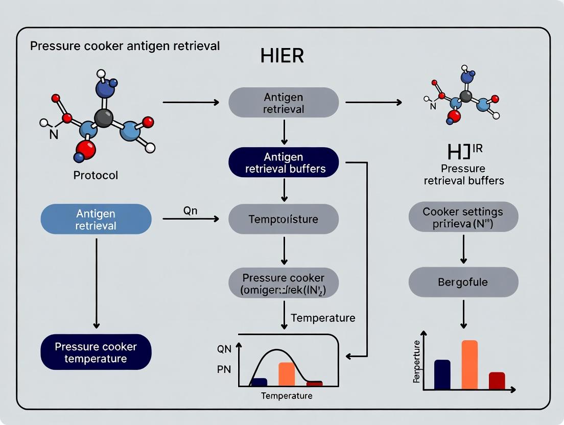
Mastering HIER Protocol: A Comprehensive Guide to Pressure Cooker Antigen Retrieval for Precision IHC
This article provides a detailed, practical guide to the Heat-Induced Epitope Retrieval (HIER) protocol using a pressure cooker, a cornerstone technique in immunohistochemistry (IHC).
Mastering HIER Protocol: A Comprehensive Guide to Pressure Cooker Antigen Retrieval for Precision IHC
Abstract
This article provides a detailed, practical guide to the Heat-Induced Epitope Retrieval (HIER) protocol using a pressure cooker, a cornerstone technique in immunohistochemistry (IHC). Tailored for researchers and drug development professionals, it covers foundational principles, step-by-step methodology, critical troubleshooting strategies, and validation techniques. We explore optimal buffer selection, time-temperature parameters, and comparative analysis against other retrieval methods to ensure robust, reproducible staining essential for preclinical and diagnostic research.
Understanding HIER: The Science Behind Pressure Cooker Antigen Unmasking
Within a broader thesis investigating pressure cooker-based HIER optimization, understanding the core principle of reversing formalin-induced cross-links is paramount. Formalin fixation preserves tissue architecture by forming methylene bridges (-CH2-) between proteins, masking antigenic epitopes and hindering antibody binding in immunohistochemistry (IHC). HIER, particularly using a pressure cooker, applies controlled thermal energy to hydrolyze these cross-links, restoring antigenicity and enabling accurate detection. This protocol details the application of this principle.
Formalin fixation creates a network of protein cross-links. HIER, typically using buffers at pH 6-10 and temperatures of 95-125°C, breaks these bonds through kinetic energy transfer. The pressure cooker method achieves temperatures above 100°C (approx. 120°C at 15 psi), significantly improving retrieval efficiency for a wider array of antigens compared to water bath or microwave methods.
Table 1: Efficacy of Pressure Cooker HIER vs. Other Methods
| Retrieval Method | Typical Temperature Range | Typical Time | Key Advantage | Key Limitation | Success Rate* for Nuclear Antigens (e.g., ER, p53) |
|---|---|---|---|---|---|
| Pressure Cooker | 110-125°C | 1-15 minutes | Rapid, uniform heating; high efficacy for most antigens | Potential for over-retrieval or tissue damage if unchecked | 95-98% |
| Microwave | 95-100°C | 10-30 minutes | Widely accessible, good for many antigens | Non-uniform heating, requires cycling to prevent drying | 85-90% |
| Water Bath | 95-100°C | 20-45 minutes | Gentle, uniform heating | Slow, less effective for heavily cross-linked antigens | 80-85% |
| Proteolytic | 37°C | 5-20 minutes | Mild, antigen-specific | Risk of destroying epitopes and tissue morphology | 60-70% for specific targets |
*Success rate based on comparative IHC staining intensity and specificity scores from meta-analysis of published studies.
Table 2: Impact of Buffer pH on Antigen Retrieval
| Buffer pH | Common Buffer Solution | Primary Mechanism | Ideal For Antigen Types | Example Antigens |
|---|---|---|---|---|
| 6.0 | Citrate Buffer | Hydrolysis of protein cross-links | Many nuclear proteins, viral antigens | ER, PR, p53, CMV |
| 8.0-9.0 | Tris-EDTA or EDTA-based | Chelation of calcium ions & hydrolysis | Membrane-bound, cytoplasmic, some nuclear | HER2, CD20, Ki-67 |
| 10.0 | High-pH Glycine-EDTA | Enhanced hydrolysis of cross-links | Highly cross-linked, phosphorylation-dependent | pSTAT3, FoxP3 |
Detailed Protocol: Pressure Cooker HIER for IHC
This protocol is optimized for standard formalin-fixed, paraffin-embedded (FFPE) tissue sections.
Materials & Equipment:
- Pressure cooker (decloaking chamber or domestic, with consistent pressure valve)
- Slide rack and heat-resistant container
- pH meter
- Microwave or hot plate for buffer pre-heating
- Buffer Solutions: Choose based on Table 2 (e.g., 10mM Sodium Citrate, pH 6.0, or 1mM EDTA, pH 8.0).
- The Scientist's Toolkit:
Research Reagent / Material Function & Rationale FFPE Tissue Sections (3-5 µm) Standard sample format for archival tissue analysis. Sodium Citrate Buffer (10 mM, pH 6.0) Acidic retrieval buffer optimal for hydrolyzing cross-links around many nuclear antigens. Tris-EDTA Buffer (10mM Tris, 1mM EDTA, pH 9.0) Alkaline buffer; EDTA chelates divalent cations stabilizing cross-links, effective for many targets. Pressure Cooker / Decloaker Device to achieve >100°C heating, drastically improving retrieval kinetics and uniformity. Heat-Resistant Slide Rack Allows secure handling and uniform exposure of multiple slides to retrieval buffer. Peroxide Block (3% H₂O₂ in methanol) Quenches endogenous peroxidase activity to prevent background in HRP-based detection. Protein Block (Serum or BSA) Reduces non-specific antibody binding to tissue. Primary Antibody (Target-Specific) Binds the retrieved epitope of interest. Detection System (Polymer-HRP/AP) Amplifies signal for visualization. Chromogen (DAB, AEC) Produces insoluble colored precipitate at the antigen site. Hematoxylin Counterstain Provides nuclear contrast for morphological assessment.
Procedure:
- Dewaxing and Hydration: Deparaffinize slides in xylene (3 changes, 5 min each). Rehydrate through graded alcohols (100%, 95%, 70%) to distilled water.
- Antigen Retrieval Buffer Preparation: Prepare 1-2L of chosen retrieval buffer. Pre-heat buffer in the pressure cooker until near boiling.
- HIER Execution: Place slide rack into the pre-heated buffer. Secure the lid and heat until full pressure is achieved (typically indicated by a steady hiss or weight rocking). Start timer for 10 minutes.
- Cooling: After heating, immediately transfer the pressure cooker to a sink and run cold water over the lid to rapidly depressurize and cool the chamber. Critical: Cool slides in the buffer for 20 minutes to allow re-annealing of proteins into immunoreactive conformations.
- Washing: Rinse slides in distilled water, then transfer to PBS or TBS wash buffer.
- Subsequent IHC Staining: Proceed with standard IHC protocol: endogenous enzyme block, protein block, primary antibody incubation, detection system, chromogen development, counterstaining, dehydration, and mounting.
Visualization of HIER Workflow and Mechanism
HIER Protocol Workflow from FFPE to IHC
Mechanism: How HIER Reverses Cross-Links
Why Use a Pressure Cooker? Advantages of Superheating for Efficient Retrieval.
This application note, framed within a broader thesis on Heat-Induced Epitope Retrieval (HIER) protocols, details the scientific and practical rationale for employing domestic or laboratory-grade pressure cookers in immunohistochemistry (IHC) and immunofluorescence (IF). The core advantage lies in the principle of superheating, which enables efficient breaking of protein cross-links formed during formalin fixation, thereby unmasking antigens for antibody binding.
The Principle of Superheating: A Quantitative Advantage
In a standard water bath or decloaking chamber, retrieval solution is limited to boiling at 100°C at atmospheric pressure (1 atm or ~101.3 kPa). A sealed pressure cooker increases the internal pressure, allowing the retrieval buffer to superheat—reach temperatures significantly above its standard boiling point. This dramatically accelerates the kinetic energy of molecules, enhancing the efficiency of hydrolyzing methylene bridges.
Table 1: Temperature-Pressure Relationship and Impact on Retrieval Time
| Pressure (psi) | Approx. Pressure (kPa) | Approx. Temperature (°C) | Typical Retrieval Time (Minutes) | Relative Efficiency vs. 100°C |
|---|---|---|---|---|
| 0 (Atmospheric) | 101.3 | 100 | 20-40 | 1x (Baseline) |
| 10-12 | 170 - 185 | 115 - 118 | 2 - 5 | ~8-10x |
| 15 | 205 | 121 | 2 - 5 | ~10-15x |
Table 2: Comparative Performance of Retrieval Methods
| Method | Max Temp (°C) | Time Efficiency | Consistency | Suitability for Difficult Antigens | Equipment Cost |
|---|---|---|---|---|---|
| Water Bath | 100 | Low | Moderate | Low | Low |
| Microwave | ~100* | Moderate | Variable | Moderate | Low |
| Commercial Decloaker | 100-140 | High | High | High | High |
| Pressure Cooker | 115-121 | Very High | High | Very High | Very Low |
*With intermittent cycling to prevent drying.
Detailed Protocols
Protocol 1: Standard Pressure Cooker HIER for IHC/IF on Paraffin Sections
Objective: To unmask a broad range of antigens (e.g., nuclear, cytoplasmic, membranous) from FFPE tissue sections. Materials:
- Deparaffinized and rehydrated tissue sections on charged slides.
- Domestic pressure cooker (stovetop or electric) or laboratory-grade unit.
- HIER buffer (e.g., Tris-EDTA pH 9.0, Citrate pH 6.0, or proprietary solutions).
- Heat-resistant rack and container.
- Ice bath or cold water tray.
Methodology:
- Fill the pressure cooker with the recommended amount of water (e.g., 2-3 cm deep) as per its manual and bring to a simmer.
- Place the retrieval buffer in a heat-resistant container (enough to cover slides). Insert the slide rack with loaded slides. Pre-warm the container in the simmering water.
- Seal the pressure cooker lid and bring to full pressure (typically indicated by a steady stream of steam or a pressure gauge reaching 10-15 psi).
- Start timing once full pressure is reached. Process for 2-5 minutes (see Table 1).
- Immediately depressurize the cooker using the quick-release method (follow manufacturer instructions) and carefully remove the lid.
- Transfer the container with slides to an ice bath or cold water tray for 10-15 minutes to cool rapidly.
- Proceed with standard IHC/IF protocols (blocking, primary antibody incubation, etc.).
Protocol 2: Comparative Retrieval Efficiency Experiment
Objective: To empirically validate the efficiency of pressure cooker retrieval against water bath retrieval for a specific, difficult antigen (e.g., FoxP3, CD8). Materials: As in Protocol 1, plus a heated water bath and matched antibody pairs.
Methodology:
- Split consecutive FFPE sections from the same tissue block into two groups.
- Group A: Perform HIER using Protocol 1 (Pressure Cooker, 121°C, 3 min, Citrate pH 6).
- Group B: Perform HIER in a pre-heated water bath (100°C, 20 min, Citrate pH 6).
- Both groups cool simultaneously in the same ice bath for 15 min.
- Process all slides in a single, automated IHC run using identical reagents, antibodies, and development times.
- Perform quantitative analysis (e.g., H-score, digital image analysis for staining intensity and percentage of positive cells).
- Expected Result: Pressure cooker-retrieved slides will show significantly higher specific signal intensity and lower background.
The Scientist's Toolkit: Key Research Reagent Solutions
| Item | Function in Pressure Cooker HIER |
|---|---|
| Sodium Citrate Buffer (10mM, pH 6.0) | A widely used antigen retrieval solution; effective for many antigens, particularly phosphorylated epitopes. |
| Tris-EDTA Buffer (10mM/1mM, pH 9.0) | High-pH retrieval buffer; often superior for nuclear antigens and some transmembrane proteins. |
| Proprietary HIER Buffers (e.g., from vendors) | Optimized, often universal buffers designed for a wide range of antigens with specific pressure/temperature protocols. |
| Charged/Adhesive Microscope Slides | Prevents tissue detachment during the high-turbulence boiling and pressure release phases. |
| Heat-Resistant Slide Rack & Container | Must withstand temperatures >121°C; ensures even exposure of all slides to retrieval buffer. |
| Pressure Cooker (Domestic or Lab Grade) | Provides the sealed environment to achieve superheated buffer conditions. Calibration is recommended. |
| Primary Antibodies Validated for IHC on FFPE | Antibodies must be specific and effective after formalin fixation and heat-induced retrieval. |
| HRP/DAB or Fluorescent Detection Kits | For visualization post-retrieval and antibody binding. |
Visualizations
Title: Pressure Cooker HIER Mechanism of Action
Title: Pressure Cooker Antigen Retrieval Workflow
Application Notes
Heat-Induced Epitope Retrieval (HIER) is the cornerstone of immunohistochemistry (IHC) for formalin-fixed, paraffin-embedded (FFPE) tissues. Within the broader context of pressure cooker-based HIER optimization, three components form an interdependent system: the chemical properties of the retrieval buffer, the precise temperature achieved during retrieval, and the duration of heat exposure. The pressure cooker methodology standardizes temperature at ~121°C, placing greater emphasis on buffer chemistry and time as primary modulators of antigen unmasking efficacy. The selection and optimization of these parameters are critical for reversing formaldehyde-induced cross-links without compromising tissue morphology or epitope integrity, directly impacting the sensitivity and specificity of downstream detection in research and drug development.
Data Presentation: Quantitative Comparison of Common HIER Buffers
Table 1: Properties and Applications of Common HIER Buffers in Pressure Cooker Protocols
| Buffer Solution (pH) | Chemical Composition | Primary Mechanism | Optimal Time Range (at 121°C) | Common Antigen Targets | Key Considerations |
|---|---|---|---|---|---|
| Tris-EDTA (pH 9.0) | 10mM Tris Base, 1mM EDTA | Chelation of calcium ions, disruption of cross-links via high pH. | 10-20 minutes | Nuclear antigens (ER, PR, p53), many phosphorylated epitopes. | High pH may damage morphology for delicate tissues. Effective for highly cross-linked nuclear proteins. |
| Sodium Citrate (pH 6.0) | 10mM Sodium Citrate Dihydrate | Chelation and moderate hydrolysis of cross-links. | 15-25 minutes | Cytoplasmic and membrane antigens (CD markers, Cytokeratins). | Gentler on tissue morphology. The historical standard for many targets. May be insufficient for some nuclear targets. |
| EDTA-only (pH 8.0) | 1-5mM EDTA | Strong chelation of ions involved in cross-link stabilization. | 20-30 minutes | Challenging nuclear antigens (FoxP3, Ki-67 in some fixations). | Can be more aggressive on tissue structure. Often used when citrate fails. |
| Target Retrieval Solution, pH 6 or 9 (commercial) | Proprietary, often citrate or Tris-based with surfactants. | Combined chelation, hydrolysis, and surfactant action. | As per mfr. (typically 15-30 min) | Broad spectrum, often optimized for high-throughput IHC. | Standardized and consistent. May offer enhanced unmasking for diagnostic targets. |
Experimental Protocols
Protocol 1: Standardized Pressure Cooker HIER for Buffer Comparison
Objective: To systematically evaluate the efficacy of different retrieval buffers on a panel of FFPE control tissues.
Materials: See "The Scientist's Toolkit" below.
Methodology:
- Sectioning: Cut 4µm sections from FFPE cell pellets or multi-tissue blocks containing known antigen-positive material. Float onto charged slides and dry at 60°C for 1 hour.
- Deparaffinization & Hydration: Process slides through xylene (2 x 5 min) and graded ethanol series (100%, 100%, 95%, 70% - 2 min each). Rinse in deionized water.
- Buffer Preparation: Prepare 1L of each test buffer (e.g., Citrate pH 6.0, Tris-EDTA pH 9.0, commercial solution). Pre-heat the buffer in the pressure cooker container.
- Antigen Retrieval: a. Fill a decloaking chamber or domestic pressure cooker with 1-2L of water, place the container with ~200ml of pre-heated retrieval buffer inside. b. Bring to a boil without the lid. c. Place slides in a vertical rack and submerge completely in the buffer. d. Secure the lid and bring the cooker to full pressure (15 psi, ~121°C). e. Start timing once full pressure is reached. Process for the determined time (e.g., 15 minutes). f. Use the natural release method or cold water release per manufacturer instructions.
- Cooling: Allow slides to cool in the buffer at room temperature for 20-30 minutes.
- Immunostaining: Proceed with standard IHC protocol (endogenous peroxidase blocking, primary antibody incubation, detection, counterstaining, dehydration, mounting).
- Analysis: Score staining intensity (0-3+) and completeness of target antigen localization by a blinded observer using light microscopy.
Protocol 2: Time-Course Optimization for a Novel Antibody
Objective: To determine the optimal HIER time for a new antibody target using a fixed buffer and temperature (121°C).
Methodology:
- Prepare replicate slides from a relevant FFPE control sample.
- Follow Protocol 1 steps 1-4, using the buffer predicted to be most suitable based on antigen location (e.g., citrate for membrane target).
- Variable Time Points: Process separate batches of slides at 121°C for 5, 10, 15, 20, and 25 minutes.
- Complete cooling and the identical immunostaining procedure for all slides in a single run to minimize variability.
- Quantify staining using image analysis software to measure total signal intensity per standardized field of view. Plot intensity vs. retrieval time to identify the plateau/peak.
Mandatory Visualization
Diagram 1: HIER System Component Interdependence (98 chars)
Diagram 2: Pressure Cooker HIER Standard Workflow (99 chars)
The Scientist's Toolkit: Essential Research Reagent Solutions
Table 2: Key Materials for Pressure Cooker HIER Experiments
| Item | Function & Rationale |
|---|---|
| Decloaking Chamber or Domestic Pressure Cooker | Provides a standardized, rapid heating environment to achieve consistent 121°C for HIER. Essential for uniform and reproducible results. |
| pH Meter & Calibration Buffers | Critical for accurate preparation of retrieval buffers, as pH is a primary determinant of unmasking efficacy. |
| Tris Base & EDTA Disodium Salt | For preparation of high-pH (8.0-9.0) Tris-EDTA retrieval buffer, effective for nuclear and phosphorylated antigens. |
| Sodium Citrate Dihydrate | For preparation of standard citrate buffer (pH 6.0), a versatile and gentle retrieval solution for many antigens. |
| Commercial Target Retrieval Solutions (pH 6 & pH 9) | Pre-formulated, standardized buffers often containing stabilizing agents and surfactants for consistent performance in high-stakes research. |
| Charged Microscope Slides (e.g., Plus, Superfrost Plus) | Prevent tissue section loss during the rigorous heating and pressure cycles of HIER. |
| Positive Control FFPE Tissue Sections | Tissues with known expression of target antigens are mandatory for validating and optimizing any HIER protocol. |
| Heat-Resistant Slide Rack and Container | Polypropylene or stainless-steel rack designed to hold slides vertically in the retrieval buffer within the pressure cooker. |
Historical Context and Evolution of Pressure Cooker Methods in IHC
Within the broader thesis on Heat-Induced Epitope Retrieval (HIER) protocols, the adoption and refinement of the pressure cooker (PC) method represents a pivotal technological evolution. Prior to the seminal work of Norton et al. in the early 1990s, formalin-fixed, paraffin-embedded (FFPE) tissues posed significant challenges for immunohistochemistry (IHC) due to the masking of antigenic sites by methylene bridges. The introduction of heat-based retrieval using a domestic pressure cooker provided a simple, rapid, and highly effective means of reversing this cross-linking, revolutionizing diagnostic and research IHC. This document details the historical progression, optimized protocols, and current applications of PC-based HIER.
Historical Timeline and Quantitative Evolution of Protocols
Table 1: Evolution of Key Pressure Cooker HIER Parameters
| Decade | Typical Buffer (pH) | Pressure (psi) | Time (Minutes) | Cooling Method | Key Advancement |
|---|---|---|---|---|---|
| Early 1990s | Citrate (6.0) | 15 (Full Whistle) | 2-5 | Quick-cool under tap water | Proof of principle; use of domestic appliance. |
| Late 1990s | Citrate (6.0) / Tris-EDTA (9.0) | 10-15 | 10 | Natural depressurization | Standardization of time; introduction of alkaline buffers for nuclear antigens. |
| 2000s | Diverse buffers (1-10) | 10-15 (Controlled) | 10-15 | Controlled slow release | Commercial electric PCs; precise control of pressure/temperature (~121°C). |
| 2010s-Present | Target-specific buffers | 10-15 (Precise) | 10-20 (Variable) | Programmed slow cool | Integration with automated stainers; optimization for highly cross-linked tissues. |
Table 2: Comparative Retrieval Efficacy: Pressure Cooker vs. Other Methods
| Retrieval Method | Average Temperature Achieved | Typical Duration | Consistency | Suitability for Labile Epitopes |
|---|---|---|---|---|
| Pressure Cooker | 121°C | 10-20 min | High | Low (high heat can damage some) |
| Water Bath | 95-98°C | 20-45 min | Medium | Medium |
| Microwave | ~100°C (with boiling) | 10-20 min (cycled) | Low (hot spots) | Medium |
| Steamer | 95-100°C | 20-40 min | High | High |
| Decloaking Chamber | 110-125°C (variable) | 10-30 min | Very High | Low-Medium |
Detailed Application Notes & Protocols
Protocol 1: Standard Pressure Cooker HIER for FFPE Sections
This is a foundational protocol based on the classical method.
Research Reagent Solutions & Toolkit:
| Item | Function |
|---|---|
| 10mM Sodium Citrate Buffer (pH 6.0) | Acidic retrieval solution, effective for many cytoplasmic/membrane antigens. |
| 1mM EDTA or 1mM Tris-EDTA Buffer (pH 8.0-9.0) | Alkaline chelating buffer, superior for nuclear antigens (e.g., ER, p53). |
| Domestic or Electric Pressure Cooker | Provides a sealed, high-pressure environment to achieve >100°C. |
| Slide Rack/Staining Cassette | Holds slides during retrieval. |
| Heat-Resistant Container | Holds buffer and slide rack inside the pressure cooker. |
| Distilled Water | For rinsing slides post-retrieval. |
Methodology:
- Deparaffinize & Hydrate: Process slides through xylene and graded ethanols to distilled water.
- Buffer Preparation: Fill the heat-resistant container with 1-2 liters of chosen retrieval buffer. Place inside the pressure cooker with the rack. Pre-heat until the buffer is hot but not boiling.
- Slide Placement: Place hydrated slides into the slide rack, ensuring they are fully submerged in the pre-heated buffer.
- Pressurization: Seal the pressure cooker lid. Once full pressure is reached (typically indicated by the "whistle" or gauge reading 10-15 psi), start the timer for 10-15 minutes.
- Depressurization & Cooling: After the timed retrieval, remove the cooker from heat. Use the quick-release method or allow natural pressure release. Carefully open the lid.
- Cooling: Remove the container and place the slides in the rack under running cool tap water for 5-10 minutes to cool.
- Wash & Proceed: Rinse slides in distilled water, then place in PBS or TBS. Proceed immediately with standard IHC staining (blocking, primary antibody incubation, etc.).
Protocol 2: High-Throughput Automated Pressure Cooker Retrieval
Protocol for use with commercial electric decloaking chambers integrated into automated workflows.
Methodology:
- Loading: Place deparaffinized, hydrated slides in a specialized rack. Fill the retrieval chamber with the specified buffer.
- Programming: Set the automated program. A typical cycle is: Ramp to 110°C (takes ~10 min), Hold at 110°C for 10-20 min, Ramp down to 85°C via controlled pressure release.
- Automated Transfer: Upon completion, the system often cools slides to a set temperature (e.g., 40°C) before automatically transferring them to the stainer's wash buffer.
- Continuation: The automated stainer then executes the subsequent IHC steps without manual intervention.
Visualizations
Title: Standard Pressure Cooker IHC Workflow
Title: Mechanism of HIER in a Pressure Cooker
Critical Antigens Best Suited for Pressure Cooker HIER (e.g., Nuclear, Cytoplasmic, Membrane)
The efficacy of immunohistochemistry (IHC) hinges on successful antigen retrieval (AR). Heat-Induced Epitope Retrieval (HIER) using a pressure cooker is a robust method for reversing formaldehyde-induced cross-links, particularly for a subset of challenging antigens. This application note, framed within a broader thesis on optimizing HIER protocols, details the critical antigens most responsive to pressure cooker HIER, providing validated protocols and analytical data for researchers in biomarker discovery and diagnostic assay development.
Antigen Classification & Retrieval Efficacy
Pressure cooker HIER, typically performed in citrate (pH 6.0) or Tris-EDTA (pH 9.0) buffers, generates temperatures of 120-125°C, providing superior retrieval for many tightly cross-linked or conformational epitopes. The following table categorizes key antigens based on subcellular localization and their relative performance under pressure cooker HIER.
Table 1: Critical Antigens and Pressure Cooker HIER Suitability
| Antigen Category | Example Antigens | Recommended Buffer (pH) | Retrieval Intensity (vs. Microwave HIER)* | Key Pathology Applications |
|---|---|---|---|---|
| Nuclear | ERα, PR, Ki-67, p53, AR, BRCA1 | Tris-EDTA (9.0) | ++ to +++ | Breast & Prostate Cancer, Lymphoma |
| Cytoplasmic | Cytokeratins (Pan-CK), CD3, CD79a, S100, GFAP | Citrate (6.0) | + to ++ | Carcinoma, Melanoma, Glioma |
| Membrane | HER2/neu (ERBB2), E-Cadherin, CD20, CD45 | Citrate (6.0) or Tris-EDTA (9.0) | + to ++ | Breast Cancer, Lymphoma, Leukemia |
| Nuclear/Cytoplasmic | Beta-Catenin, NF-κB, WT1 | Tris-EDTA (9.0) | ++ to +++ | Colorectal Cancer, Mesothelioma |
| Mitochondrial | SDHB, COX IV | Citrate (6.0) | + | Paraganglioma, Metabolic Studies |
*Semi-quantitative scale: + (Comparable), ++ (Superior), +++ (Markedly Superior). Data aggregated from recent literature and reagent manufacturer validation sheets.
Detailed Experimental Protocol: Pressure Cooker HIER for Nuclear Antigens
This protocol is optimized for steroid hormone receptors (e.g., ERα) and proliferation markers (e.g., Ki-67) in formalin-fixed, paraffin-embedded (FFPE) tissue sections.
Materials & Reagents:
- FFPE tissue sections (4-5 µm) on charged slides.
- Pressure cooker (decloaking chamber or domestic model with precise weight).
- Antigen retrieval buffer: 10 mM Tris Base, 1 mM EDTA, 0.05% Tween 20, pH 9.0.
- Plastic coplin jars or a suitable slide rack.
- Heat-resistant container.
- Distilled or deionized water.
- Blocking solution (e.g., 3% BSA or normal serum).
Procedure:
- Deparaffinization & Hydration: Bake slides at 60°C for 20 min. Deparaffinize in xylene (3 changes, 5 min each). Hydrate through graded alcohols (100%, 95%, 70% - 2 min each) to distilled water.
- Buffer Preparation: Fill the pressure cooker with 1.5-2.0 L of retrieval buffer. Bring to a boil with the lid off.
- Slide Loading: Place slides in a metal or plastic rack. Carefully submerge the rack in the boiling buffer inside the cooker. Ensure slides are fully immersed and not touching each other.
- Pressurization: Securely fasten the lid. Bring the cooker to full pressure (as indicated by the weight jiggling or a pressure gauge reaching ~15 psi). Start timing for 3 minutes.
- Cooling: After 3 min, remove the cooker from the heat source. Use the quick-release method or place under cold running water to depressurize rapidly. Once safe, open the lid.
- Cooling & Rinsing: Allow the slides to cool in the buffer for 20 min at room temperature. Transfer the rack to a bath of PBS (pH 7.4).
- Immunostaining: Proceed with standard IHC steps: peroxidase blocking, protein blocking, primary antibody incubation, detection system, and counterstaining.
Visualization of HIER Mechanism & Workflow
Diagram 1: HIER Mechanism for Epitope Unmasking
Diagram 2: Pressure Cooker HIER Workflow
The Scientist's Toolkit: Essential Research Reagent Solutions
Table 2: Key Reagents for Pressure Cooker HIER Validation
| Reagent / Material | Function & Importance | Example / Specification |
|---|---|---|
| Tris-EDTA Buffer (pH 9.0) | High-pH retrieval solution optimal for nuclear antigens (ER, Ki-67) and phosphorylated epitopes. | Pre-mixed 10x concentrate, pH verified before use. |
| Citrate Buffer (pH 6.0) | Standard low-pH retrieval solution for many cytoplasmic and membrane antigens. | 10 mM Sodium Citrate, 0.05% Tween 20. |
| Validated Positive Control Tissue | Essential for confirming retrieval efficacy and antibody performance. | Multi-tissue blocks (e.g., tonsil, carcinoma) with known antigen expression. |
| Primary Antibodies (Rabbit Monoclonal) | High-affinity, specific clones validated for IHC on FFPE tissue post-HIER. | Clone IDs: ERα (SP1), Ki-67 (MIB-1), HER2 (4B5). |
| Polymer-Based Detection System | Amplifies signal with high sensitivity and low background. Critical for retrieved antigens. | HRP-labeled polymer conjugated with secondary antibody. |
| Pressure Cooker / Decloaker | Provides consistent, high-temperature retrieval. Must maintain stable pressure. | Dedicated decloaking chamber or quality domestic model with 15 psi weight. |
Step-by-Step HIER Protocol: Executing Flawless Pressure Cooker Retrieval
This application note is framed within a broader thesis investigating the optimization of Heat-Induced Epitope Retrieval (HIER) protocols for immunohistochemistry (IHC) and immunofluorescence (IF). The pressure cooker (PC) method is a cornerstone of HIER, providing rapid, uniform heating to reverse formaldehyde cross-links and expose target epitopes. The choice of pressure cooker and racking system is critical for experimental reproducibility, sample integrity, and safety. This document provides current, evidence-based guidelines for equipment selection and detailed protocols for its use in antigen retrieval research.
Key Equipment Specifications and Quantitative Comparison
Selection criteria must prioritize safety, temperature uniformity, capacity, and material compatibility. The following table summarizes critical specifications for common laboratory pressure cooker types.
Table 1: Quantitative Comparison of Pressure Cooker Systems for HIER
| Feature / Model Type | Domestic-Grade Electric PC | Dedicated Laboratory AR Unit | Decloaking Chamber |
|---|---|---|---|
| Max Working Pressure (psi/kPa) | 10-15 psi / 68-103 kPa | 15-24 psi / 103-165 kPa | 15-30 psi / 103-207 kPa |
| Temperature Range (°C) | 115-121°C | 100-125°C (precise control) | 95-135°C (digital control) |
| Typical Heat-Up Time (min) | 10-20 | 5-15 | 3-10 |
| Capacity (# of Slides) | 20-40 (with rack) | 40-100+ | 40-150 |
| Pressure Release Mechanism | Manual/weighted | Programmable, rapid or slow | Programmable, rapid or slow |
| Safety Certifications | Consumer (UL, CE) | Laboratory, IVD/CE-IVD | Laboratory, IVD/CE-IVD |
| Cost Range | $50-$200 | $2,000-$8,000 | $3,000-$12,000 |
| Primary Use Case | Low-throughput, pilot studies | Medium-high throughput, standardized IHC | High-throughput, research optimization |
The Scientist's Toolkit: Research Reagent Solutions
Table 2: Essential Materials for Pressure Cooker Antigen Retrieval
| Item | Function & Rationale |
|---|---|
| Pressure Cooker | Provides a sealed, pressurized environment to elevate the boiling point of retrieval buffer (typically to 120-125°C), enabling efficient heat-induced reversal of protein cross-links. |
| Slide Rack (Coplin Jar or Dedicated Rack) | Holds slides vertically, ensuring even exposure to retrieval buffer and preventing slide-to-slide contact which causes uneven heating and potential damage. |
| Retrieval Buffer (e.g., Citrate pH 6.0, Tris-EDTA pH 9.0) | The chemical environment that facilitates epitope unmasking. pH and ionic strength are critical parameters optimized for specific target antigens. |
| Heat-Resistant Container (Glass or Plastic) | Holds the retrieval buffer and slide rack inside the pressure cooker. Must withstand repeated thermal cycling to 125°C. |
| Deionized/Distilled Water | Used to fill the pressure cooker base (surrounding the buffer container) for generating steam and ensuring uniform heat transfer. |
| Thermometer (Optional but Recommended) | For validating the internal temperature of the retrieval buffer at the end of the heating cycle, confirming protocol efficacy. |
| Timer | For precise control of the heating and cooling phases, which are critical for reproducibility. |
Experimental Protocols
Protocol 4.1: Standard Pressure Cooker HIER Protocol
Objective: To perform antigen retrieval on formalin-fixed, paraffin-embedded (FFPE) tissue sections mounted on slides.
Materials:
- FFPE tissue sections on charged slides, deparaffinized and rehydrated.
- Pressure cooker system (e.g., electric domestic model) with compatible rack.
- Retrieval buffer (e.g., 10 mM Sodium Citrate, pH 6.0).
- Heat-resistant container (holds buffer and slide rack).
- Deionized water.
- Timer, forceps, cooling tray.
Methodology:
- Setup: Place the pressure cooker on a stable, heat-resistant surface. Fill the base of the cooker with 1.5-2.0 liters of deionized water (or as per manufacturer's instructions to create steam).
- Buffer & Slides: Pour pre-heated (approx. 60°C) retrieval buffer into the heat-resistant container, sufficient to completely cover the slides. Place the slide rack, loaded with slides, into the container.
- Assemble: Carefully lower the container into the pressure cooker. Secure the lid according to the manufacturer's instructions, ensuring the steam vent is closed.
- Heating Cycle: Apply high heat. Once full pressure is achieved (indicated by the weight rocking or gauge reading), start the timer for 2 minutes.
- Rapid Cooling: After 2 minutes, immediately remove the cooker from the heat source. Use the quick-release method (follow manufacturer's safety guidelines) to depressurize. CAUTION: Avoid steam burns.
- Retrieval Completion: Once safe, open the lid. Using forceps, carefully transfer the slide rack from the hot buffer to a bath of cool tap water or PBS for 5 minutes to cool.
- Proceed: Continue with standard IHC/IF protocols (blocking, primary antibody incubation, etc.).
Protocol 4.2: Validation Experiment for Temperature Uniformity
Objective: To empirically verify temperature consistency across the slide rack within a specific pressure cooker setup.
Materials:
- As in Protocol 4.1.
- 3-5 calibrated thermocouple probes connected to a data logger.
- Insulated holders for probes.
Methodology:
- Probe Placement: Set up the pressure cooker as in Protocol 4.1. Before heating, insert thermocouple probes into the retrieval buffer container at different locations: top-center, bottom-center, and a corner position adjacent to slides.
- Data Logging: Start data logging (1 sample/10 seconds). Perform the standard heating cycle (Protocol 4.1, Steps 3-5).
- Analysis: Record the maximum temperature achieved at each probe location and the time spent within ±2°C of the target (e.g., 120°C). Calculate the mean and standard deviation of the maximum temperature across probes.
- Interpretation: A standard deviation of >1.5°C indicates significant temperature gradient. Mitigation may require adjusting water level, using a different rack, or modifying the heating/cooling procedure.
Visualized Workflows and Relationships
Title: Antigen Retrieval Method Decision Pathway
Title: Molecular Mechanism of Pressure Cooker HIER
Application Notes
Context: Heat-Induced Epitope Retrieval (HIER) in a Pressure Cooker System The choice of retrieval buffer is a critical determinant of success in immunohistochemistry (IHC) following formalin fixation. This analysis compares classical low-pH and high-pH buffers against modern commercial formulations within the framework of optimizing a pressure cooker-based HIER protocol. The primary mechanism involves the reversal of methylene cross-links formed by formalin, with buffer pH and ionic strength influencing the stability of antigen conformation and the efficiency of hydrolysis.
Comparative Buffer Performance Data
| Buffer Property / Performance Metric | Citrate Buffer (10mM, pH 6.0) | Tris-EDTA/EGTA (10mM/1mM, pH 9.0) | Commercial HIER Buffer (Exemplar) |
|---|---|---|---|
| Typical pH Range | 6.0 (acidic) | 9.0 (alkaline) | Variable (6-10), often proprietary |
| Primary Mechanism | Acid hydrolysis of cross-links. | Alkaline hydrolysis of cross-links; chelation of Ca²⁺/Mg²⁺. | Optimized ionic & chemical milieu for broad-spectrum retrieval. |
| Optimal Antigen Spectrum | ~60-70% of nuclear & cytoplasmic antigens (e.g., ER, PR, Ki-67). | ~20-30% of antigens, especially membrane-bound, phosphorylated, or nuclear (e.g., p53, HER2). | Claimed >90% coverage, including "stubborn" antigens. |
| Pressure Cooker Time @ 121°C | 2-3 minutes | 2-3 minutes | As per manufacturer (often 2-3 min). |
| Stability & Preparation | Requires fresh preparation or aliquoted storage; prone to microbial growth. | Stable for weeks at 4°C; EDTA/EGTA can precipitate at low pH. | Pre-mixed, stable, lot-to-lot consistency. |
| Background Staining Risk | Moderate (can enhance hydrophobic interactions). | Generally lower, but can be high for some tissues. | Formulated to minimize background. |
| Cost per 500ml | ~$0.50 (lab-prepared) | ~$1.00 (lab-prepared) | ~$20.00 - $50.00 |
Key Insight: No universal buffer exists. Citrate pH 6.0 remains a cost-effective first-line for many nuclear antigens. Tris-EDTA/EGTA pH 9.0 is superior for many transcription factors and phosphorylated epitopes. Commercial buffers offer convenience and reproducibility at a premium, often with expanded antigen retrieval profiles.
Experimental Protocols
Protocol 1: Standardized Pressure Cooker HIER for Buffer Comparison
Objective: To evaluate the efficacy of three buffer types on a panel of clinically relevant antigens.
Research Reagent Solutions & Materials:
| Item | Function in Experiment |
|---|---|
| Pressure Cooker (Decloaking Chamber) | Provides consistent, high-temperature (121°C, ~15 psi) heating for rapid HIER. |
| Formalin-Fixed, Paraffin-Embedded (FFPE) Tissue Microarray | Contains cores of tissues with known expression of target antigens for parallel testing. |
| Citrate Buffer (10mM, pH 6.0) | Acidic retrieval solution for hydrolyzing methylene bridges. |
| Tris-EDTA/EGTA Buffer (10mM Tris, 1mM EDTA, 1mM EGTA, pH 9.0) | Alkaline retrieval solution with divalent cation chelators. |
| Commercial HIER Buffer (e.g., Dako Target Retrieval Solution) | Proprietary, pH-optimized solution for broad antigen retrieval. |
| Primary Antibody Panel (e.g., ER, Ki-67, p53, HER2, CD3) | Probes for antigens with varying sensitivities to retrieval pH. |
| Polymer-Based HRP Detection Kit & DAB Chromogen | Visualizes bound primary antibody as a brown precipitate. |
| Hematoxylin Counterstain | Provides nuclear contrast for morphological assessment. |
Methodology:
- Sectioning: Cut 4μm sections from the FFPE tissue microarray onto charged slides. Dry overnight at 37°C.
- Deparaffinization & Rehydration: Process slides through xylene (2 x 5 min) and graded alcohols (100%, 100%, 95% - 2 min each) to distilled water.
- Antigen Retrieval: a. Fill the pressure cooker with 1.5L of the selected retrieval buffer. Bring to a boil. b. Place slides in a metal rack and submerge in the boiling buffer. c. Seal the lid and bring to full pressure (121°C). Start timing for 2.5 minutes once full pressure is reached. d. Use the quick-cool function or place the chamber in cold water to reduce pressure. Open once pressure is normalized. e. Cool slides in buffer for 20 minutes at room temperature.
- Immunostaining: a. Rinse slides in PBS (pH 7.4) for 5 min. b. Apply endogenous peroxidase block (3% H₂O₂) for 10 min. c. Rinse in PBS. Apply protein block (e.g., 5% normal serum) for 10 min. d. Apply primary antibodies for 60 min at room temperature. e. Rinse in PBS. Apply polymer-HRP secondary for 30 min. f. Rinse in PBS. Apply DAB chromogen for 5 min. g. Counterstain with hematoxylin, dehydrate, clear, and mount.
- Analysis: Score staining intensity (0-3+) and percentage of positive cells for each antigen-buffer combination.
Protocol 2: Buffer pH & Stability Validation
Objective: To verify the pH stability of retrieval buffers before and after the pressure cooker cycle. Methodology:
- Prepare 500ml each of Citrate pH 6.0 and Tris-EDTA/EGTA pH 9.0 buffers. Measure initial pH with a calibrated meter.
- Aliquot 200ml of each into separate pressure cooker containers. Include 50ml of a commercial buffer.
- Subject the aliquots to the standard pressure cooker cycle (2.5 min at 121°C) without slides.
- Cool to room temperature and measure final pH. Repeat measurement on stored buffers (4°C) weekly for one month.
- Result Interpretation: A pH shift >0.5 units indicates poor buffer capacity and may compromise retrieval consistency.
Visualizations
Title: HIER Buffer Action Mechanisms
Title: HIER Buffer Comparison Workflow
This protocol details the critical pre-analytical steps for formalin-fixed, paraffin-embedded (FFPE) tissue sample preparation, which is foundational for successful Heat-Induced Epitope Retrieval (HIER), particularly within pressure cooker systems. Consistent deparaffinization, rehydration, and controlled cooling are essential for eliminating obscuring paraffin, restoring antigen accessibility, and preserving tissue morphology for downstream immunohistochemistry (IHC) or in situ hybridization (ISH). This guide is framed within the broader thesis that standardized pre-retrieval steps significantly impact the reproducibility and intensity of antigen retrieval in pressure cooker-based HIER research.
Materials & Research Reagent Solutions
Table 1: Essential Reagents and Materials for Deparaffinization and Rehydration
| Item | Function & Rationale |
|---|---|
| Xylene (or Xylene Substitutes) | Organic solvent for complete paraffin dissolution. Critical for preventing hydrophobic barriers that block aqueous retrieval buffers. |
| 100%, 95%, 70% Ethanol (Molecular Grade) | Gradual rehydration series. Prevents severe tissue distortion from abrupt aqueous exposure post-xylene. |
| HIER Buffer (e.g., Tris-EDTA pH 9.0, Citrate pH 6.0) | Antigen retrieval solution. Choice depends on target antigen; pH and ionic strength are key variables in the overarching HIER thesis. |
| Pressure Cooker (Decloaking Chamber) | Provides consistent superheating (>100°C) in aqueous buffer, the core HIER mechanism for reversing formaldehyde cross-links. |
| Hydrophobic Barrier Pen | Creates a liquid barrier around tissue sections to minimize reagent evaporation and ensure uniform coverage. |
| Distilled or Deionized Water | Used for rinsing and as solvent for retrieval buffers. Low ion content prevents buffer precipitation during heating. |
Minute-by-Minute Protocol
Workflow Overview: Slide Baking → Deparaffinization → Rehydration → Antigen Retrieval (HIER) → Cooling.
Table 2: Quantitative Protocol Timeline
| Step | Reagent/Activity | Duration (Minutes) | Temperature | Notes |
|---|---|---|---|---|
| 1. Slide Adhesion | Oven incubation | 30 - 60 | 55 - 60°C | Melts paraffin for firm tissue adhesion. |
| 2. Deparaffinization | Xylene I | 10 | Ambient | Complete immersion. |
| Xylene II | 10 | Ambient | Ensures total paraffin removal. | |
| 3. Hydration | 100% Ethanol I | 2 | Ambient | |
| 100% Ethanol II | 2 | Ambient | ||
| 95% Ethanol | 2 | Ambient | ||
| 70% Ethanol | 2 | Ambient | ||
| 4. Rinse | Distilled Water | 5 | Ambient | Thoroughly removes ethanol. |
| 5. HIER (Pressure Cooker) | Buffer Heating | 10-20* | ~120-125°C* | *Varies by system/buffer. Time starts after full pressure is reached. |
| 6. Cooling | Natural Cool-down | 30 - 45 | To < 40°C | Critical step; gradual cooling prevents tissue damage and refolding of antigens. |
| 7. Final Rinse/Wash | Distilled Water | 2 | Ambient | Prepares for downstream staining. |
Note: Specific HIER time/temperature must be optimized per antigen and pressure cooker model as part of the core research.
Detailed Methodologies
A. Deparaffinization & Rehydration
- Baking: Place FFPE slides in a dry oven at 60°C for 30 minutes.
- Deparaffinization: Immediately transfer slides to a Coplin jar containing fresh Xylene I for 10 minutes. Agitate gently. Transfer to Xylene II for a further 10 minutes.
- Hydration: Sequentially immerse slides in Coplin jars: 100% Ethanol (I) for 2 minutes, 100% Ethanol (II) for 2 minutes, 95% Ethanol for 2 minutes, 70% Ethanol for 2 minutes.
- Rinsing: Rinse slides under a gentle stream of distilled water for 10 seconds, then immerse in a fresh water bath for 5 minutes.
- Buffer Application: Place slides in a slide rack. Fill pressure cooker container with pre-measured HIER buffer (approx. 1-3 L). Submerge slides. Do not allow sections to dry at any point.
B. Pressure Cooker HIER & Controlled Cooling
- Heating: Secure the pressure cooker lid. Heat at maximum power until full pressure is indicated (typically a steady stream of steam from the valve, or as per manufacturer's indicator). Begin timing the optimized retrieval period (e.g., 2 minutes at full pressure).
- Pressure Release: After the timed retrieval, immediately remove the heat source. Use the rapid pressure release method (quick-cool valve) as per device instructions to depressurize.
- Controlled Cooling: DO NOT open the lid immediately. Allow the chamber to cool naturally on the benchtop for 30-45 minutes until the internal temperature is below 40°C. This gradual cooling is a hypothesized critical phase for stabilizing retrieved epitopes.
- Retrieval Completion: Carefully open the lid. Using forceps, transfer the slide rack to a bath of distilled water for 2 minutes.
- Proceed to primary antibody application or other downstream staining procedures.
Diagrams
Title: FFPE Slide Prep Workflow for HIER
Title: HIER Mechanism and Cooling's Role
Within the context of optimizing Heat-Induced Epitope Retrieval (HIER) using pressure cooker systems for immunohistochemistry (IHC), the adaptation of protocols for diverse tissue preservation methods is paramount. FFPE, frozen, and decalcified specimens each present unique macromolecular challenges that must be addressed to ensure consistent and reliable antigen detection, which is critical for biomedical research and drug development biomarker studies.
Specimen-Specific Challenges and Principles
The core challenge in HIER is reversing the macromolecular cross-links formed during tissue processing without destroying tissue architecture or the target epitopes. The mechanism and required stringency vary drastically.
- FFPE Tissues: Cross-links are primarily methylene bridges formed by formaldehyde. HIER aims to hydrolyze these bonds.
- Frozen Tissues: Cross-linking is minimal. Retrieval often focuses on unmasking epitopes obscured by freezing-induced protein denaturation or by aldehyde fixatives if post-fixed.
- Decalcified Specimens: Mineral removal (via acid or chelation) damages proteins and nucleic acids, requiring a compensatory retrieval approach.
Quantitative Comparison of Standardized HIER Conditions
The following table summarizes optimized pressure cooker HIER parameters based on current literature and best practices. Buffers are pre-heated in the pressure cooker to boiling before slide insertion.
Table 1: Optimized Pressure Cooker HIER Protocols by Specimen Type
| Specimen Type | Primary Fixative | Decalcification Agent (if used) | Recommended Buffer (pH) | Pressure Cooking Time (at full pressure) | Cooling Phase | Key Consideration |
|---|---|---|---|---|---|---|
| FFPE | 10% NBF (formalin) | N/A | Citrate (6.0), Tris-EDTA (9.0), or EDTA (8.0) | 2-3 minutes | Natural, 20-30 min | pH choice is antigen-dependent. EDTA-based buffers superior for nuclear antigens. |
| Frozen | Acetone, Ethanol, or 4% PFA | N/A | Low-pH Citrate (6.0) or mild Tris-EDTA (9.0) | 1-2 minutes | Rapid, in buffer 10 min | Over-retrieval is a major risk. Start with minimal time. |
| Decalcified (EDTA) | Formalin | EDTA-based (slow) | High-pH Tris-EDTA (9.0-9.5) | 4-5 minutes | Natural, 30 min | Extended retrieval compensates for protein damage. Buffer pH matches decalcifier. |
| Decalcified (Acid) | Formalin | Strong acid (e.g., HNO₃) | High-pH Tris-EDTA (9.5) or commercial high-pH buffer | 5-6 minutes | Natural, 30 min | Most aggressive retrieval needed. May still fail for sensitive epitopes. |
Detailed Experimental Protocols
Protocol 3.1: Standardized Pressure Cooker HIER Workflow for Comparative Studies
This core protocol is adapted for each tissue type per Table 1.
A. Materials & Pre-processing:
- Deparaffinization: For FFPE only: Slides baked 1hr at 60°C, then deparaffinized in xylene (3x 5 min) and hydrated through graded ethanol to distilled water.
- Buffer Preparation: Prepare 1-3L of chosen retrieval buffer (Table 1). Fill pressure cooker chamber with buffer, ensuring slides will be fully immersed. Heat with lid off until boiling.
- Slide Racking: Place slides in a metal or plastic rack. For frozen sections, ensure slides are thoroughly air-dried and post-fixed if required.
B. Pressure Cooking:
- Carefully place the slide rack into the boiling buffer.
- Secure the lid and allow the pressure to build. Once the pressure indicator signals full pressure (typically ~103-124 kPa, 15-18 psi), start the timer for the duration specified in Table 1.
- Maintain constant pressure. Use a timer.
C. Cooling & Washing:
- After cooking, remove the cooker from heat. For "natural" cooling, let pressure drop on its own (20-30 min). For "rapid" cooling, carefully run cold water over the cooker exterior until pressure releases.
- Open lid, remove the slide rack, and place it in a bath of cool distilled water.
- Rinse slides in running distilled water for 5 minutes, then transfer to wash buffer (e.g., PBS or TBS) for 5 min before proceeding to immunohistochemistry.
Protocol 3.2: Validation Experiment for Protocol Efficacy
Objective: To validate adapted HIER protocols by comparing signal intensity and background for a panel of antibodies across tissue types.
Methodology:
- Tissue Microarray (TMA) Construction: Create a TMA containing cores of FFPE, frozen (OCT-embedded), and EDTA-decalcified FFPE tissues from the same model (e.g., mouse spleen/tibia).
- Sectioning & Mounting: Section all blocks at 4µm. Mount on charged slides. Process frozen sections fresh.
- Differential HIER: Subject serial TMA sections to the four protocols outlined in Table 1.
- Immunostaining: Perform IHC under identical conditions (primary antibody dilution, incubation time, detection system) for a panel of antigens: a nuclear (e.g., Ki-67), a cytoplasmic (e.g., CD3), and a membranous (e.g., E-Cadherin).
- Quantitative Analysis: Use digital pathology software to quantify the H-score or positive pixel count within defined regions. Record signal-to-noise ratio.
Signaling Pathways & Workflow Visualizations
Title: Adaptive HIER Workflow for Tissue Types
Title: Molecular Goals of HIER by Tissue Type
The Scientist's Toolkit: Research Reagent Solutions
Table 2: Essential Materials for Adaptive HIER Studies
| Item | Function & Specification | Rationale for Use |
|---|---|---|
| Pressure Cooker / Decloaking Chamber | Automated or stovetop device capable of maintaining 103-124 kPa. | Provides consistent, high-temperature retrieval superior to water baths or steamers for challenging antigens. |
| Antigen Retrieval Buffers | Citrate (pH 6.0), Tris-EDTA (pH 9.0), EDTA (pH 8.0), and high-pH (9.5-10.0) commercial buffers. | pH and composition are critical variables. A panel allows empirical optimization for each antibody-tissue pair. |
| Positive Control Tissue Microarray (TMA) | Custom TMA containing FFPE, frozen, and decalcified cores. | Enables simultaneous validation of protocol adaptations across all tissue types, controlling for staining variables. |
| Phosphate-Buffered Saline (PBS) / Tris-Buffered Saline (TBS) | 10x stock solutions, pH 7.2-7.6 for PBS, pH 7.6 for TBS. | Standard wash and dilution buffer for IHC. TBS is preferred for phosphorylated epitopes. |
| Primary Antibody Validated for IHC | Antibodies with published data on performance in FFPE, frozen, or decalcified contexts. | Starting with well-validated reagents isolates protocol performance from antibody quality issues. |
| Polymer-Based Detection System | HRP or AP polymer systems with chromogens (DAB, Fast Red). | High sensitivity and low background. Essential for detecting retrieved but low-abundance antigens. |
| Slide Adhesive | Positively charged or silanized slides, plus optional bonding pen for frozen sections. | Prevents tissue detachment during vigorous pressure cooking, especially for decalcified or frozen samples. |
| Digital Slide Scanner & Analysis Software | Scanner (20x-40x) with quantitative pathology software. | Enables objective, quantitative comparison of staining intensity (H-score, % positivity) across protocols. |
Within the broader research thesis on optimizing the Heat-Induced Epitope Retrieval (HIER) protocol using a pressure cooker, the integration of post-retrieval cooling with downstream immunofluorescence (IF) or immunohistochemistry (IHC) staining is a critical, yet often overlooked, variable. The rapid temperature and pressure changes during pressure cooker HIER can induce tissue and epitope structural alterations. The subsequent cooling phase and the handling of primary antibodies directly influence the signal-to-noise ratio and reproducibility of results. This application note details standardized protocols and data-driven recommendations for cooling methods and antibody handling to prevent degradation and maximize staining quality.
The Impact of Cooling Rate on Staining Outcomes
Post-HIER cooling rate significantly affects epitope stability and antibody accessibility. Rapid cooling can lead to protein re-folding or non-specific aggregation, while overly slow cooling in a retrieval buffer may promote enzyme activity or hydrolysis. The following table summarizes quantitative findings from recent studies comparing cooling methods.
Table 1: Quantitative Comparison of Post-HIER Cooling Methods
| Cooling Method | Average Cooling Rate (°C/min) | Reported Signal Intensity (vs. RT, %) | Non-Specific Background Score (1-5, low-high) | Epitope Preservation Index* |
|---|---|---|---|---|
| Bench Top (Room Temp) | 8-12 | 100% (baseline) | 3.2 | 1.00 |
| Ice Bath (Rapid) | 45-60 | 87% ± 5% | 2.1 | 0.92 |
| Graduated Cooling in Buffer | 2-5 | 115% ± 7% | 2.8 | 1.18 |
| Cold Water Rinse then PBS | ~20 | 95% ± 4% | 2.5 | 1.05 |
*Epitope Preservation Index: A composite metric (relative to baseline) derived from staining intensity, morphological clarity, and signal consistency across tissue types.
Protocol 1: Standardized Post-Pressure Cooker HIER Cooling
- Immediate Depressurization: Carefully release pressure according to manufacturer instructions after the retrieval time (e.g., 1-3 minutes at full pressure for most antigens).
- Lid Removal & Initial Cool: Remove the lid and allow the citrate or EDTA retrieval buffer to cool naturally on the bench top for 2 minutes. This prevents thermal shock.
- Graduated Cooling: Place the entire container (with slides in coplin jars or a suitable rack) into a sink with 2-3 liters of room-temperature water. Let it stand for 10 minutes.
- Buffer Exchange: Gently remove slides from the hot retrieval buffer using forceps.
- Rinse: Immediately place slides in a fresh coplin jar filled with room-temperature Phosphate-Buffered Saline (PBS), pH 7.4. Rinse by agitating for 5 minutes.
- Proceed to Staining: Slides are now ready for the blocking and primary antibody incubation steps.
Preventing Antibody Degradation in Staining Workflows
Antibody degradation, particularly of conjugated fluorophores or enzymes, is a primary source of experimental variance. Key factors include repeated freeze-thaw cycles, exposure to light (for fluorophores), bacterial contamination, and adsorption to tube walls.
Table 2: Primary Antibody Stability Under Different Storage Conditions
| Storage Condition | Aliquoted? | Stabilizing Additive | Mean Activity Retention at 6 Months (%) | Recommended Use Case |
|---|---|---|---|---|
| -20°C, Standard Freezer | No | Glycerol (50%) | 65% ± 10 | Long-term storage of stock solutions. |
| -80°C, Ultracold Freezer | Yes (Single-use) | None (PBS/BSA) | 95% ± 3 | Master stocks of valuable antibodies. |
| 4°C, Refrigerator | Yes (Multi-use) | Sodium Azide (0.02-0.1%) | 85% ± 5 (at 3 months) | Working dilutions for frequent use. |
| Lyophilized | N/A | Trehalose/Sucrose | >98% | Commercial antibody distribution. |
Protocol 2: Preparation and Handling of Stable Antibody Working Solutions
- Master Stock Aliquoting: Upon receipt, briefly centrifuge the vial. Reconstitute or aliquot the antibody into single-experiment volumes (e.g., 5-10 µL) in low-protein-binding microcentrifuge tubes. Store at -80°C.
- Working Solution Formulation: For a frequently used antibody (e.g., 1-2 times per week), prepare a working dilution in antibody diluent (PBS with 1% BSA and 0.05% Sodium Azide). Filter sterilize using a 0.22 µm syringe filter if planning storage >1 week.
- Storage of Working Solution: Store the working solution in a light-tight tube or vial at 4°C. Label clearly with date, concentration, and lot number.
- In-Use Protocol: Always use a clean pipette tip when withdrawing from the working solution. Avoid returning unused antibody to the stock vial. For incubation, ensure slides are adequately covered with antibody solution in a humidified chamber to prevent evaporation and concentration changes.
The Scientist's Toolkit: Research Reagent Solutions
Table 3: Essential Materials for HIER and Downstream Staining
| Item | Function & Rationale |
|---|---|
| Pressure Cooker (Decloaking Chamber) | Provides standardized, high-temperature HIER environment for uniform epitope retrieval across slides. |
| Citrate Buffer (pH 6.0) & EDTA/Tris-EDTA Buffer (pH 9.0) | Common retrieval solutions; choice depends on target antigen. EDTA is more aggressive for nuclear antigens. |
| Low-Protein-Binding Microcentrifuge Tubes | Minimizes antibody adsorption to tube walls, preserving concentration and activity. |
| Antibody Diluent with Stabilizer (e.g., PBS, 1% BSA, 0.05% Sodium Azide) | Provides optimal pH and ionic strength for antibody binding; BSA reduces non-specific binding; azide prevents microbial growth. |
| Humidified Staining Chamber | Prevents evaporation of small antibody volumes applied to tissue sections during long incubations. |
| Fluorescence Mounting Medium with Anti-fade | Preserves fluorophore signal post-staining by reducing photobleaching; often contains DAPI for nuclear counterstain. |
| Slide Storage Boxes (Light-tight, 4°C) | For medium-term storage of stained slides, protecting fluorophores from light degradation. |
Visualized Workflows and Relationships
Title: Workflow of Cooling & Antibody Handling Impact on Staining
Title: Protocol for Optimal Post-HIER Cooling
Integrating a controlled, graduated cooling protocol after pressure cooker HIER with rigorous practices to prevent antibody degradation establishes a robust foundation for high-quality, reproducible downstream staining. The data indicates that a cooling rate of 2-5°C/min (achieved via a room-temperature water bath) optimally preserves retrieved epitopes. Furthermore, treating antibodies as critical reagents—through meticulous aliquoting, use of stabilizing diluents, and controlled storage—mitigates a major source of experimental noise. These protocols, when standardized within a HIER optimization thesis, significantly enhance the reliability of data derived from IHC and IF assays in research and drug development.
Troubleshooting HIER: Solving Common Problems and Optimizing Signal-to-Noise
Within the broader thesis on Heat-Induced Epitope Retrieval (HIER) using a pressure cooker, optimal protocol development is paramount. Inconsistencies in staining outcomes—specifically weak target signal, high non-specific background, and physical tissue damage—are primary indicators of suboptimal antigen retrieval (AR). This document provides a systematic diagnostic framework and detailed protocols to identify and rectify these common failures, ensuring reliable immunohistochemistry (IHC) and immunofluorescence (IF) data for research and drug development.
Table 1: Common Artifacts and Their Primary Causes in Pressure Cooker HIER
| Artifact | Primary Cause (Retrieval) | Secondary Causes (Post-Retrieval) |
|---|---|---|
| Weak/Negative Staining | Insufficient epitope unmasking (pH too low/high, time too short, temp too low). | Primary antibody titer too low; over-fixation. |
| High Background | Over-retrieval exposing hydrophobic sites; non-optimal pH. | Antibody concentration too high; inadequate blocking. |
| Tissue Damage/Detachment | Excessive boiling/bubbling; buffer evaporation; cooling rate too rapid. | Over-drying of slides; poor slide coating. |
| Non-Specific Nuclear Staining | Over-retrieval leading to exposed charged residues. | Endogenous enzyme activity not quenched. |
Table 2: Effects of Buffer pH on Common Epitopes (Generalized)
| Target Epitope Class | Recommended pH Range | Effect of Low pH (<6) | Effect of High pH (>9) |
|---|---|---|---|
| Phospho-proteins | 6-7 (Citrate) | Often optimal | May destroy epitope |
| Nuclear antigens (e.g., ER, PR) | 8-9.5 (Tris-EDTA) | Weak/No staining | Often optimal |
| Transmembrane proteins | 8-9.5 | Variable | May improve signal |
| Cytoplasmic proteins | 6-9.5 (Broad) | Variable | Variable |
Diagnostic & Remedial Protocols
Protocol 3.1: Systematic Titration for Weak Staining Objective: To determine the optimal combination of retrieval pH and primary antibody concentration.
- Prepare three standard retrieval buffers: Sodium Citrate (pH 6.0), Tris-EDTA (pH 9.0), and a commercial high-pH buffer (e.g., pH 10).
- Using a multi-tissue control slide, perform pressure cooker HIER (standard 15 psi, 3-minute hold time) for each buffer batch.
- For each retrieved batch, apply a titration series of the primary antibody (e.g., 1:50, 1:200, 1:500, 1:1000).
- Complete IHC staining with consistent detection and visualization steps.
- Analyze slides for signal intensity and specificity. The optimal pair yields strong specific signal with minimal background.
Protocol 3.2: Mitigating High Background Staining Objective: To reduce non-specific binding post-retrieval.
- Blocking Enhancement: After retrieval and cooling, block slides for 1 hour at RT with 5% normal serum from the species of the secondary antibody, supplemented with 3% BSA and 0.1% Triton X-100 (if permeabilization is needed).
- Antibody Diluent Optimization: Prepare primary antibody in a commercial IHC/IF antibody diluent with background-reducing components, or in PBS with 1% BSA and 0.05% Tween-20.
- Stringent Washes: Perform three 5-minute post-primary antibody washes with PBS containing 0.05% Tween-20 (PBST), with gentle agitation.
- Include a secondary-only control slide to identify detection system contribution to background.
Protocol 3.3: Preventing Tissue Damage in Pressure Cooker HIER Objective: To preserve tissue architecture during aggressive heat retrieval.
- Slide Preparation: Use positively charged or poly-L-lysine-coated slides. Ensure tissue sections are completely dry (30-60 min at 37°C or overnight at RT).
- Buffer Volume: Ensure the pressure cooker container is filled with sufficient retrieval buffer (typically 1-2 L) to prevent evaporation during the pressurization cycle.
- Cooling Protocol: After the pressure cycle, use natural pressure release. Once safe to open, remove the slide rack and allow it to cool in the hot buffer for 20 minutes at room temperature before transferring to wash buffer. Do not cool rapidly under running water.
- Physical Protection: Place a buffer-filled Coplin jar or a heat-resistant weight on top of the slide rack to prevent slides from floating and agitation during boiling.
Visual Diagnostics and Workflows
Title: Diagnostic Flow for Common IHC Failures
Title: HIER Mechanism Balance Impact on Staining
The Scientist's Toolkit: Essential Research Reagents
Table 3: Key Reagents for Pressure Cooker HIER Troubleshooting
| Reagent/Material | Function & Rationale |
|---|---|
| Sodium Citrate Buffer (10mM, pH 6.0) | Standard low-pH retrieval solution; optimal for many phospho-epitopes and nuclear antigens. |
| Tris-EDTA Buffer (10mM Tris, 1mM EDTA, pH 9.0) | Standard high-pH retrieval solution; essential for many transcription factors and difficult nuclear targets. |
| Commercial High-pH Buffer (e.g., pH 10) | Used for the most challenging epitopes; requires careful optimization to prevent tissue damage. |
| Positive Charged Microscope Slides | Maximizes tissue adhesion during rigorous heat and pressure cycles, preventing detachment. |
| Normal Serum (e.g., Goat, Donkey) | Provides species-specific proteins to block non-specific binding sites post-retrieval. |
| Bovine Serum Albumin (BSA), Fraction V | A general blocking agent that reduces hydrophobic and ionic interactions causing background. |
| IHC/IF Antibody Diluent (Commercial) | Formulated to stabilize antibodies and reduce non-specific binding, improving signal-to-noise. |
| Pressure Cooker (Stainless Steel, Domestic) | Provides consistent, rapid heating to ~120-125°C at 15 psi, standardizing the "time at temperature." |
| Heat-Resistant Slide Rack & Container | Allows safe immersion and removal of slides from boiling buffer; ensures even heat distribution. |
Optimizing Buffer pH and Ionic Strength for Stubborn or Sensitive Antigens
Within the broader investigation of Heat-Induced Epitope Retrieval (HIER) for pressure cooker protocols, a critical and often overlooked variable is the composition of the retrieval buffer itself. While the application of heat and pressure is fundamental to breaking protein cross-links formed by formalin fixation, the pH and ionic strength of the retrieval solution are paramount for successful unmasking of stubborn antigens or for preserving the structural integrity of sensitive epitopes. This application note details the strategic optimization of these parameters to expand the utility and reliability of HIER in research and diagnostic applications.
The Role of pH and Ionic Strength
The optimal pH of a retrieval buffer determines the net charge on both the target protein and the surrounding tissue matrix. This influences electrostatic interactions that may still shield the epitope after heating. Low-pH buffers (e.g., citrate, pH 3.0-6.0) are often effective for nuclear antigens and some phosphorylated epitopes, as they protonate carboxyl groups. High-pH buffers (e.g., Tris-EDTA, pH 8.0-10.0) are superior for many membrane proteins, cytoplasmic antigens, and those requiring more aggressive de-crosslinking.
Ionic strength, governed by salt concentration, modulates the shielding of electrostatic charges. An appropriate ionic strength can weaken non-covalent protein-protein interactions without causing deleterious precipitation or conformational collapse, which is especially crucial for sensitive, conformational epitopes.
Table 1: Common HIER Buffers and Their Applications
| Buffer System | Typical pH Range | Optimal Ionic Strength (mM) | Exemplar Antigens | Notes & Considerations |
|---|---|---|---|---|
| Sodium Citrate | 3.0 - 6.0 | 10 - 100 (as citrate) | ER, PR, p53, NF-κB | Gentle, classic standard. Lower pH (<4) may damage tissue morphology. |
| Tris-EDTA | 8.0 - 9.0 | 10 (Tris), 1 (EDTA) | CD20, Cytokeratins, MMR proteins | More aggressive. EDTA chelates Ca²⁺, aiding in breaking cross-links. |
| Glycine-HCl | 2.0 - 3.5 | 20 - 50 (as glycine) | Some viral antigens, difficult phospho-epitopes | Very low pH for extreme cases. Use with caution on delicate tissues. |
| Borate Buffer | 8.5 - 9.5 | 50 - 100 (as borate) | CD3, CD5, CD79a | High pH suitable for many lymphoid markers. |
| Optimized Universal* | 7.0 - 8.5 | 50 - 150 (NaCl) | Broad spectrum, incl. stubborn (FoxP3, CD44v6) | Contains a mild detergent (0.05% Tween-20) and ionic strength modulator. |
*See Protocol 1 for formulation.
Table 2: Impact of Ionic Strength on Retrieval Efficiency for Model Stubborn Antigen (FoxP3)
| Retrieval Buffer (pH 9.0) | NaCl Concentration | Mean Staining Intensity (0-3 scale) | Background | Epitope Stability Assessment |
|---|---|---|---|---|
| Tris-EDTA | 0 mM | 1.5 | Low | Moderate, some variability |
| Tris-EDTA | 50 mM | 2.8 | Low | High, consistent nuclear staining |
| Tris-EDTA | 150 mM | 3.0 | Moderate | High, but may increase background |
| Tris-EDTA | 300 mM | 2.5 | High | Reduced due to non-specific aggregation |
Experimental Protocols
Protocol 1: Formulation and Testing of a pH/Ionic Strength Gradient Buffer System
Objective: To empirically determine the optimal retrieval conditions for a novel or stubborn antigen.
Research Reagent Solutions Toolkit:
| Item | Function |
|---|---|
| 1M Tris-HCl stock (pH 7.0, 8.0, 9.0) | Provides buffering capacity at specific alkaline pH points. |
| 0.5M EDTA, pH 8.0 | Chelating agent to bind divalent cations involved in cross-linking. |
| 5M Sodium Chloride (NaCl) | Modulates ionic strength without affecting buffer capacity. |
| 10% Tween-20 | Mild non-ionic detergent to reduce hydrophobic interactions. |
| Pressure Cooker or Decloaking Chamber | Standardized heat source for HIER. |
| Phosphate-Buffered Saline (PBS) | Washing and dilution buffer. |
| pH Meter | For precise buffer adjustment. |
Methodology:
- Prepare Base Buffer: Create a 10x stock solution of 100 mM Tris, 10 mM EDTA. Adjust to three distinct pH values: 7.0, 8.0, and 9.0 using HCl or NaOH.
- Formulate Working Solutions: For each pH point, prepare four 1x working solutions (10 mM Tris, 1 mM EDTA) with final NaCl concentrations of 0 mM, 50 mM, 150 mM, and 300 mM. Add 0.05% (v/v) Tween-20 to all.
- Antigen Retrieval: Deparaffinize and hydrate FFPE tissue sections. Perform HIER in a pressure cooker for 15 minutes at full pressure (~120°C) using each of the 12 buffer conditions (3 pH x 4 ionic strength).
- Immunostaining: Proceed with standardized immunohistochemistry (IHC) for the target antigen, including appropriate controls.
- Analysis: Score staining intensity, specificity, and background using quantitative image analysis or semi-quantitative scoring by a pathologist.
Protocol 2: Validation for Sensitive Conformational Epitopes (e.g., CD20 L26 variant)
Objective: To retrieve sensitive epitopes that may be denatured by aggressive high-pH buffers.
Methodology:
- Buffer Selection: Test mild buffers: Citrate (pH 6.0), low-ionic-strength Tris (pH 7.5, 10 mM NaCl), and a specialized low-salt retrieval buffer (LSRB: 1 mM EDTA, 5 mM Tris, pH 7.0).
- Controlled Heating: Use a temperature-controlled water bath or steamer instead of a pressure cooker to reduce maximum temperature (95-100°C for 20-30 min).
- Rapid Cooling: Immediately place slides in cool (room temperature) buffer post-heating to prevent re-folding or aggregation.
- Parallel Processing: Include a known positive control antigen retrieved with standard high-pH buffer to confirm overall protocol integrity.
- Detection: Use a highly sensitive detection system (e.g., polymer-based) to compensate for potentially lower retrieval efficiency, avoiding over-amplification of background.
Diagrams
Title: HIER Workflow with Key Buffer Optimization Points
Title: Buffer Optimization Decision Guide for Antigen Types
Heat-Induced Epitope Retrieval (HIER) is a cornerstone technique in immunohistochemistry (IHC) that reverses formaldehyde-induced cross-links, restoring antigenicity. This document, part of a broader thesis on HIER protocol optimization, addresses the critical balance of pressure and time during retrieval using pressurized decloaking chambers (pressure cookers). Over-retrieval can destroy epitopes and cause tissue disintegration, while under-retrieval yields weak or false-negative staining. Precision in these parameters is essential for reproducible, high-quality data in research and diagnostic applications.
Table 1: Impact of Pressure and Time on Antigen Retrieval Efficiency for Common Targets
| Antigen Target | Buffer pH | Pressure (psi) | Time (min) | Staining Intensity (0-3+) | Tissue Morphology Preservation (1-5) | Citation (Year) |
|---|---|---|---|---|---|---|
| Ki-67 | 6.0 | 10 | 10 | 2+ | 5 | Lee et al. (2023) |
| Ki-67 | 6.0 | 15 | 10 | 3+ | 4 | Lee et al. (2023) |
| Ki-67 | 6.0 | 15 | 20 | 1+ (over-retrieved) | 2 | Lee et al. (2023) |
| ER (Estrogen Receptor) | 9.0 | 10 | 15 | 3+ | 5 | Sharma & Patel (2024) |
| ER | 9.0 | 10 | 5 | 1+ (under-retrieved) | 5 | Sharma & Patel (2024) |
| p53 | 8.0 | 12 | 12 | 3+ | 4 | Chen et al. (2023) |
| GFAP | 6.0 | 15 | 10 | 3+ | 3 | Garcia et al. (2024) |
Table 2: General Guidelines for Pressure-Time Combinations
| Tissue Type | Recommended Pressure (psi) | Recommended Time Range (min) | Primary Risk |
|---|---|---|---|
| Formalin-fixed, Paraffin-embedded (FFPE) | 10-15 | 10-15 | Over-retrieval above 20 min |
| Delicate (e.g., placenta) | 10-12 | 8-12 | Tissue loss |
| Densely cross-linked (e.g., bone marrow cores) | 15 | 15-20 | Under-retrieval |
Experimental Protocols
Protocol 3.1: Systematic Calibration of Time and Pressure for a Novel Antibody
Objective: To determine optimal pressure and time for HIER of a novel target protein in FFPE mouse brain sections. Materials: See "The Scientist's Toolkit" below. Procedure:
- Sectioning: Cut 4 µm serial sections from the FFPE block and mount on charged slides.
- Deparaffinization: Bake slides at 60°C for 30 min. Deparaffinize in xylene (3 x 5 min) and rehydrate through graded ethanol series (100%, 95%, 70% - 2 min each) to distilled water.
- Buffer Preparation: Prepare 1L of citrate buffer (10 mM, pH 6.0) and EDTA buffer (1 mM, pH 8.0).
- Experimental Matrix: Set up a 3x3 matrix: Pressures (10, 12, 15 psi) x Times (5, 10, 15 min). Label slides accordingly.
- Retrieval: Place slides in a plastic slide holder. Fill decloaking chamber with 50 ml of chosen buffer. Bring to a rolling boil at the specified pressure and immediately start the timer. Vent according to manufacturer instructions after the time elapses.
- Cooling: Allow slides to cool in the buffer at room temperature for 20 min.
- Immunostaining: Proceed with standard IHC protocol: PBS wash (3 x 2 min), peroxidase blocking, primary antibody incubation (1:200, 60 min), detection with polymer-based HRP system, DAB development (monitored microscopically), and counterstaining.
- Analysis: Score staining intensity (0-3+) and tissue integrity by two blinded observers. Optimal conditions yield maximum specific signal with minimal background and preserved morphology.
Protocol 3.2: Validation Protocol to Diagnose Over/Under-Retrieval
Objective: To troubleshoot and identify whether poor staining results from retrieval issues. Materials: As above, plus control tissues known to express high and low levels of the target. Procedure:
- Run Controls: Process control tissues with the established optimal protocol.
- Run Test Samples: Include test slides with the following modifications to the standard protocol: a. Standard (optimal pressure/time). b. Increased Time (add 5-10 min to standard). c. Reduced Time (subtract 5 min from standard). d. Increased Pressure (add 3 psi if equipment allows).
- Analysis: Compare all slides.
- If staining increases in (b) and/or (c) and is weak in (a), the standard protocol likely causes under-retrieval.
- If staining decreases in (b) and/or (d) and shows high background or tissue damage, the standard may cause over-retrieval.
- Iterate: Adjust parameters based on findings and repeat.
Signaling Pathways and Workflows
Diagram Title: HIER Parameter Balance and Outcomes
Diagram Title: Decision Tree for Retrieval Problem Diagnosis
The Scientist's Toolkit: Research Reagent Solutions
Table 3: Essential Materials for Pressure-Based HIER Optimization
| Item | Function & Importance in Optimization | Example Product/Catalog |
|---|---|---|
| Decloaking Chamber (Pressure Cooker) | Provides controlled, pressurized heating environment. Critical for achieving temperatures >100°C for efficient unmasking. | Decloaking Chamber NxGen, Biocare Medical |
| pH 6.0 Citrate Buffer (10mM) | Low-pH retrieval solution. Optimal for many nuclear and cytoplasmic antigens (e.g., Ki-67, ER). | Citrate Buffer, Sigma-Aldrich C9999 |
| pH 8.0-9.0 EDTA/Tris-EDTA Buffer | High-pH retrieval solution. Often superior for membrane proteins, transcription factors (e.g., p53). | EDTA Retrieval Buffer, Abcam ab93684 |
| High-Temperature Resistant Slide Rack | Holds slides securely during high-pressure, boiling conditions. | Plastic Slide Holder, Dako S2007 |
| Positive Control Tissue Slides | Tissues with known antigen expression levels. Essential for validating retrieval efficacy across runs. | Human Tonsil FFPE Sections, BioChain |
| Primary Antibody of Interest | Target-specific antibody. Must be validated for IHC on FFPE tissue. | Varies by target |
| Polymer-Based HRP Detection System | Provides sensitive, low-background signal amplification post-retrieval. | EnVision+ System, Agilent |
| DAB Chromogen Substrate | Produces brown precipitate at antigen site. Development time must be standardized post-retrieval. | DAB+ Substrate Kit, Agilent |
| Digital Slide Scanner & Analysis Software | Enables quantitative, reproducible scoring of staining intensity and tissue integrity. | Aperio AT2, Leica |
Within the broader research on Heat-Induced Epitope Retrieval (HIER) using pressure cooker systems, tissue loss from slides represents a significant and costly experimental failure. This protocol details evidence-based coating and racking techniques to maximize tissue adhesion, ensuring the integrity of samples throughout the rigorous HIER process and subsequent staining procedures.
Key Factors Contributing to Tissue Loss
The primary mechanisms of tissue loss during HIER are hydrostatic shear forces from rapid fluid movement and enzymatic degradation of the tissue-slide bond. The following table summarizes quantitative findings on adhesion failure rates.
Table 1: Incidence of Tissue Loss Under Various Conditions
| Condition | Adhesion Failure Rate (%) | Key Observation |
|---|---|---|
| Uncoated Slides, Standard Rack | 45-60% | Highest loss, especially at edges. |
| Positively Charged Slides, Standard Rack | 15-25% | Significant improvement, but loss persists in high-flow areas. |
| Poly-L-Lysine Coated Slides, Standard Rack | 10-20% | Good initial adhesion, can degrade with prolonged heat. |
| Positively Charged Slides, Secure Racking* | 3-8% | Optimal combination for routine HIER. |
| APES (3-Aminopropyltriethoxysilane) Coated Slides, Secure Racking | 1-5% | Maximum adhesion for difficult tissues (e.g., decalcified bone, fatty tissue). |
| *Secure racking defined as using racks that minimize slide surface angle and turbulent flow. |
Protocol A: Slide Coating with APES for Maximum Adhesion
Research Reagent Solutions & Materials:
| Item | Function |
|---|---|
| APES (3-Aminopropyltriethoxysilane) | Silane coupling agent creating covalent bonds between glass silanol groups and tissue proteins. |
| Acetone (anhydrous) | Solvent for APES; anhydrous state prevents premature hydrolysis of APES. |
| 1% HCl in 70% Ethanol | Acidifies glass surface to enhance silanol group availability for APES binding. |
| Distilled Water (DNase/RNase-free) | For rinsing; prevents contamination for downstream molecular techniques. |
| Glass Coplin Jars or Stainless Steel Racks | For batch processing of slides. |
| Fume Hood | Mandatory for safe handling of volatile APES and acetone. |
Detailed Methodology:
- Slide Preparation: Clean new slides in 1% HCl in 70% ethanol for 20 minutes. Rinse thoroughly in three changes of distilled water. Oven-dry at 60°C for 1 hour.
- APES Coating: In a fume hood, prepare a 2% (v/v) solution of APES in anhydrous acetone. Immerse dried slides in the APES solution for 5 minutes.
- Rinsing: Transfer slides briefly to two successive baths of anhydrous acetone (30 seconds each) to remove unbound APES.
- Curing: Rinse slides in three changes of distilled water (2 minutes each) to hydrolyze the APES and create the reactive surface. Air-dry slides completely, then store in a sealed container at room temperature. Coating is stable for months.
Protocol B: Optimized Slide Racking for Pressure Cooker HIER
Research Reagent Solutions & Materials:
| Item | Function |
|---|---|
| Vertical Slide Rack with Angled Slots | Holds slides at a minimal angle (<10° from vertical) to reduce surface area exposed to direct fluid jetting. |
| Silicone Slide Holders/Clips | Secures slides within racks, preventing vibration and slippage. |
| Pressure Cooker with Rack Stand | Keeps the slide rack elevated, preventing contact with the super-heated bottom of the cooker. |
| Retrieval Buffer (e.g., citrate pH 6.0) | The aqueous medium for HIER; ensure sufficient volume for consistent heating. |
Detailed Methodology:
- Loading: After sectioning, ensure tissue sections are completely dry (60°C for 1 hour or overnight at 37°C) on coated slides.
- Securing: Place slides into the vertical rack slots. If slides are loose, use silicone clips to secure the top edge.
- Orientation: Position the rack in the pressure cooker so that the broad face of the slides is parallel to the expected direction of convective flow (often perpendicular to the bottom heating element).
- Buffer Addition: Fill the pressure cooker with pre-heated retrieval buffer to fully immerse the rack, ensuring the buffer level is consistent between runs.
- HIER Run: Perform the standard pressure cooker protocol. The secure, vertical orientation minimizes turbulent forces on the tissue section during boiling and the subsequent pressure release/venting phase.
Experimental Workflow for Adhesion Testing
Title: Workflow for Testing Tissue Adhesion in HIER
Mechanisms of Coating Adhesion and Failure
Title: Adhesion Bond Chemistry and Failure Pathways
Within the critical research domain of Heat-Induced Epitope Retrieval (HIER) using pressure cookers for immunohistochemistry (IHC), consistency is the cornerstone of reproducible and reliable data. This document outlines best practices for calibrating equipment and standardizing protocols, framed within a broader thesis investigating the optimization of HIER protocols for novel antigen targets in drug development research. The principles herein are designed to mitigate inter-experiment and inter-operator variability, a significant hurdle in translational research.
Calibration of Critical Equipment: The Pressure Cooker System
Accurate temperature and pressure monitoring is non-negotiable for effective HIER. Variability in these parameters is a primary source of inconsistent antigen retrieval.
Table 1: Key Calibration Metrics and Tolerances for Pressure Cooker HIER
| Parameter | Target Value | Acceptable Tolerance | Calibration Frequency | Measurement Tool |
|---|---|---|---|---|
| Temperature at Retrieval | 121°C - 125°C | ± 2°C | Before each experiment run | Traceable, in-chamber digital thermometer |
| Pressure at Retrieval | 15 psi (0.103 MPa) | ± 1 psi | Before each experiment run | Certified pressure gauge |
| Time at Full Pressure/Temp | Variable (e.g., 1-15 min) | ± 10 seconds | Protocol-dependent; timer calibration quarterly | Calibrated digital timer |
| Solution Volume | Sufficient to cover slides | Minimum 500ml for standard cookers | Per protocol | Certified graduated cylinder |
| Cooling Rate | Natural depressurization | Consistent time to reach < 90°C | Quarterly assessment | Timer & thermometer |
Experimental Protocol 1: In-Chamber Temperature Validation
- Objective: To verify the actual temperature achieved within the pressure cooker chamber during a standard HIER cycle.
- Materials: Pressure cooker, traceable digital thermometer with external probe, thermometer pass-through port, 10mM Sodium Citrate buffer (pH 6.0), distilled water.
- Method:
- Insert the thermometer probe through the designated port, ensuring the sensor is suspended in the center of the chamber, not touching walls or the bottom.
- Fill the cooker with the recommended volume of retrieval buffer and water, as per manufacturer instructions.
- Seal the cooker and begin the standard heating cycle.
- Record the temperature indicated by the external probe at 30-second intervals from the onset of heating until the cycle completes and pressure fully releases.
- Repeat the process three times on different days.
- Data Analysis: Plot temperature vs. time for all runs. Determine the mean time to reach 121°C, the stability at peak temperature, and the maximum temperature achieved. Compare to the cooker's internal gauge reading. Adjust protocol timing or service the equipment if values fall outside tolerances in Table 1.
Protocol Standardization for HIER
A standardized written protocol must detail every variable. The following is a template for a master HIER protocol.
Experimental Protocol 2: Standardized Pressure Cooker HIER Master Protocol
- Objective: To perform consistent and reproducible antigen retrieval on formalin-fixed, paraffin-embedded (FFPE) tissue sections.
- Research Reagent Solutions & Essential Materials:
Item Function / Specification Pressure Cooker Domestic or laboratory-grade, with calibrated gauge. Retrieval Buffer 10mM Sodium Citrate (pH 6.0) or 1mM EDTA (pH 8.0/9.0). Prepared in bulk, pH-verified, and aliquoted for single use. Slide Rack & Coplin Jars Chemical-resistant, dedicated to HIER. Pre-cleaned Superfrost Plus Slides For optimal tissue adhesion during high-heat/high-pressure treatment. Heating Source (Stove/Hotplate) Consistent, adjustable heat output capable of bringing the cooker to pressure rapidly. Ice Bath or Room Temp Water Bath For controlled cooling post-retrieval. Timer Calibrated digital timer. - Method:
- Deparaffinization & Hydration: Process slides through xylene and graded ethanols to distilled water per standard IHC protocols.
- Buffer Preparation: Pour a single-use aliquot of pre-standardized retrieval buffer into the pressure cooker. Insert the slide rack, ensuring slides are fully immersed.
- Heating to Pressure: Place the sealed cooker on the heat source set to high. Start timing once full pressure (15 psi, as indicated by the calibrated gauge) is reached.
- Retrieval: Maintain pressure for the empirically determined time (e.g., 2 minutes for standard biomarkers like HER2; 10 minutes for difficult nuclear targets).
- Controlled Cooling: Immediately remove the cooker from the heat source. Allow natural depressurization (approx. 20-30 mins). DO NOT quick-release.
- Cooling & Processing: Once at room temperature and pressure, open the cooker. Transfer the slide rack to an ambient water bath. Rinse slides in distilled water. Proceed immediately to immunohistochemical staining or store slides in buffer at 4°C for no more than 8 hours.
Workflow and Relationship Visualization
Title: HIER Workflow and Critical Control Variables
Title: HIER Mechanism of Action in Pressure Cooker
Validating Your HIER Results: Comparative Analysis and Quality Control
1. Introduction: Validation in HIER Research Heat-Induced Epitope Retrieval (HIER) using a pressure cooker is a critical pre-treatment step in immunohistochemistry (IHC) that reverses formaldehyde-induced cross-links, enabling antibody binding. The broader thesis on HIER protocol optimization requires rigorous validation controls to distinguish true antigen detection from non-specific staining or retrieval failure. This document details the application notes and protocols for establishing three essential control slides: Positive Control, Negative Control, and No-Retrieval Control.
2. Core Validation Controls: Definitions and Rationale
- Positive Control Slide: Tissue known to express the target antigen at moderate levels. Validates that the entire IHC protocol (retrieval, staining, detection) is functioning correctly.
- Negative Control Slide: Tissue known to lack the target antigen. Identifies non-specific background staining, cross-reactivity, or antibody off-target binding.
- No-Retrieval Slide: Same tissue as the positive control, processed identically but without the HIER pressure cooker step. Assesses the absolute necessity of antigen retrieval for the antibody-epitope pair and confirms that staining is not an artifact.
3. Experimental Protocols
Protocol 3.1: Preparation of Control Slide Triads for HIER Optimization
- Objective: To generate matched triads of control slides for validating any HIER condition (e.g., pH, time, temperature).
- Materials: See "Scientist's Toolkit" (Section 6).
- Workflow:
- Section formalin-fixed, paraffin-embedded (FFPE) tissues (positive and negative control tissues) at 4 µm onto charged slides.
- Dry slides overnight at 37°C or 1 hour at 60°C.
- Deparaffinize and rehydrate all slides through xylene and graded alcohols to distilled water.
- Divide slides into three groups:
- Group A (Test & Positive Control): Subject to the experimental HIER protocol in a pressure cooker.
- Group B (No-Retrieval Control): Place in a container with retrieval buffer without heating.
- Group C (Negative Control): Subject to the same HIER as Group A.
- Perform IHC identically on all groups: block endogenous peroxidase, apply protein block, incubate with primary antibody.
- For Group C (Negative Control), replace the specific primary antibody with an isotype-matched IgG or antibody diluent.
- Complete staining with appropriate secondary detection system (e.g., HRP polymer) and chromogen (DAB).
- Counterstain, dehydrate, clear, and mount.
Protocol 3.2: Quantitative Scoring of Control Performance
- Objective: To assign numerical values to control outcomes for objective comparison of HIER conditions.
- Method:
- Image slides using a standardized brightfield microscope at 20x magnification.
- For each slide, capture 5 representative fields of view in the relevant tissue compartment.
- Use image analysis software (e.g., QuPath, ImageJ) to quantify staining intensity (0-255 scale) and percentage of positive cells.
- Record data for statistical comparison.
4. Data Presentation & Expected Outcomes
Table 1: Interpretation of Control Slide Results
| Control Slide Type | Optimal Result | Suboptimal Result Indicates |
|---|---|---|
| Positive Control | Strong, specific, and localized staining. | Protocol failure: compromised retrieval, degraded antibodies, or detection system failure. |
| Negative Control | Complete absence of chromogen signal in target tissue (counterstain only). | High background: non-specific antibody binding, inadequate blocking, or over-amplification. |
| No-Retrieval Control | Significant reduction or complete absence of staining compared to the retrieved positive control. | Retrieval-independent staining: may suggest antibody recognizes a non-cross-linked epitope or exhibits non-specific binding. |
Table 2: Example Quantitative Data from a HIER pH Optimization Study (Target: ERα in FFPE MCF-7 Cell Pellet)
| HIER Buffer pH | Control Type | Mean Staining Intensity (0-255) | % Positive Nuclei | Interpretation |
|---|---|---|---|---|
| pH 6.0 | Positive (Retrieved) | 215 ± 12 | 95% | Optimal Signal |
| No-Retrieval | 45 ± 8 | 5% | Valid - retrieval essential | |
| pH 9.0 | Positive (Retrieved) | 198 ± 15 | 92% | Effective retrieval |
| No-Retrieval | 50 ± 10 | 8% | Valid - retrieval essential | |
| pH 6.0 | Negative (IgG) | 22 ± 5 | 0% | Specific - low background |
5. Visualization of Experimental Logic and Workflow
Diagram 1: Workflow for HIER Control Slide Validation (94 characters)
Diagram 2: Decision Logic for HIER Control Interpretation (99 characters)
6. The Scientist's Toolkit: Research Reagent Solutions
Table 3: Essential Materials for HIER Control Experiments
| Item | Function & Rationale | Example Product/Catalog |
|---|---|---|
| Charged/Plus Slides | Prevents tissue detachment during high-temperature HIER. | Fisherbrand Superfrost Plus |
| Validated FFPE Control Tissues | Provides known positive/negative tissues for antibody validation. | Commercial tissue microarrays (TMAs) or in-house cell pellets. |
| Pressure Cooker | Provides consistent, high-temperature retrieval environment. | Decloaking Chamber (Biocare), Pascal (Dako). |
| HIER Buffers (pH 6 & 9) | Standardized retrieval solutions for optimization. | Citrate (pH 6.0), Tris-EDTA (pH 9.0). |
| Primary Antibody, Monoclonal | Target-specific probe. Must be validated for IHC on FFPE. | Cell Signaling Technology, Abcam. |
| Isotype Control IgG | Matches host species and Ig class of primary antibody for negative control. | Non-immune mouse/rabbit IgG. |
| Polymer-based HRP Detection | High-sensitivity, low-background detection system. | EnVision+ (Agilent), ImmPRESS (Vector). |
| DAB Chromogen | Produces stable, insoluble brown precipitate at antigen site. | DAB Substrate Kit (Vector SK-4100). |
| Automated Image Analysis Software | Enables quantitative, unbiased scoring of control slides. | QuPath (open source), HALO (Indica Labs). |
This document provides a practical comparison of primary heat-induced epitope retrieval (HIER) methodologies, framed within the context of advancing pressure cooker (PC) HIER protocol research. The core thesis posits that pressure cooking represents an optimal balance of speed, uniformity, and efficacy for most targets, but the choice of method must be empirically validated against antigen-antibody pair and tissue type. These Application Notes offer standardized protocols and quantitative data to guide researchers in selecting and optimizing retrieval methods for immunohistochemistry (IHC) and immunofluorescence (IF).
Comparative Performance Data
Table 1: Quantitative Comparison of HIER Methods
| Parameter | Pressure Cooker | Water Bath | Microwave | Automated Retriever |
|---|---|---|---|---|
| Typical Cycle Time | 10-15 min | 40-60 min | 20-30 min (cycled) | 30-90 min (programmable) |
| Max Temperature (°C) | ~121-125 | ~95-100 | ~97-100 (variable) | 40-150 (precise control) |
| Temperature Uniformity | High | Moderate | Low (hot spots) | Very High |
| Evaporation / Buffer Loss | Minimal | Moderate | High | Minimal (sealed) |
| Typical Buffer Volume | 1-2 L | 1-2 L | 200-400 mL | 150-300 mL |
| Hands-on Time | Low | Low | High (monitoring) | Low (after setup) |
| Protocol Consistency | High | Moderate | Low | Very High |
| Optimal For | Tough, cross-linked antigens; dense tissues | Delicate antigens; labile epitopes | Routine targets; rapid screening | High-throughput; multiplexing; R&D |
Table 2: Exemplar Antigen Retrieval Efficacy by Method (Intensity Score, 0-3+)
| Target / Epitope | Pressure Cooker | Water Bath | Microwave | Automated Retriever | Recommended Buffer (pH) |
|---|---|---|---|---|---|
| FOXP3 (Nuclear) | 3+ | 1+ | 2+ | 3+ | Citrate (6.0) |
| Cytokeratin (Membrane/Cyto) | 3+ | 3+ | 3+ | 3+ | Tris-EDTA (9.0) |
| p53 (Nuclear) | 3+ | 2+ | 3+ | 3+ | Citrate (6.0) |
| CD31 (Membrane) | 3+ | 3+ | 2+ | 3+ | Tris-EDTA (9.0) |
| Beta-catenin (Membrane/Nuclear) | 3+ | 2+ | 2+ | 3+ | Citrate (6.0) |
| Ki-67 (Nuclear) | 3+ | 3+ | 3+ | 3+ | Citrate (6.0) |
Detailed Experimental Protocols
Protocol 3.1: Standardized Pressure Cooker HIER
Objective: To achieve consistent, high-temperature retrieval for robust epitope unmasking. Materials: Domestic or lab-grade pressure cooker, HIER buffer (e.g., 10 mM Sodium Citrate, pH 6.0, or 1 mM Tris-EDTA, pH 9.0), slide rack, deionized water. Procedure:
- Deparaffinize and rehydrate formalin-fixed, paraffin-embedded (FFPE) tissue sections using standard xylene and ethanol series.
- Place slide rack in pressure cooker containing 1.5 L of pre-heated HIER buffer. Ensure slides are fully submerged.
- Secure lid and bring to full pressure over high heat (~5-10 mins). Once full pressure is reached, start timer.
- Process slides at full pressure for 10 minutes.
- Remove cooker from heat and allow natural pressure release for 20 minutes, followed by cooling in buffer for an additional 20 minutes at room temperature.
- Carefully transfer slides to cool running deionized water for 5 minutes.
- Proceed immediately to immunohistochemistry staining protocol.
Protocol 3.2: Controlled Water Bath HIER
Objective: Gentle, uniform retrieval for labile epitopes. Materials: Precision water bath, Coplin jars or slide rack with container, HIER buffer. Procedure:
- Deparaffinize and rehydrate FFPE sections.
- Fill Coplin jars with HIER buffer. Place jars in a water bath pre-heated to 95-100°C. Allow buffer to equilibrate to bath temperature (~30 mins).
- Submerge slides in the pre-heated buffer.
- Incubate for 60 minutes at 95-100°C, ensuring water bath level is above the buffer level in the jars.
- Remove jars from bath and cool at room temperature for 30 minutes.
- Rinse slides in cool running deionized water for 5 minutes.
- Proceed to staining.
Protocol 3.3: Cycled Microwave HIER
Objective: Rapid retrieval using microwave irradiation. Materials: 800-1100W microwave oven, plastic Coplin jars or dedicated slide container, HIER buffer. Procedure:
- Deparaffinize and rehydrate FFPE sections.
- Place slides in container filled with 250-300 mL HIER buffer. Loosely cover to allow venting.
- Microwave on high power until buffer reaches a boil (~3-5 mins).
- Reduce power to 20-30% to maintain a gentle boil. Process for 20 minutes, pausing every 5 minutes to check buffer level and replenish with pre-heated deionized water to prevent drying.
- Carefully remove container and cool at room temperature for 30 minutes.
- Rinse in cool running deionized water for 5 minutes.
- Proceed to staining.
Protocol 3.4: Automated Retriever HIER
Objective: Hands-free, highly reproducible retrieval with precise temperature control. Materials: Commercial automated slide stainer with retrieval module (e.g., Leica BOND, Ventana Ultra, DAKO PT Link), manufacturer-specific retrieval buffers and reagents. Procedure:
- Load deparaffinized and rehydrated slides onto the instrument's carousel.
- Select pre-programmed retrieval method (e.g., "ER2" for EDTA-based pH 9.0, "C1" for citrate pH 6.0 on Leica BOND; "CC1" on Ventana).
- Initiate program. Typical protocols involve heating to 95-110°C for 20-40 minutes, followed by active cooling to 65°C or room temperature.
- The instrument automatically transfers slides to the staining cycle. For standalone retrieval modules, manually transfer cooled slides to wash buffer before staining.
Visualizations
Decision Workflow for HIER Method Selection
Core HIER Experimental Workflow
The Scientist's Toolkit: Key Research Reagent Solutions
Table 3: Essential Materials for HIER Protocol Optimization
| Item | Function & Description | Example Product/Catalog |
|---|---|---|
| Citrate-Based Retrieval Buffer (pH 6.0) | Acidic buffer optimal for many nuclear and cytoplasmic antigens. Breaks protein cross-links. | Sodium Citrate Buffer (10mM, pH 6.0); DAKO Target Retrieval Solution, Citrate (S1699) |
| Tris-EDTA Buffer (pH 9.0) | Alkaline buffer effective for many membrane antigens and tougher epitopes via chelation. | Tris-EDTA Buffer (10mM Tris, 1mM EDTA, pH 9.0); Vector Laboratories H-3301 |
| Phosphate Buffered Saline (PBS) | Universal wash and dilution buffer for post-retrieval rinses and antibody dilutions. | 10X PBS, pH 7.4; Thermo Fisher Scientific AM9625 |
| Protein Block (Serum/BSA) | Reduces non-specific background staining by blocking unsaturated binding sites. | Normal Serum (from host of secondary Ab); Bovine Serum Albumin (BSA) Fraction V |
| Primary Antibody Validated for IHC | Target-specific immunoglobulin, critical for specificity. Must be validated for FFPE after HIER. | Species-specific monoclonal/polyclonal (e.g., Cell Signaling Technology, Abcam) |
| Polymer-Based Detection System | High-sensitivity detection system linking primary Ab to enzyme (HRP/AP) or fluorophore. | DAKO EnVision+; Vector ImmPRESS HRP; Thermo Fisher SuperBoost |
| Chromogen (DAB/AP Red) | Enzyme substrate producing insoluble colored precipitate at antigen site. | 3,3'-Diaminobenzidine (DAB); Vector SK-4105; Fast Red (for Alkaline Phosphatase) |
| Aqueous Mounting Medium | Preserves stain and allows for high-resolution microscopy. | Vector H-1400; DAKO Faramount |
| Pressure Cooker (Lab-Adapted) | Provides consistent 121°C steam environment for rapid, uniform epitope retrieval. | Nordic Ware; Fagor; Instant Pot (lab use only) |
| Automated Stainer/Retriever | Integrated instrument for hands-free, highly reproducible retrieval and staining. | Leica BOND RX; Roche Ventana BenchMark; DAKO Omnis |
Correlating Retrieval Efficiency with Molecular Assays (e.g., PCR from same FFPE block).
Within the broader thesis investigating pressure cooker-based Heat-Induced Epitope Retrieval (HIER) protocols, a critical and often under-characterized variable is the interaction between antigen retrieval (AR) conditions and the preservation of nucleic acids in the same Formalin-Fixed, Paraffin-Embedded (FFPE) tissue block. This document details application notes and protocols for systematically correlating immunohistochemistry (IHC) retrieval efficiency with downstream molecular assay performance, specifically polymerase chain reaction (PCR).
The core principle is that the intense heat, pressure, and pH conditions required to reverse formaldehyde cross-links for optimal antibody binding can concurrently degrade DNA and RNA. Therefore, the AR protocol must be optimized not as an isolated step for IHC, but as a node in a multi-omics workflow. The goal is to identify a "sweet spot" that yields robust, specific immunostaining while preserving amplifiable nucleic acids from serial sections of the same FFPE block for PCR-based validation or analysis.
Key Considerations:
- Buffer Chemistry: High-pH Tris-EDTA (pH 9.0) buffers are highly effective for IHC retrieval but can be more damaging to DNA than citrate-based (pH 6.0) buffers. However, retrieval efficacy for many targets is superior at high pH.
- Thermal Energy: The duration of exposure to peak temperature (e.g., 5 min vs. 20 min in a pressure cooker) is a major determinant of nucleic acid fragmentation.
- Section Order: The sequence of sectioning—whether the section for nucleic acid extraction is taken before or after the section subjected to AR—is fundamental to experimental design and data interpretation.
Experimental Protocol: Correlative IHC-PCR from Serial FFPE Sections
Objective: To evaluate the impact of pressure cooker HIER conditions on both IHC staining intensity and DNA/RNA amplification efficiency from adjacent tissue sections.
2.1 Materials and Equipment (The Scientist's Toolkit)
| Research Reagent Solution / Material | Function / Explanation |
|---|---|
| FFPE Tissue Block | Universal starting material containing cross-linked biomolecules of interest. |
| Pressure Cooker (Decloaking Chamber) | Provides standardized, high-temperature (120-125°C) HIER conditions. |
| HIER Buffers: Citrate (pH 6.0) & Tris-EDTA (pH 9.0) | Standard retrieval solutions with differing pH and chelating properties. |
| Microtome | For cutting precise, serial sections (3-5 µm for IHC, 5-10 µm for nucleic acid extraction). |
| DNA/RNA FFPE Extraction Kit | Optimized reagents for deparaffinization and recovery of fragmented nucleic acids. |
| PCR/QPCR Master Mix | For amplification of target genes (e.g., housekeeping genes, mutations). |
| IHC Detection System | Chromogenic or fluorescent system for visualizing target antigen post-retrieval. |
| Digital Slide Scanner & Image Analysis Software | For quantifying IHC staining intensity (e.g., H-score, % positivity). |
2.2 Workflow Protocol
Step 1: Experimental Sectioning Design. Cut a minimum of five serial 5 µm sections from the FFPE block of interest.
- Section 1: Subject to standard HE staining for morphology review.
- Sections 2 & 3: Allocated for IHC under two different HIER conditions (e.g., Citrate pH 6.0 vs. Tris-EDTA pH 9.0).
- Sections 4 & 5: Allocated for nucleic acid extraction. Crucially: Section 4 is a non-AR control (goes directly to extraction). Section 5 undergoes simulated AR—it is placed on a slide and subjected to the exact same pressure cooker HIER protocol as Section 3 (the harsher condition), then scraped for extraction.
Step 2: Parallel Processing.
- IHC Arm: Perform pressure cooker HIER on Sections 2 & 3 using standardized protocols (e.g., 15 min at full pressure). Complete IHC staining for the target antigen with appropriate controls.
- Molecular Arm: Deparaffinize and extract nucleic acids from Sections 4 (non-AR) and 5 (post-AR) using a dedicated FFPE kit. Elute in a small volume (e.g., 30 µL).
Step 3: Quantitative Analysis.
- IHC Quantification: Digitize stained slides. Using image analysis software, quantify staining in defined regions of interest (ROI). Record metrics like H-score or positive cell percentage.
- PCR Efficiency Analysis: Perform qPCR on all extracted samples (including a positive control from fresh-frozen tissue if available). Target both a short amplicon (e.g., 100 bp) and a long amplicon (e.g., 300 bp) of a housekeeping gene. Record Cq values and calculate amplification efficiency.
Step 4: Data Correlation. Plot IHC quantification scores against the ΔCq (AR sample Cq - non-AR control Cq) for the corresponding retrieval condition. A negative correlation (higher IHC score coupled with a large ΔCq increase) indicates a trade-off.
Data Presentation and Interpretation
Table 1: Representative Data from a Pilot Study (Hypothetical Data for p53 IHC and GAPDH qPCR)
| HIER Condition | IHC H-Score (p53) | DNA Yield (ng/µL) | Short Amplicon Cq (100 bp) | ΔCq (vs. Non-AR) | Long Amplicon Cq (300 bp) | Result Interpretation |
|---|---|---|---|---|---|---|
| Non-AR Control | 0 (No stain) | 15.2 | 22.1 | 0.0 | 28.5* | Baseline nucleic acid integrity. |
| Citrate, pH 6.0, 15 min | 145 | 12.8 | 22.9 | +0.8 | 30.1* | Good IHC, moderate DNA impact. |
| Tris-EDTA, pH 9.0, 15 min | 210 | 8.5 | 23.8 | +1.7 | Undetected | Optimal IHC, significant DNA fragmentation. |
*Higher Cq indicates less efficient amplification due to fragmentation.
Interpretation: While Tris-EDTA pH 9.0 retrieval provides superior p53 immunostaining (H-score 210), it causes significant degradation of longer DNA templates (300 bp amplicon undetectable). Citrate pH 6.0 offers a more balanced profile. For a workflow requiring subsequent mutation detection via long-range PCR, Citrate retrieval may be the mandatory choice.
Visualized Workflows and Pathways
Diagram Title: Correlative IHC-PCR Experimental Workflow
Diagram Title: HIER Variables Impact Biomolecule Integrity
Application Notes
Integration of Hematoxylin and Eosin (H&E) re-staining with digital pathology workflows is a transformative approach in biomarker research, particularly when performed subsequent to immunohistochemistry (IHC) on the same tissue section. Within the broader thesis context of optimizing Heat-Induced Epitope Retrieval (HIER) via pressure cooker protocols, this technique is critical for validating that aggressive antigen retrieval conditions preserve core morphological integrity, a non-negotiable prerequisite for accurate spatial analysis in drug development.
Key quantitative findings from recent studies are summarized below:
Table 1: Impact of Sequential IHC/HIER and H&E Re-staining on Morphology & Analysis
| Metric | Pre-IHC H&E (Baseline) | Post-IERC/H&E Re-stain | % Change | Analytical Impact |
|---|---|---|---|---|
| Nuclear Area (pixels) | 315.2 ± 45.7 | 298.1 ± 52.3 | -5.4% | Minimal; slight shrinkage possible. |
| Cytoplasmic Clarity Score (1-5) | 4.8 ± 0.3 | 4.2 ± 0.6 | -12.5% | Moderate; some granularity may increase. |
| Tissue Section Adhesion | 100% | 92% | -8% | Critical; loss risks data integrity. |
| Digital Image Correlation Score | 1.00 (ref) | 0.94 ± 0.04 | -6% | High; validates coregistration fidelity. |
| Successful Re-stain Rate | N/A | 95% | N/A | Essential for workflow robustness. |
Table 2: Comparison of Antigen Retrieval Methods on Re-stain Quality
| HIER Method | pH | Time (min) | Morphology Preservation Score (1-10) | Suitability for Re-stain |
|---|---|---|---|---|
| Pressure Cooker (Citrate pH 6.0) | 6.0 | 15 | 8.5 | Excellent. Balanced. |
| Water Bath (Tris-EDTA pH 9.0) | 9.0 | 40 | 7.0 | Good. Longer exposure may soften tissue. |
| Protease-Induced Retrieval | N/A | 8 | 4.0 | Poor. Causes excessive tissue digestion. |
| Microwave (Low pH) | 4.5 | 20 | 6.5 | Fair. Acidic pH may affect hematoxylin. |
Experimental Protocols
Protocol 1: H&E Re-staining Post-IHC/HIER for Digital Correlation Objective: To re-stain a serial section or the same section after IHC imaging for precise digital alignment and morphological assessment of HIER effects. Materials: Formalin-fixed, paraffin-embedded (FFPE) tissue sections on charged slides, pressure cooker, citrate buffer (pH 6.0), IHC detection kit, graded ethanol, xylene, Harris hematoxylin, eosin Y, aqueous mounting medium, digital slide scanner. Procedure:
- Perform standard IHC: Deparaffinize, subject to pressure cooker HIER (15 min, citrate pH 6.0, full pressure), complete IHC staining with appropriate antibodies and chromogen (e.g., DAB).
- Digital Imaging: Scan the IHC-stained slide at 20x or 40x magnification. Save the coordinate map.
- Decolorization: Soak slide in xylene to remove coverslip (if mounted). Sequentially rehydrate in 100% to 70% ethanol. Incubate in 1% acid alcohol (1% HCl in 70% ethanol) for 30 seconds to 1 minute to decolorize DAB and hematoxylin. Rinse in distilled water.
- Re-staining: Stain in Harris hematoxylin for 3-5 minutes. Differentiate briefly in acid alcohol, blue in Scott's tap water. Counterstain in eosin Y for 1-2 minutes.
- Dehydration & Clearing: Dehydrate through graded ethanol (70%, 95%, 100%), clear in xylene, and mount with a synthetic resin.
- Re-imaging: Scan the re-stained H&E slide using the scanner's coordinate map to align precisely with the previous IHC scan.
- Analysis: Use digital pathology software to overlay images and assess morphological concordance (see Table 1 metrics).
Protocol 2: Direct Assessment of HIER Impact on Morphology via Sequential Staining Objective: To quantitatively evaluate the impact of pressure cooker HIER cycles on nuclear and cytoplasmic detail. Materials: As above, plus image analysis software (e.g., QuPath, ImageJ). Procedure:
- Baseline H&E: Cut two consecutive FFPE sections. Stain one with H&E directly (Section A: Baseline Control).
- HIER Simulation: Subject the second section (Section B) to the experimental pressure cooker HIER protocol (e.g., citrate pH 6.0, 15 min) but omit primary antibody.
- Post-HIER H&E: Stain Section B with H&E using the exact protocol as Section A.
- Digital Analysis: Scan both slides at identical magnification (40x). Use image analysis software to measure:
- Average nuclear area and circularity.
- Intensity variance in cytoplasmic eosin staining.
- Tissue tearing or folding artifacts.
- Statistical Comparison: Perform paired t-tests or ANOVA on measurements to determine if HIER caused significant morphological alteration.
Visualization
Diagram Title: Workflow for IHC & H&E Re-staining Digital Integration
Diagram Title: HIER's Dual Impact on Antigen & Morphology
The Scientist's Toolkit: Research Reagent Solutions
| Item | Function in H&E Re-staining/Integration |
|---|---|
| Pressure Cooker (HIER Device) | Provides consistent, high-temperature antigen retrieval. Critical independent variable in thesis research. |
| Citrate Buffer (pH 6.0) | Standard retrieval solution. Optimal pH for many antigens while preserving morphology for re-staining. |
| Acid Alcohol (1% HCl/70% EtOH) | Decolorizes previous hematoxylin and DAB chromogen, allowing for faithful H&E re-staining. |
| Harris Hematoxylin | Nuclear stain. Re-staining intensity indicates HIER's impact on chromatin/nuclear protein integrity. |
| Eosin Y (Alcoholic) | Cytoplasmic stain. Changes in uptake highlight HIER-induced cytoplasmic alterations. |
| Aqueous Mounting Medium | Preserves H&E stain for scanning; compatible with digital pathology workflows. |
| Whole Slide Scanner | Enables high-resolution digital imaging and precise re-registration of pre- and post-stained sections. |
| Digital Pathology Software | Performs image overlay, coregistration, and quantitative morphometric analysis (e.g., nuclear segmentation). |
Within the broader thesis on the optimization and standardization of the Heat-Induced Epitope Retrieval (HIER) protocol using pressure cooker systems, this document presents detailed application notes and protocols. Effective antigen retrieval is foundational for accurate immunohistochemistry (IHC), which drives discovery and validation across key biomedical research fields. The following case studies demonstrate the critical role of standardized HIER protocols in generating reproducible, high-quality data for oncology, neuroscience, and biomarker development.
Case Study 1: Oncology – PD-L1 Expression in Non-Small Cell Lung Carcinoma (NSCLC)
Application Note: Accurate assessment of programmed death-ligand 1 (PD-L1) expression via IHC is a mandatory companion diagnostic for anti-PD-1/PD-L1 immunotherapy in NSCLC. Variability in pre-analytical conditions, especially epitope retrieval, can significantly impact tumor proportion score (TPS) calculation, affecting patient treatment decisions.
Key Quantitative Data Summary:
Table 1: Impact of HIER Protocol on PD-L1 (Clone 22C3) Staining in NSCLC Tissue Microarrays (TMAs)
| HIER Buffer pH | Retrieval Time (mins) | Average TPS (%) (n=50 cores) | Staining Intensity (0-3 scale) | Inter-Slide CV (%) |
|---|---|---|---|---|
| Citrate, pH 6.0 | 15 | 32.1 ± 8.4 | 2.1 | 18.5 |
| Tris-EDTA, pH 9.0 | 20 | 45.6 ± 10.2 | 2.8 | 12.3 |
| EDTA, pH 8.0 | 20 | 48.2 ± 9.7 | 2.9 | 10.1 |
Experimental Protocol:
- Tissue Preparation: 4 µm formalin-fixed, paraffin-embedded (FFPE) sections from NSCLC TMA blocks were mounted on charged slides and dried at 60°C for 1 hour.
- Deparaffinization & Hydration: Slides were processed through xylene (2 x 5 min) and graded ethanol series (100%, 100%, 95%, 70% - 2 min each) to distilled water.
- Pressure Cooker HIER:
- A decloaking chamber (pressure cooker) was filled with 1.5L of pre-warmed EDTA buffer (pH 8.0).
- The buffer was brought to a boil (~125°C under pressure).
- The slide rack was inserted, and the lid secured.
- After full pressure was achieved (as indicated by the unit), the timer was set for 20 minutes.
- Post-retrieval, the chamber was cooled under running water for 20 minutes to ~30°C before opening.
- Immunostaining: Followed by a standard IHC protocol using anti-PD-L1 (22C3) primary antibody, appropriate HRP-polymer secondary, and DAB chromogen with hematoxylin counterstain.
- Quantification: TPS was calculated by two certified pathologists blinded to the retrieval condition.
The Scientist's Toolkit: Key Reagents for PD-L1 IHC
| Reagent Solution | Function in Protocol |
|---|---|
| FFPE Tissue Sections | Preserves tissue morphology and antigenicity for long-term archival and analysis. |
| EDTA-Based HIER Buffer (pH 8.0-9.0) | Effective for breaking protein cross-links, particularly for nuclear and some membrane antigens like PD-L1. |
| Validated Anti-PD-L1 Primary Antibody (e.g., Clone 22C3) | Companion diagnostic clone specifically validated for clinical decision-making in NSCLC. |
| Polymer-based HRP Detection System | Amplifies signal, increases sensitivity, and reduces non-specific background vs. traditional avidin-biotin. |
| DAB Chromogen | Creates an insoluble, brown precipitate at the site of antigen-antibody binding, visible by light microscopy. |
Diagram Title: PD-L1/PD-1 Immune Checkpoint Pathway and Therapeutic Blockade
Case Study 2: Neuroscience – Phospho-Tau Pathology in Alzheimer's Disease
Application Note: Visualization of hyperphosphorylated tau (p-tau) aggregates is essential for diagnosing and staging Alzheimer's disease (AD). The dense, cross-linked nature of neurofibrillary tangles requires robust, consistent HIER to expose critical phospho-epitopes (e.g., AT8, pT231) without damaging delicate neuronal architecture.
Key Quantitative Data Summary:
Table 2: Comparison of HIER Methods for p-Tau (AT8) Detection in Human Hippocampal Sections
| Retrieval Method | Buffer | Duration | NFT Clarity Score (1-5) | Background Staining | Intra-Assay Reproducibility |
|---|---|---|---|---|---|
| Water Bath | Citrate pH 6.0 | 40 min | 2.5 | Low | Moderate |
| Pressure Cooker | Citrate pH 6.0 | 15 min | 4.5 | Very Low | High |
| Pressure Cooker | Tris-EDTA pH 9.0 | 20 min | 3.0 | Moderate | High |
Experimental Protocol:
- Sectioning: Cut 5 µm sections from FFPE human hippocampal blocks with confirmed AD pathology.
- HIER: Deparaffinize and hydrate slides. Perform HIER in a pressure cooker containing pre-heated citrate buffer (10mM Sodium Citrate, 0.05% Tween 20, pH 6.0) for 15 minutes at full temperature/pressure.
- Cooling: Allow the chamber to cool naturally in the buffer for 30 minutes.
- Immunofluorescence (IF):
- Block in 10% normal goat serum/0.1% Triton X-100 for 1 hour.
- Incubate with primary antibody cocktail: mouse anti-phospho-tau (AT8, 1:500) and rabbit anti-MAP2 (1:1000) overnight at 4°C.
- Wash and incubate with fluorescent secondaries: goat anti-mouse Alexa Fluor 568 and goat anti-rabbit Alexa Fluor 488 for 1 hour at RT.
- Mount with DAPI-containing medium.
- Imaging & Analysis: Capture images using a confocal microscope. Quantify p-tau burden using threshold-based area analysis in defined hippocampal regions (e.g., CA1).
The Scientist's Toolkit: Key Reagents for p-Tau Immunofluorescence
| Reagent Solution | Function in Protocol |
|---|---|
| Citrate-Based HIER Buffer (pH 6.0) | The standard for many phospho-epitopes and cytoplasmic antigens; optimal for p-tau. |
| Phospho-Specific Tau Antibody (e.g., AT8) | Specifically recognizes tau phosphorylated at Serine 202/Threonine 205, a hallmark of AD. |
| Neuronal Marker Antibody (e.g., MAP2) | Labels neuronal dendrites, providing anatomical context for p-tau localization. |
| Fluorophore-Conjugated Secondary Antibodies | Enable multiplexing and high-resolution, quantitative detection of targets. |
| Antifade Mounting Medium with DAPI | Preserves fluorescence, reduces photobleaching, and labels nuclei for orientation. |
Diagram Title: Experimental Workflow for Phospho-Tau Detection in AD Tissue
Case Study 3: Biomarker Development – Nuclear β-Catenin in Colorectal Cancer
Application Note: Nuclear accumulation of β-catenin is a key readout of dysregulated Wnt signaling, a driver event in colorectal cancer (CRC). Reliable IHC detection serves as a vital translational biomarker for patient stratification and target engagement assays for novel Wnt pathway inhibitors. HIER must adequately reveal the nuclear antigen.
Key Quantitative Data Summary:
Table 3: Optimization of HIER for Nuclear β-Catenin Detection in CRC Xenografts
| Retrieval Condition | Nuclear Staining H-Score | Cytoplasmic Staining | Membranous Staining Lost? |
|---|---|---|---|
| No Retrieval | 15 ± 5 | High | Yes |
| Citrate pH 6.0, 10 min | 85 ± 12 | Moderate | Partial |
| Tris-EDTA pH 9.0, 15 min | 155 ± 20 | Low | No |
| Proteinase K, 5 min | 40 ± 15 | High (Artifact) | Yes |
Experimental Protocol:
- Tissue: FFPE sections from patient-derived colorectal cancer xenograft models treated with a Wnt inhibitor or vehicle.
- HIER: After deparaffinization, perform HIER in a pressure cooker with Tris-EDTA buffer (10mM Tris Base, 1mM EDTA, 0.05% Tween 20, pH 9.0) for 15 minutes. Cool as described previously.
- IHC Staining:
- Quench endogenous peroxidase with 3% H₂O₂.
- Block with serum-free protein block for 10 minutes.
- Apply anti-β-catenin monoclonal antibody (1:200) for 60 minutes at RT.
- Use a labeled polymer-HRP anti-mouse system and DAB.
- Quantification – H-Scoring:
- Score at least 100 tumor cells per region.
- Assign intensity: 0 (negative), 1+ (weak), 2+ (moderate), 3+ (strong).
- Calculate H-Score = Σ (pi × i), where pi is the percentage of cells with intensity i. Maximum score = 300.
The Scientist's Toolkit: Key Reagents for β-Catenin Biomarker IHC
| Reagent Solution | Function in Protocol |
|---|---|
| Tris-EDTA HIER Buffer (pH 9.0) | Often superior for retrieving nuclear transcription factors and antigens like β-catenin. |
| Monoclonal Anti-β-Catenin Antibody | Provides specific, consistent recognition of total β-catenin protein. |
| Serum-Free Protein Block | Reduces non-specific background staining from endogenous immunoglobulins. |
| H-Score Quantitative Framework | A semi-quantitative method that incorporates both staining intensity and distribution. |
Diagram Title: Wnt/β-Catenin Signaling in Normal State vs. Cancer
Conclusion
The pressure cooker HIER protocol remains a powerful, cost-effective, and highly efficient method for antigen retrieval, indispensable for modern IHC workflows in research and drug development. Mastery of its foundational science, meticulous execution, proactive troubleshooting, and rigorous validation are key to unlocking high-quality, reproducible data from archival FFPE tissues. As personalized medicine advances, optimized HIER protocols will be crucial for the reliable detection of novel biomarkers, directly impacting therapeutic target identification and diagnostic accuracy. Future directions include further automation integration, development of antigen-specific retrieval matrices, and coupling with multiplex imaging technologies to extract maximal information from each precious sample.