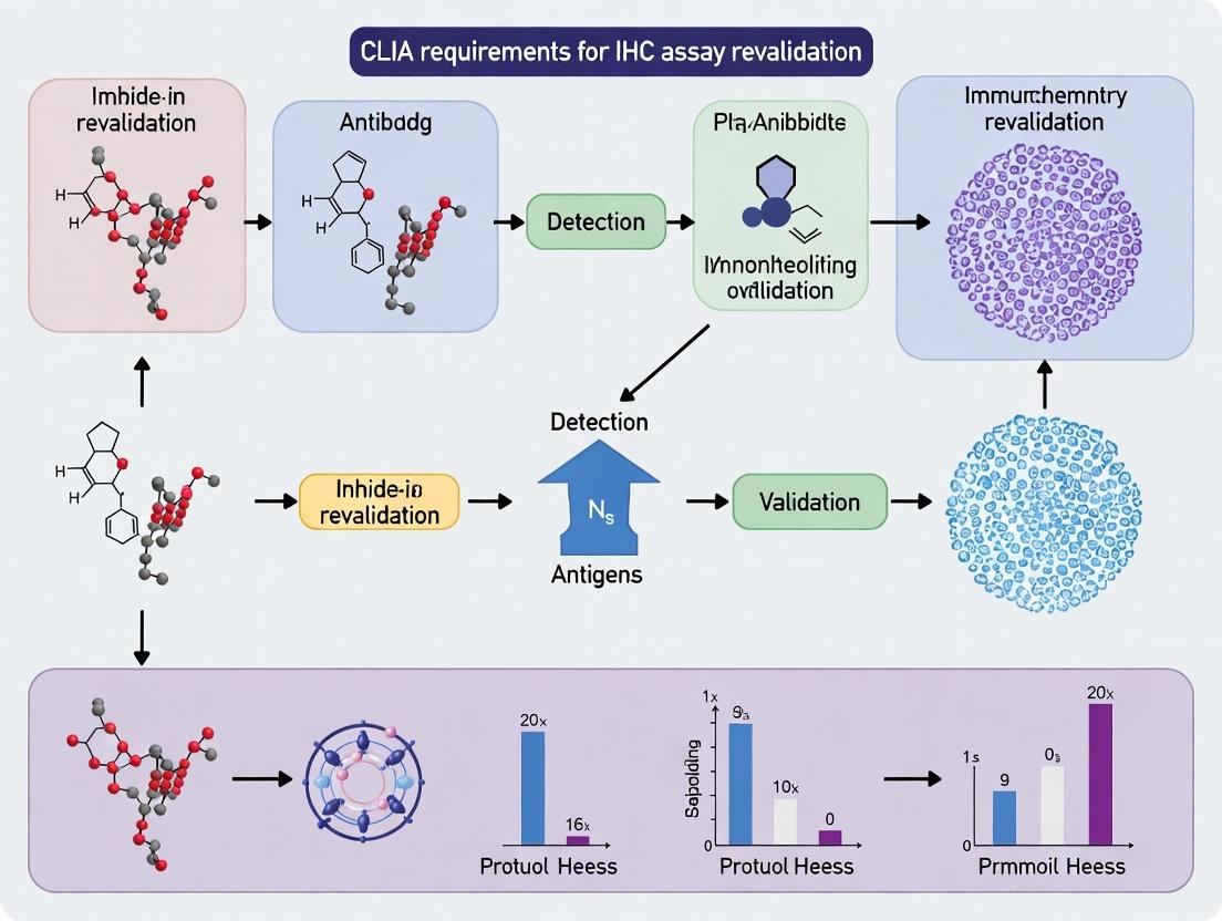
Navigating CLIA Compliance: A Complete Guide to IHC Assay Revalidation for Clinical Labs
This comprehensive guide demystifies CLIA regulations for Immunohistochemistry (IHC) assay revalidation.
Navigating CLIA Compliance: A Complete Guide to IHC Assay Revalidation for Clinical Labs
Abstract
This comprehensive guide demystifies CLIA regulations for Immunohistochemistry (IHC) assay revalidation. Targeted at laboratory directors, researchers, and drug development professionals, it provides a step-by-step framework from foundational regulatory requirements to advanced troubleshooting. Readers will learn the critical triggers mandating revalidation, discover compliant methodological workflows, master strategies to overcome common assay challenges, and understand how to execute comparative validation studies to ensure patient safety and data integrity in a CLIA-certified environment.
Understanding CLIA: The Non-Negotiable Framework for IHC Revalidation
Defining CLIA and its Jurisdiction over Clinical IHC Testing
The Clinical Laboratory Improvement Amendments of 1988 (CLIA) establish the federal regulatory standards applicable to all clinical laboratory testing performed on humans in the United States, with the primary objective of ensuring the analytical validity, reliability, and clinical utility of test results. The Centers for Medicare & Medicaid Services (CMS) enforces CLIA regulations in partnership with the Food and Drug Administration (FDA) and the Centers for Disease Control and Prevention (CDC). Jurisdiction over clinical immunohistochemistry (IHC) testing is squarely under the CLIA umbrella, as IHC is classified as a high-complexity test, mandating compliance with stringent personnel, quality control (QC), proficiency testing (PT), and validation requirements.
The FDA categorizes IHC tests as either laboratory-developed tests (LDTs), which are designed, manufactured, and used within a single CLIA-certified laboratory, or in vitro diagnostic (IVD) kits, which are commercially distributed. For LDTs, CLIA laboratories bear full responsibility for establishing test performance specifications. The FDA regulates commercially marketed IHC IVD kits (e.g., companion diagnostics) through pre-market review, but once such a kit is in use in a lab, its operation falls under CLIA oversight. The core mandate is that any IHC test used for patient diagnosis, prognosis, or treatment selection must be performed in a CLIA-certified laboratory that meets all applicable standards.
CLIA Requirements for IHC Assay Validation and Revalidation
Under CLIA §493.1253, laboratories must establish and verify performance specifications for all high-complexity testing, including IHC. For a new IHC assay, full validation is required. Revalidation is mandated when there is a change in pre-analytical, analytical, or post-analytical conditions that could affect test performance. This is a cornerstone of the broader thesis on CLIA requirements for IHC assay revalidation research, which posits that systematic, evidence-based revalidation protocols are critical for maintaining diagnostic integrity in the face of evolving methodologies and reagents.
Key Triggers for IHC Assay Revalidation:
- Change in antibody clone, vendor, or lot (if not standardized through bridging studies).
- Change in antigen retrieval method (e.g., pH, time, epitope retrieval buffer).
- Change in detection system or visualization chemistry.
- Change in staining platform (automated stainers).
- Change in specimen type (e.g., moving from resection to biopsy samples) or fixative.
- Implementation of a new scoring algorithm or digital image analysis.
Quantitative Data on CLIA Laboratory Performance (2023)
Table 1: Summary of CLIA Laboratory Deficiencies and PT Performance (2023 Data)
| Category | Metric | Value | Source/Notes |
|---|---|---|---|
| CLIA Labs | Total CLIA-certified labs | ~260,000 | CMS Data |
| IHC PT | Major PT failures (all tests) | ~1.2% of challenges | CMS CLIA Database |
| Common Deficiencies | QC procedures (Tag: §493.1256) | 18% of labs cited | Top citation in inspections |
| Common Deficiencies | Test validation (Tag: §493.1253) | 15% of labs cited | CMS CLIA Database |
| Revalidation Focus | Antibody lot-to-lot verification | Required per CAP checklist | CAP ANP.22950 |
Table 2: Core CLIA Validation/Revalidation Parameters for IHC (with Target Benchmarks)
| Performance Parameter | CLIA Requirement | Typical IHC Benchmark | Experimental Protocol Summary |
|---|---|---|---|
| Accuracy | Agreement with a reference method or material. | >95% concordance with known positive/negative. | See Protocol 1. |
| Precision | Repeatability (within-run) and Reproducibility (between-run, day, operator). | ≥95% intra-assay concordance. | See Protocol 2. |
| Analytical Sensitivity | Detection limit (lowest antigen level detectable). | Consistent staining at expected dilutions of control tissues. | Titration of primary antibody on control tissue. |
| Analytical Specificity | Includes interference (e.g., necrosis, edge artifact) and cross-reactivity. | No staining in known negative tissue types. | Stain tissue microarray with known negative tissues. |
| Reportable Range | All anticipated staining intensities (0 to 3+) are distinguishable. | Clear differential staining across control set. | Use controls with graded expression levels. |
| Reference Range | Interpretation criteria (positive/negative, scoring thresholds). | Clearly defined, clinically validated cut-offs. | Correlate staining intensity with clinical outcome. |
Detailed Experimental Protocols for Revalidation
Protocol 1: Accuracy/Concordance Study for IHC Revalidation
Objective: To determine the agreement between the new/test condition (e.g., new antibody lot) and the established condition. Materials: 20 previously characterized patient specimens (10 positive, 5 negative, 5 variable/low expression). Slides from the same blocks. Method:
- Stain all cases in parallel using the established protocol and the modified (test) protocol.
- Perform blinded, independent evaluation by at least two qualified pathologists using the laboratory's standard scoring criteria.
- Calculate positive, negative, and overall percent agreement. Statistical analysis (e.g., Cohen's kappa for inter-rater reliability) should be performed. Kappa >0.90 indicates excellent agreement. Acceptance Criterion: ≥95% overall agreement with the established method.
Protocol 2: Precision (Reproducibility) Study for IHC Revalidation
Objective: To assess inter-run and intra-run reproducibility of the modified IHC assay. Materials: A set of 3 control tissues (strong positive, weak positive, negative). Multiple slides from the same blocks. Method (Inter-run):
- Run the assay on the control set across 5 separate days, using the same stainer but different operators if applicable.
- Evaluate staining intensity and distribution.
- Calculate the coefficient of variation (if using quantitative image analysis) or concordance rate. Method (Intra-run):
- Place the same 3 controls in different positions on a single slide run.
- Evaluate uniformity of staining across the slide. Acceptance Criterion: ≥95% concordance across all runs and positions.
Signaling Pathway and Workflow Visualizations
Diagram 1: CLIA IHC Test Lifecycle with Revalidation Trigger
Diagram 2: CLIA Agency Jurisdiction and Roles
The Scientist's Toolkit: Essential Research Reagents & Materials for IHC Revalidation
Table 3: Key Research Reagent Solutions for IHC Revalidation Studies
| Item | Function in Revalidation Research | Example/Notes |
|---|---|---|
| Formalin-Fixed, Paraffin-Embedded (FFPE) Tissue Microarrays (TMAs) | Contain multiple tissue cores on one slide for simultaneous staining of positive, negative, and variable controls under identical conditions. Essential for assessing specificity and sensitivity. | Commercial or laboratory-constructed TMAs with validated expression status for target antigen. |
| Cell Line Xenografts or Control Slides | Provide consistent, homogeneous antigen expression for precision studies (inter/intra-run). Useful for titrating new antibody lots. | Pellet blocks of cell lines with known antigen expression levels (e.g., HER2 0, 1+, 2+, 3+). |
| Reference Standard Antibodies | The previously validated antibody clone used as the comparator in accuracy/concordance studies. Serves as the benchmark. | Should be from a defined lot, aliquoted and stored to maintain stability throughout the revalidation period. |
| Isotype Controls / Negative Control Ig | Monoclonal antibodies of the same isotype but irrelevant specificity. Critical for demonstrating staining specificity and background. | Used at the same concentration as the primary antibody. |
| Automated Stainer Calibration & QC Kits | Reagents and slides provided by stainer manufacturers to ensure optical, fluidic, and thermal systems are performing within specification. | Mandatory for revalidation after instrument maintenance or relocation. |
| Digital Image Analysis Software & Controls | Enables quantitative, objective assessment of staining intensity and percentage for precision and reportable range studies. | Use validated algorithms and standardized scanning settings. Paired with appropriate software calibration slides. |
Within the regulatory framework for clinical laboratory testing in the United States, the Clinical Laboratory Improvement Amendments (CLIA) establish the quality standards for all laboratory testing performed on human specimens. For researchers and drug development professionals, particularly those engaged in Immunohistochemistry (IHC) assay revalidation research, a precise understanding of CLIA's core requirements for assay validation is paramount. This guide unpacks these requirements, translating regulatory mandates into actionable technical protocols to ensure data integrity, reproducibility, and compliance in a research and development context.
The CLIA regulations, particularly under 42 CFR §493.1253, specify standards for establishing and verifying test performance specifications. The following table summarizes the key quantitative parameters required for a robust assay validation.
Table 1: Core CLIA Assay Validation Parameters & Targets
| Validation Parameter | CLIA Requirement / Standard Target | Key Consideration for IHC Revalidation |
|---|---|---|
| Accuracy | Comparison to a reference method or clinical truth. | For IHC, this often involves comparison to a gold-standard assay (e.g., PCR for biomarkers), orthogonal method, or well-characterized patient cohort outcomes. |
| Precision | Within-run and between-run reproducibility must be established. | Includes evaluation of staining intensity and heterogeneity across slides, days, operators, and reagent lots. A CV <20% is often a pragmatic target for semi-quantitative scores. |
| Reportable Range | The span of results the method can reliably produce. | In IHC, this relates to antigen expression levels. Defined by testing samples with known expression levels from negative to strongly positive. |
| Reference Range | "Normal" or expected values for the population. | Critical for IHC markers with prognostic or diagnostic cut-offs. Established using a sufficient number of confirmed negative/normal tissue samples. |
| Analytical Sensitivity (Limit of Detection) | The lowest amount of analyte reliably detected. | Determined by testing serial dilutions of cell lines or tissues with known low expression. The lowest dilution yielding specific, reproducible staining. |
| Analytical Specificity | Includes Interference and Cross-reactivity. | Testing for endogenous enzymes, biotin, or other pre-analytical factors. Specificity confirmed via isotype controls, peptide blockade, and testing on tissues known to express related antigens. |
| Robustness / Ruggedness | Resistance to changes in pre-analytical & analytical conditions. | Essential for IHC revalidation. Tests impact of variable fixation times, antigen retrieval conditions, primary antibody incubation time/temperature, and reagent lot changes. |
Detailed Experimental Protocols for IHC Assay Validation
The following methodologies provide a framework for meeting CLIA-level validation rigor in a research setting, particularly for assay revalidation.
Protocol 1: Precision (Reproducibility) Testing
Objective: To determine within-run (repeatability) and between-run (intermediate precision) variability of the IHC assay.
- Sample Selection: Select a minimum of 3 tissue specimens spanning the assay's dynamic range (negative, weak positive, strong positive). Use formalin-fixed, paraffin-embedded (FFPE) tissue blocks.
- Experimental Design:
- Within-Run: For each sample, cut 5 consecutive serial sections. Process all 5 sections in a single IHC run using the same reagent lots, equipment, and operator.
- Between-Run: Repeat the entire IHC assay for the same 3 samples across 5 separate runs. Introduce expected variables: different days, different reagent lots (if applicable), and multiple qualified operators.
- Analysis: All slides are scored independently by at least two qualified pathologists using the standardized scoring method (e.g., H-score, Allred score). Calculate the Coefficient of Variation (CV%) for each sample group across runs.
Protocol 2: Analytical Specificity & Cross-Reactivity
Objective: To confirm the primary antibody's specificity for the target epitope.
- Peptide Blockade (Competition Assay):
- Pre-incubate the optimized dilution of the primary antibody with a 10-fold molar excess of the target immunizing peptide (blocked condition) versus a non-relevant peptide (control condition) for 1 hour at room temperature.
- Perform IHC using both pre-absorbed antibody solutions on a known positive tissue.
- Validation Criterion: Significant reduction or complete abolition of staining in the blocked condition confirms specificity.
- Cross-Reactivity Panel:
- Perform IHC using the validated protocol on a tissue microarray (TMA) containing a wide range of normal human tissues.
- Evaluate staining patterns for expected on-target expression and any unexpected, off-target staining that may indicate cross-reactivity with homologous proteins.
Protocol 3: Robustness Testing for Revalidation
Objective: To evaluate the assay's reliability when minor, deliberate changes are made to procedural parameters.
- Identify Critical Variables: These typically include antigen retrieval time (± 5 minutes), primary antibody incubation time (± 25%), and incubation temperature (room temp vs. 4°C).
- Experimental Design: Use a 2^3 factorial design. Test all combinations of high/low values for the three chosen variables on a positive and a weak positive sample.
- Analysis: Score results and use statistical analysis (e.g., ANOVA) to determine which variables, if any, have a statistically significant and clinically relevant impact on the staining outcome. This defines the acceptable operational tolerances for the standard operating procedure (SOP).
Diagram: IHC Assay Validation & Revalidation Workflow
The Scientist's Toolkit: Essential Reagents & Materials for IHC Validation
Table 2: Key Research Reagent Solutions for IHC Assay Validation
| Item | Function in Validation | Critical Consideration |
|---|---|---|
| Well-Characterized FFPE Tissue Controls | Positive, negative, and low-expressing controls for precision, sensitivity, and specificity runs. | Must be validated themselves. Should include tissues with known genetic status (e.g., HER2 FISH-characterized). |
| Isotype Control Antibody | Matched immunoglobulin of the same species, subclass, and concentration as the primary antibody. | Distinguishes specific staining from non-specific background or Fc receptor binding. |
| Immunizing Peptide (for blockade) | Synthetic peptide corresponding to the primary antibody's epitope. | The gold-standard for confirming antibody specificity during validation. |
| Cell Line Microarrays (CLMA) | FFPE blocks containing pellets of cell lines with known target expression levels. | Provides a consistent, renewable resource for precision and sensitivity testing across multiple runs. |
| Automated Staining Platform | Provides consistent reagent application, incubation times, and temperatures. | Essential for meeting CLIA requirements for robust, reproducible assay performance. |
| Validated Detection System | A polymer- or enzyme-based visualization system with known sensitivity and low background. | The key reagent for establishing the Limit of Detection. Lot-to-lot consistency is crucial. |
| Digital Image Analysis Software | Enables quantitative or semi-quantitative scoring of staining intensity and percentage. | Reduces observer variability, providing objective, reproducible data for precision studies. |
A disciplined approach to assay validation, grounded in the core requirements of CLIA, is not merely a regulatory hurdle but a foundational pillar of rigorous scientific research. For professionals engaged in IHC assay development and revalidation, translating these requirements into detailed experimental protocols—assessing precision, specificity, and robustness—ensures that subsequent research data and conclusions are reliable. This process, supported by appropriate controls, reagents, and tools, forms the critical bridge between exploratory biomarker research and the generation of clinically actionable data in drug development.
What Triggers IHC Revalidation? A Checklist of Mandatory Events.
Within the regulatory framework of the Clinical Laboratory Improvement Amendments (CLIA), the validation and revalidation of immunohistochemistry (IHC) assays are critical to ensuring analytical accuracy and clinical utility. This guide serves as an in-depth technical whitepaper, framed within a broader thesis on CLIA compliance, to delineate the mandatory events that trigger a full or partial revalidation of an IHC assay. It is designed for researchers, scientists, and drug development professionals tasked with maintaining rigorous quality standards in diagnostic and pre-clinical testing.
Part 1: Mandatory Revalidation Triggers & Regulatory Context
Revalidation is not discretionary; it is a mandated response to specific changes that could affect assay performance. The core principle, per CLIA and guidelines from the College of American Pathologists (CAP), is that any modification to a previously validated assay requires an assessment of its impact and, typically, revalidation.
The quantitative thresholds for required action are summarized in the table below, synthesizing current regulatory guidance and industry standards.
Table 1: IHC Revalidation Triggers and Required Actions
| Trigger Event | Scope of Change | Recommended Validation Type | Key Parameters to Assess |
|---|---|---|---|
| Change in Primary Antibody | Clone, species, vendor, or conjugation. | Full Revalidation | Sensitivity, specificity, staining pattern, intensity, optimal dilution. |
| Change in Detection System | New kit, polymer, chromogen, or automation platform. | Full Revalidation | Sensitivity, background, signal-to-noise ratio, incubation times. |
| Change in Antigen Retrieval | Method (heat vs. enzyme), pH, time, or buffer. | Partial/Focused Revalidation | Staining intensity, uniformity, cellular localization. |
| Change in Tissue Processing | Fixative type (e.g., NBF to PAXgene), fixation time. | Full Revalidation | Epitope preservation, morphology, staining intensity & specificity. |
| Instrument/Platform Change | New stainer, slide scanner, or imaging system. | Parallel Testing & Verification | Reproducibility, staining uniformity, quantitative image analysis outputs. |
| New Tissue Type | Applying assay to a previously untested tissue or cell type. | Full Revalidation | Specificity, background, expected staining pattern in new context. |
| Critical Reagent Lot Change | New lot from same vendor (esp. antibody). | Abridged Verification | Comparison of staining intensity and pattern to established positive/negative controls. |
| Updated Clinical Guidelines | New cut-offs or scoring criteria (e.g., HER2). | Analytical Revalidation | Precision (reproducibility), accuracy against reference standard. |
Part 2: Experimental Protocols for Core Revalidation Studies
A robust revalidation protocol must objectively compare the new condition to the established, validated method.
Protocol 1: Protocol for Parallel Testing of a New Primary Antibody Objective: To establish equivalence or superiority of a new antibody clone.
- Slide Selection: Assemble a tissue microarray (TMA) containing 20-50 cases representing the expected spectrum of antigen expression (negative, weak, moderate, strong) and relevant morphological variants.
- Staining: Stain paired serial sections from the TMA using the old (validated) protocol and the new protocol (with the antibody change) in the same batch to minimize inter-run variability.
- Blinded Review: Have at least two board-certified pathologists score all slides in a blinded fashion using the laboratory's established scoring system (e.g., H-score, percentage positivity).
- Data Analysis: Calculate inter-observer concordance (Cohen's kappa) and perform statistical correlation (e.g., Pearson's r) between scores from the old and new protocols. A correlation coefficient >0.90 and kappa >0.80 typically indicate acceptable concordance.
- Specificity Check: Evaluate known internal negative controls within the tissues for any non-specific staining.
Protocol 2: Protocol for Verification of a New Detection System/Lot Objective: To ensure new detection reagents do not alter assay sensitivity or background.
- Control Tissues: Use a defined set of positive controls (cell line pellets with known antigen expression levels) and negative controls.
- Cross-Staining: Perform staining with both the old and new detection systems on consecutive sections. Include a no-primary antibody control for each system.
- Quantitative Analysis: For assays with digital image analysis, extract quantitative metrics (e.g., average optical density, percentage of positive pixels) from identical regions of interest.
- Acceptance Criteria: Define pre-set acceptance criteria (e.g., ≤10% coefficient of variation in quantitative metrics, no increase in background staining in negative controls).
Part 3: Visualization of Key Concepts
Title: IHC Revalidation Decision Pathway
Title: IHC Workflow with Revalidation Triggers
Part 4: The Scientist's Toolkit
Table 2: Essential Research Reagent Solutions for IHC Revalidation
| Item | Function in Revalidation |
|---|---|
| Formalin-Fixed, Paraffin-Embedded (FFPE) Tissue Microarray (TMA) | Contains multiple tissue types/controls on one slide, enabling high-throughput, comparative analysis of staining performance under old vs. new conditions. |
| Cell Line Pellet Controls | Provide a consistent, renewable source of tissue with known, homogeneous antigen expression levels for quantitative assessment of sensitivity and reproducibility. |
| Multitissue Blocks (e.g., Spleen, Tonsil, Tumor) | Serve as comprehensive positive and negative control tissues to assess specificity and staining patterns across a diverse antigen landscape. |
| Isotype Control Antibodies | Critical for distinguishing specific signal from non-specific background staining, especially when validating a new primary antibody. |
| Digital Image Analysis Software | Enables objective, quantitative measurement of staining intensity (optical density) and percentage positivity, reducing scorer bias for robust comparison. |
| Reference Standard Slides | A set of archived slides stained with the original validated protocol, serving as the "gold standard" for visual and quantitative comparison during revalidation. |
Within the framework of CLIA (Clinical Laboratory Improvement Amendments) requirements, the revalidation of Immunohistochemistry (IHC) assays is not merely a regulatory formality but a critical safeguard. This technical guide examines the severe consequences of non-compliance, detailing the technical, clinical, and legal ramifications for laboratories and the patients who depend on accurate diagnostic results. The imperative for rigorous revalidation is underscored by its role in ensuring assay accuracy, reproducibility, and clinical utility in drug development and personalized medicine.
The Regulatory and Clinical Imperative: CLIA and Beyond
CLIA regulations (42 CFR Part 493) establish quality standards for laboratory testing. For IHC, a high-complexity test, compliance requires rigorous validation and revalidation under specific conditions. These conditions include changes in reagent lots, equipment, or test procedures. Non-compliance triggers a cascade of risks.
Table 1: Documented Consequences of IHC Assay Non-Compliance
| Consequence Category | Specific Impact | Quantitative Data / Incidence |
|---|---|---|
| Analytical Performance Failure | Shift in assay sensitivity/specificity | Up to 15-20% variance in staining intensity with uncontrolled reagent lot changes. |
| Misdiagnosis & Patient Harm | False Positive/Negative Results | Studies indicate non-validated IHC contributes to ~5% of significant diagnostic errors in surgical pathology. |
| Clinical Trial Compromise | Invalid Biomarker Data | Can lead to incorrect patient stratification, potentially invalidating trial phases and costing $500k-$5M per incident in lost R&D. |
| Regulatory & Legal Penalties | CLIA certification revocation, fines, litigation | CMS penalties can exceed $10,000 per day of non-compliance; malpractice suit settlements average $500,000. |
| Operational & Reputational | Lab accreditation loss, increased scrutiny | 85% of labs with major CLIA citations report significant patient volume loss within one year. |
Core Experimental Protocol: IHC Assay Revalidation
The following protocol outlines a standard revalidation methodology mandated after a critical change, such as a new primary antibody lot.
Protocol: Revalidation of a Predictive IHC Assay (e.g., HER2)
- Define Scope & Acceptance Criteria: Based on initial validation. For a HER2 assay, criteria may be ≥95% concordance with prior valid lot and reference method (e.g., FISH) for scored samples.
- Selection of Tissue Samples: Use a well-characterized tissue microarray (TMA) or full sections.
- Positive Controls (n≥10): Tissues with known, varying levels of antigen expression (weak, moderate, strong).
- Negative Controls (n≥5): Tissues known to lack the target antigen.
- Comparator: Archival slides stained with the previously validated antibody lot.
- Staining Procedure: Perform IHC staining using the new reagent lot alongside the old lot on serial sections under identical, optimized conditions (automated platform, retrieval method, incubation times).
- Blinded Evaluation: Two qualified pathologists independently score slides (e.g., for HER2: 0, 1+, 2+, 3+) without knowledge of the lot or other scorer's results.
- Data Analysis:
- Calculate concordance rates (positive, negative, overall percentage agreement) between new lot and old lot.
- Assess inter-observer agreement using Cohen's kappa statistic.
- Compare scores to reference method data if available.
- Decision Point: If acceptance criteria are met, the new lot is approved. If not, root cause analysis is initiated, and the lot is rejected.
Key Signaling Pathways in IHC Biomarkers
Accurate IHC detection hinges on understanding the target pathway.
Title: HER2 Signaling Pathway & IHC Detection Target
IHC Revalidation Workflow
A systematic process is essential for compliant revalidation.
Title: IHC Assay Revalidation Compliance Workflow
The Scientist's Toolkit: Research Reagent Solutions
Critical materials for robust IHC revalidation studies.
Table 2: Essential Reagents & Materials for IHC Revalidation
| Item | Function in Revalidation | Key Consideration |
|---|---|---|
| Validated Primary Antibody (Old Lot) | Serves as the gold standard comparator for the new lot. | Must be from a previously CLIA-compliant validation run. Store aliquots for this purpose. |
| New Primary Antibody Lot | The test article requiring performance verification. | Ensure clone, host species, and conjugation are identical to the old lot. |
| Multitissue Control Microarray (TMA) | Provides a range of positive, negative, and borderline tissues in one slide. | Must be well-characterized with orthogonal method confirmation (e.g., FISH, PCR). |
| IHC Detection Kit | Enzymatic (HRP/AP) or polymer-based system for signal amplification. | Keep constant between validation runs. Changing this requires a full revalidation. |
| Antigen Retrieval Buffer | Unmasks epitopes formalin-fixed, paraffin-embedded tissue. | pH and heating method must be rigorously controlled and documented. |
| Automated IHC Stainer | Provides consistent, hands-off processing of slides. | Calibration and maintenance logs are critical for CLIA inspection. |
| Reference Standard Slides | Archived slides from prior clinical cases with confirmed results. | Essential for bridging studies and demonstrating longitudinal accuracy. |
The stakes of non-compliance in IHC assay revalidation are prohibitively high, directly impacting diagnostic integrity, patient outcomes, and the validity of clinical research. Adherence to a structured, documented revalidation protocol—as outlined in this guide—is the principal defense against these risks. For researchers and drug development professionals, this process is not an obstacle but a foundational component of ethical, reliable, and legally defensible laboratory science.
Differentiating Revalidation from Initial Validation and Verification
In the regulated environment of clinical diagnostics, compliance with the Clinical Laboratory Improvement Amendments (CLIA) is paramount. For Immunohistochemistry (IHC) assays, a cornerstone of pathology and drug development, understanding the distinct processes of verification, initial validation, and revalidation is critical. This whitepaper delineates these processes, framed explicitly within the requirements for IHC assay revalidation research, a necessary activity when modifications occur in the pre-analytic, analytic, or post-analytic phases of testing.
Core Definitions: Verification vs. Validation vs. Revalidation
| Term | Definition | CLIA Context for IHC Assays | Primary Objective |
|---|---|---|---|
| Verification | Confirming that a procedure or test system performs according to its stated specifications. | Required for FDA-cleared/approved IHC tests. The lab must demonstrate it can achieve the manufacturer's performance claims. | Ensure test performs as expected in the user's environment. |
| Initial Validation | Establishing the performance specifications of a laboratory-developed test (LDT) or a modified FDA-cleared test. | Required for IHC LDTs. A comprehensive study to define analytical performance (accuracy, precision, sensitivity, specificity, etc.). | Establish robust, evidence-based performance characteristics. |
| Revalidation | Re-establishing performance specifications after a change to an already validated test system. | Triggered by changes affecting IHC assay performance (e.g., new antibody clone, antigen retrieval method, detection system, instrument). | Document that the modified test maintains its validated performance. |
Quantitative Comparison of Key Activities
Table 1: Scope and Scale of Performance Studies
| Activity | Accuracy/Comparison Study (n) | Precision (Runs/Days/Operators) | Reportable Range | Reference Interval |
|---|---|---|---|---|
| Verification | 20-30 samples | 2-3 runs, 1-3 days, 1-2 operators | Confirm manufacturer's range | Confirm manufacturer's interval |
| Initial Validation | 50-100+ samples (disease & normal) | 5-20 runs, 5-20 days, 2-3 operators | Establish for LDT | Establish for intended population |
| Revalidation | 20-50 samples (scope depends on change impact) | 3-10 runs, 3-10 days, 2 operators | Re-confirm or re-establish if affected | Re-confirm if affected |
Table 2: Common Triggers and Required Revalidation Extent for IHC Assays
| Trigger Category | Example Change | Likely Revalidation Activities |
|---|---|---|
| Pre-analytic | New tissue fixative, change in fixation time | Accuracy (comparison), Precision, Staining Intensity Assessment |
| Analytic - Critical Reagent | New antibody vendor or clone, new detection kit | Full analytical validation (Accuracy, Precision, Sensitivity, Specificity) |
| Analytic - Equipment | New automated staining platform | Precision, Accuracy (comparison), Process workflow verification |
| Analytic - Protocol | Change in antigen retrieval method (e.g., pH, time) | Accuracy (comparison), Precision, Optimization for staining intensity |
| Post-analytic | New scoring criteria or digital image analysis algorithm | Inter-observer precision, Comparison to original method, Software verification |
Experimental Protocols for IHC Revalidation Studies
Protocol 1: Accuracy/Comparison Study
Objective: To compare results of the modified IHC assay against the previously validated method or a gold standard. Methodology:
- Select a panel of 20-50 formalin-fixed, paraffin-embedded (FFPE) tissue samples representing the full reportable range (negative, weak, moderate, strong expression).
- Cut serial sections from each block.
- Stain one section with the established (old) assay protocol and the adjacent section with the modified (new) protocol.
- Employ blinded evaluation by at least two qualified pathologists. Use a semi-quantitative scoring system (e.g., H-score, Allred score).
- Perform statistical analysis (e.g., Cohen's kappa for agreement, Pearson correlation for continuous scores, Deming regression).
Protocol 2: Precision (Reproducibility) Assessment
Objective: To evaluate the assay's repeatability (within-run) and reproducibility (between-run, between-day, between-operator). Methodology:
- Select 3-5 FFPE samples covering low, medium, and high antigen expression levels.
- Design a nested experiment where each sample is stained in duplicate across 5-10 separate runs, over 5-10 different days, by 2 different operators.
- All staining and evaluation should be performed blinded.
- Calculate the coefficient of variation (CV) for continuous data or kappa statistics for categorical scores. Analyze variance components to identify major sources of variability.
Protocol 3: Limit of Detection (LOD) / Sensitivity
Objective: To confirm the minimum amount of target antigen detectable after a reagent change. Methodology:
- Use cell line microarrays with known antigen expression levels or a dilution series of a positive tissue extract titrated into a negative matrix.
- Stain with the modified assay protocol.
- The LOD is the lowest concentration/level where the stain is consistently detectable above background (≥95% of replicates).
Visualizing the Decision and Workflow Logic
Diagram Title: CLIA IHC Assay Change Control Decision Pathway
Diagram Title: IHC Assay Phases & Revalidation Triggers
The Scientist's Toolkit: Key Research Reagent Solutions for IHC Revalidation
Table 3: Essential Materials for IHC Revalidation Studies
| Item | Function in Revalidation | Example/Notes |
|---|---|---|
| Characterized FFPE Tissue Microarray (TMA) | Serves as the primary sample set for accuracy/precision studies. Contains cores of known positive, negative, and graded expression levels. | Commercial or lab-constructed TMAs with validated staining patterns. |
| Reference Control Slides | Provides run-to-run consistency monitoring for precision studies. | Commercially available multi-tissue control slides or in-house pooled tissue controls. |
| Calibrated Digital Pathology Scanner | Enables quantitative, reproducible assessment of staining intensity for objective comparison. | Systems with consistent light intensity and calibration slides. |
| Image Analysis Software | Quantifies stain intensity (H-score, % positivity) objectively, reducing observer bias in comparison studies. | HALO, Visiopharm, QuPath, or Aperio's ImageScope. |
| Cell Line Pellet Arrays | Provide a consistent source of biologic material with defined antigen expression for sensitivity/LOD studies. | Arrays constructed from cell lines with known knockout/knockdown of target. |
| New vs. Old Critical Reagents | The direct comparison of the changed component (antibody, detection kit) against the previously validated material. | Must be sourced with full traceability (lot number, clone, concentration). |
| Statistical Analysis Software | Performs essential agreement statistics (kappa, correlation, CV, regression). | R, SPSS, GraphPad Prism, or MedCalc. |
Within the CLIA framework, revalidation is not a repetition of initial validation but a targeted, risk-based exercise to ensure continued reliability of modified IHC assays. Its scope is directly proportional to the potential impact of the change on the assay's analytical performance. A rigorous, protocol-driven approach anchored in comparison studies and precision assessment, supported by well-characterized reagents and controls, is essential for maintaining diagnostic integrity and compliance in both clinical and drug development research.
The Role of CAP Guidelines in Complementing CLIA Standards for IHC
Within the framework of CLIA requirements for IHC assay revalidation research, understanding the interplay between regulatory standards is critical. The Clinical Laboratory Improvement Amendments (CLIA) establish the federal quality standards for all laboratory testing. For immunohistochemistry (IHC), CLIA mandates non-specific quality control (QC) measures but does not prescribe detailed analytical validation or revalidation protocols. The College of American Pathologists (CAP) accreditation program, particularly through its laboratory general (GEN) and anatomic pathology (ANP) checklists, provides the specific, evidence-based guidelines that operationalize and exceed CLIA's broader mandates, ensuring analytical rigor, reproducibility, and clinical validity.
Quantitative Comparison: CLIA vs. CAP Requirements for IHC
The following table summarizes the core quantitative and qualitative differences between CLIA and CAP as they pertain to IHC validation and quality assurance.
Table 1: Comparison of CLIA Standards and CAP Guidelines for IHC
| Aspect | CLIA Requirement | CAP Guideline (ANP.22800, GEN.40396, etc.) | Complementary Role of CAP |
|---|---|---|---|
| Regulatory Nature | Federal law (42 CFR Part 493). Minimum baseline. | Voluntary accreditation program with detailed checklists. | Provides the specific, actionable pathway to meet and exceed CLIA. |
| Test Validation | Requires test validation but lacks IHC-specific details. | ANP.22800: Defines required validation parameters (e.g., sensitivity, specificity, precision). Specifies minimum case numbers (20-60 positive, 20 negative). | Transforms CLIA's general mandate into a standardized, quantitative experimental protocol. |
| Revalidation Triggers | Implied upon significant change. | Explicitly lists triggers: antibody lot change, instrument change, protocol change, fixative change (>12 hrs), tissue processor change. | Provides a clear, pre-defined research framework for revalidation studies mandated by CLIA's "condition change." |
| Daily QC | Requires calibration and control procedures. | ANP.22815: Mandates daily/run use of multi-tissue control blocks with reactive tissues and external negatives. Requires documentation. | Specifies the material (control blocks) and frequency, ensuring QC is fit-for-purpose for IHC. |
| Proficiency Testing (PT) | Requires successful PT twice annually. | GEN.80000+: Adherence to CAP's IHC-specific PT programs (e.g., PHC, PNK) is required. Peer-group grading. | Provides standardized, graded challenges that fulfill CLIA's PT requirement and benchmark performance. |
| Antibody Validation | No specific guidance. | ANP.22800: Requires verification of vendor-stated performance on in-house equipment. Positive and negative controls must be run. | Closes a critical CLIA gap, ensuring reagent performance is objectively confirmed in the local testing environment. |
Detailed Experimental Protocols for IHC Revalidation
A core thesis in CLIA-driven revalidation research is that CAP guidelines provide the definitive methodological blueprint. The following protocols are derived from CAP checklist requirements.
Protocol 1: Initial Analytic Validation (CAP ANP.22800)
Objective: To establish performance characteristics of a new IHC assay. Methodology:
- Case Selection: Assemble a retrospective cohort of 40-60 formalin-fixed, paraffin-embedded (FFPE) specimens with known status (positive/negative) via a gold-standard method (e.g., molecular, prior IHC with different epitope).
- Staining Procedure: Perform the new IHC assay according to the optimized protocol on all cases.
- Blinded Evaluation: Two qualified pathologists independently score stains for intensity (0-3+) and distribution (% positive cells). Discrepancies are resolved by consensus or a third reviewer.
- Data Analysis:
- Calculate Sensitivity: (True Positives / (True Positives + False Negatives)) * 100.
- Calculate Specificity: (True Negatives / (True Negatives + False Positives)) * 100.
- Assess Precision (Reproducibility): Run a subset of cases (n=20) across different days, with different operators, and/or different instrument lots. Calculate inter-run and inter-observer concordance (Cohen's kappa, >0.85 target).
Protocol 2: Revalidation for Antibody Lot Change (CAP ANP.22800)
Objective: To ensure consistency of performance between outgoing and incoming lots. Methodology:
- Control Tissue Selection: Select 5-10 previously characterized FFPE cases encompassing expected expression levels (strong positive, weak positive, negative).
- Parallel Staining: Stain all selected cases in a single run using identical protocols except for the antibody lot. Include multi-tissue control blocks.
- Comparative Analysis: Perform blinded side-by-side comparison of staining intensity, pattern, and background. Use digital image analysis or semi-quantitative scoring.
- Acceptance Criterion: The new lot is acceptable if the staining results are clinically concordant (no shift from positive to negative or vice versa) and show no significant qualitative or quantitative deviation (>20% change in scoring or H-score) from the old lot.
Protocol 3: Ongoing Precision Verification (CAP GEN.40396)
Objective: To monitor the long-term stability and reproducibility of the assay. Methodology:
- Establish a Longitudinal Control: A stable, well-characterized control tissue (e.g., cell line pellet, tissue microarray) is included in every routine staining batch.
- Data Tracking: The staining result (e.g., intensity score, percentage positivity) for this longitudinal control is recorded in a quality control chart (e.g., Levey-Jennings chart).
- Statistical Process Control: Establish mean and control limits (±2SD, ±3SD) from an initial baseline period.
- Monitoring and Action: Investigate any out-of-trend or out-of-control signals, which trigger corrective action and potential revalidation.
Visualizing the Complementary Relationship
Diagram 1: CLIA and CAP Relationship in IHC QA
Diagram 2: CAP-Defined Revalidation Decision Pathway
The Scientist's Toolkit: Essential Reagents & Materials for IHC Revalidation
Table 2: Key Research Reagent Solutions for IHC Validation Studies
| Item | Function in Validation/Revalidation | Key Consideration |
|---|---|---|
| Multi-Tissue Control Blocks | Contain known positive (varying levels) and negative tissues. Used for daily QC and lot-to-lot comparisons. Essential for CAP ANP.22815 compliance. | Must be well-characterized, stable over time, and representative of test specimens. |
| Cell Line Pellet Arrays | Provide a source of homogeneous, reproducible material for longitudinal precision tracking and quantitative calibration. | Select cell lines with known, stable expression of the target antigen. |
| Reference Antibodies (Clones) | Serve as the comparator ("gold standard") for validating a new antibody's specificity and sensitivity. | Should be well-established in literature and directed against a different epitope if possible. |
| Tissue Microarrays (TMAs) | Enable high-throughput validation across dozens of tissues/cases on a single slide, ensuring staining uniformity assessment. | Critical for initial validation (CAP-specified case numbers) and efficient revalidation studies. |
| Digital Image Analysis Software | Provides objective, quantitative assessment of staining intensity and percentage positivity, reducing observer bias. | Essential for generating quantitative data for statistical comparison (e.g., H-score deviation between lots). |
| PT Slides (CAP PHC/PNK) | External, graded proficiency testing materials that fulfill CLIA PT requirements and benchmark lab performance against peers. | The cornerstone of external quality assurance mandated by both CLIA and CAP. |
The CLIA-Compliant Blueprint: Step-by-Step IHC Revalidation Protocol
Revalidation of an Immunohistochemistry (IHC) assay within a CLIA-certified laboratory is a critical, structured process mandated by the Clinical Laboratory Improvement Amendments to ensure the continued accuracy, reliability, and clinical validity of test results. Pre-revalidation planning and risk assessment form the foundational stage, determining the scope, strategy, and resources required for successful revalidation. This phase is triggered by defined changes per CLIA §493.1253(b) (standard: "establish and follow written procedures for monitoring and evaluating the test system") and is integral to a broader quality management system. Failure to conduct rigorous planning risks non-compliance, erroneous patient results, and subsequent patient harm.
Risk Assessment Methodology: A Systematic Approach
A formal risk assessment, aligned with principles from ISO 14971 and CLIA's quality systems, is employed to identify and prioritize variables requiring evaluation during revalidation.
Process:
- Assay Deconstruction: Map the entire IHC testing process, from pre-analytical (tissue fixation, processing) to analytical (staining protocol) and post-analytical (interpretation, reporting).
- Change Identification: Clearly define the change initiating revalidation (e.g., new antibody lot, instrument replacement, protocol modification).
- Risk Identification: For each process step, brainstorm potential failure modes introduced by the change.
- Risk Analysis: Evaluate each failure mode for its Severity (impact on patient result) and Likelihood of occurrence. Use a quantitative scoring matrix (Table 1).
- Risk Evaluation & Control: Prioritize risks based on their score. High-priority risks dictate the design and extent of revalidation experiments.
Table 1: Risk Priority Number (RPN) Scoring Matrix
| Severity (S) | Score | Likelihood (L) | Score |
|---|---|---|---|
| Negligible impact on result | 1 | Remote (<1% of tests) | 1 |
| Low impact (minor quantitative shift) | 2 | Unlikely (~5% of tests) | 2 |
| Moderate impact (affects equivocal zone) | 3 | Possible (~10% of tests) | 3 |
| Major impact (false negative/positive) | 4 | Probable (~25% of tests) | 4 |
| Critical impact (direct patient harm) | 5 | Frequent (>33% of tests) | 5 |
RPN = S x L. RPN ≥ 8 mandates explicit experimental control in revalidation.
Defining Revalidation Scope and Strategy
Based on the risk assessment, the revalidation scope is defined as full, partial, or cross-validation.
Table 2: Revalidation Strategy Based on Change Type & Risk
| Change Trigger | Recommended Scope | Key Risk-Based Experiments |
|---|---|---|
| New lot of primary antibody | Partial | Parallel staining of critical cases, titration check, limit of detection. |
| Replacement of automated staining platform | Full | Complete precision, accuracy, and robustness study comparing old vs. new. |
| Change in antigen retrieval method | Partial/Full | Staining intensity comparison, affected epitope evaluation. |
| New tissue type (specimen) | Full | Analytical validation on new matrix, establishing reference ranges. |
| Update to digital scoring algorithm | Cross-validation | Concordance study between old and new scoring methods on a representative set. |
Experimental Protocol for Key Revalidation Experiments
Protocol 1: Parallel Staining for Comparative Analysis (Accuracy)
- Objective: To demonstrate equivalence between the old (validated) and new (changed) assay conditions.
- Methodology:
- Select a minimum of 20 representative, retrospective clinical specimens spanning the assay's dynamic range (negative, weak positive, moderate positive, strong positive). Ensure samples are from different blocks to cover biological variability.
- Cut consecutive sections from each block at the defined thickness.
- Stain one section with the established (control) protocol and the adjacent section with the modified (test) protocol.
- Perform blinded, independent scoring by at least two qualified pathologists using the laboratory's standard scoring criteria (e.g., H-score, % positivity, intensity).
- Analyze data for concordance using appropriate statistics (e.g., Cohen's kappa for categorical data, intraclass correlation coefficient for continuous data, Passing-Bablok regression for comparison).
Protocol 2: Intra-run and Inter-run Precision (Repeatability & Reproducibility)
- Objective: To assess the assay's variability under the new conditions.
- Methodology:
- Select 3 samples: low positive, medium positive, and high positive.
- Intra-run: Stain each sample 5 times within a single staining run (same day, same operator, same reagent batch).
- Inter-run: Stain each sample once per day over 5 separate days, using different reagent preparations and operators if applicable.
- Score all slides and calculate the coefficient of variation (%CV) for quantitative scores. For semi-quantitative scores, report the percentage agreement within a pre-defined tolerance (e.g., ±1 intensity score).
Protocol 3: Limit of Detection (LOD) Verification
- Objective: To confirm the minimum amount of analyte detectable by the modified assay.
- Methodology:
- Utilize a cell line microarray with known, titrated expression levels of the target antigen or a patient tissue sample with a heterogeneity of expression.
- Stain the LOD material with the modified protocol.
- The LOD is defined as the lowest expression level where all replicates (n≥3) are consistently scored as positive by all readers, with a pre-defined acceptable staining intensity.
The Scientist's Toolkit: Essential Research Reagent Solutions
Table 3: Key Materials for IHC Revalidation
| Item | Function in Revalidation |
|---|---|
| Cell Line Tissue Microarray (TMA) | Contains cell lines with calibrated, known expression levels. Serves as a precision and LOD control for quantitative assays. |
| Multi-tissue Control Block | A single paraffin block containing cores of tissues with known negative, low, and high expression. Essential for daily run validation and inter-run precision. |
| Isotype Control Antibody | Matched to the primary antibody's host species and immunoglobulin class. Critical for confirming staining specificity. |
| Antigen Retrieval Buffer (pH 6 & pH 9) | Validates that the chosen retrieval method remains optimal for the target epitope under the changed conditions. |
| Reference Slides | Archival slides from the original validation, scored by consensus. Gold standard for comparative accuracy studies. |
| Digital Image Analysis Software | Provides objective, quantitative scoring for continuous data, reducing observer bias in comparative studies. |
Visualization of Key Processes
Pre-Revalidation Risk Assessment Workflow
IHC Process Map Highlighting Critical Change Points
Within the framework of CLIA compliance for immunohistochemistry (IHC) assay revalidation, the study design is the critical foundation determining regulatory acceptance. This step demands a statistically sound and biologically relevant plan for sample size determination and specimen selection. A poorly designed revalidation study, even with perfect execution, can lead to non-compliance and the rejection of clinical data. This guide details the methodologies and considerations for this pivotal phase.
Determining Sample Size: A Statistical and Regulatory Imperative
The sample size for an IHC revalidation study must satisfy both statistical power requirements and CLIA's mandate for a "sufficient" number of samples to ensure test reliability. The goal is to demonstrate adequate agreement between the new assay conditions (e.g., new antibody lot, instrument, or protocol) and the established conditions.
Key Statistical Parameters for Agreement Studies
For revalidation studies comparing a modified IHC assay to a validated reference method, sample size calculations are typically based on demonstrating a predefined level of concordance (e.g., ≥95%) with a specified statistical confidence.
The following formula, based on the Wilson Score Interval, is often applied for such binary agreement (Positive/Negative) studies:
n = (Z^2 * p * (1-p)) / E^2
Where:
n= required sample sizeZ= Z-value for the desired confidence level (e.g., 1.96 for 95% CI)p= expected proportion of agreement (e.g., 0.95)E= margin of error (precision)
This calculation provides a starting point, but CLIA-driven revalidation often requires stratification based on clinical prevalence and result distribution.
Stratified Sample Size Framework
CLIA guidelines emphasize that the sample population must be representative of the clinical patient population. Therefore, a stratified approach is mandatory. The following table outlines a minimum sample framework for a revalidation study aiming to demonstrate ≥95% overall agreement with a 95% confidence level.
Table 1: Stratified Sample Size Matrix for IHC Assay Revalidation
| Stratum (Clinical Relevance) | Minimum Sample Number | Justification & CLIA Alignment |
|---|---|---|
| Previous Positive Results | 40 - 50 | Ensures sufficient power to estimate sensitivity and positive percent agreement (PPA). Must include a range of staining intensities (1+, 2+, 3+). |
| Previous Negative Results | 20 - 30 | Ensures sufficient power to estimate specificity and negative percent agreement (NPA). Should include true negatives and potential cross-reactive tissues. |
| Borderline/Low Positive | 10 - 20 | Critical for assessing assay robustness and precision at the clinical decision point. |
| Biologically Relevant Normal Tissues | 5 - 10 per organ system | Assesses assay specificity and identifies non-specific staining. Required by CLIA for "establishing performance specifications." |
| Total Minimum N | 75 - 110 | Provides a reasonable estimate of overall agreement with a confidence interval width of ~±5-7%. |
Note: Final numbers must be justified by a formal statistical power analysis for the primary endpoint (e.g., Lower Bound of 95% CI for Overall Percent Agreement > 90%).
Specimen Selection Criteria
Selection criteria ensure the revalidation study tests the assay under conditions reflecting real-world clinical use.
Table 2: Core Specimen Selection Criteria for IHC Revalidation
| Criterion | Detailed Requirement | Rationale |
|---|---|---|
| Pre-Analytical Diversity | Must include specimens fixed in 10% NBF for varying durations (6-72 hours) and processed with different processors. | CLIA holds the laboratory responsible for assay performance under its specific pre-analytical conditions. |
| Tissue Type & Morphology | Must encompass the full range of tissue architectures (biopsy, resection, cell blocks) and pathologies intended for clinical testing. | Verifies staining performance across heterogeneous samples. |
| Antigen Expression Spectrum | Must include specimens with known expression levels: negative, heterogeneously positive, homogenously weak (1+), moderate (2+), and strong (3+). | Demonstrates the dynamic range and clinical sensitivity of the assay. |
| Interfering Substances | Should include specimens with known potential interferents (e.g., necrosis, hemorrhage, mucin, pigment). | Evaluates assay robustness and diagnostic specificity. |
| Age of Specimen | Should include a subset of archival specimens (e.g., 1-5 years old) if they will be tested clinically. | Validates staining performance on older material where antigen integrity may be compromised. |
Detailed Experimental Protocol: The Revalidation Comparison Study
Protocol Title: Parallel Staining and Blinded Scoring for IHC Assay Revalidation
Objective: To determine the concordance between the established IHC assay (Reference Method) and the modified IHC assay (Test Method) under revalidation.
Materials & Workflow:
Title: IHC Revalidation Experimental Workflow
Procedure:
- Specimen Preparation: Using selected FFPE blocks, cut serial sections (3-4 μm) and mount on charged slides. Label slides with a unique, blinded study ID.
- Randomization: Randomize the order of all slides (both Reference and Test sets) to eliminate batch scoring bias.
- Staining: Perform IHC staining according to the validated Reference Method on one set of slides and the Test Method (subject of revalidation) on the sister sections. Staining runs should be performed within the same week using calibrated instruments.
- Digitalization: Scan all stained slides at 20x magnification using a whole slide scanner.
- Blinded Scoring: Two board-certified pathologists, blinded to the method, specimen identity, and each other's scores, evaluate each digital image.
- Scoring Criteria: Score using the laboratory's validated scoring algorithm (e.g., H-Score, Allred, % positive cells, intensity). Record results in a standardized electronic case report form (eCRF).
- Discrepancy Resolution: Any scoring discrepancy (e.g., positive vs. negative, major score difference) between the two pathologists is resolved through a joint consensus review.
- Unblinding & Analysis: After all scoring is complete, unblind the data to associate Reference and Test results for each specimen. Perform statistical analysis.
The Scientist's Toolkit: Research Reagent Solutions
Table 3: Essential Materials for IHC Revalidation Studies
| Item | Function in Revalidation | Critical Specification |
|---|---|---|
| FFPE Tissue Microarray (TMA) | Provides a compact platform for staining dozens of diverse tissue specimens under identical run conditions, ideal for initial robustness testing. | Must include cores with known, pre-validated staining results for the target antigen. |
| Multitissue Control Slides | Commercial slides containing arrays of control tissues run concurrently with test slides to monitor staining run performance. | Should include strong positive, weak positive, and negative tissues for the target. |
| Reference Standard Antibody | A retained aliquot of the antibody lot used in the original validation. Serves as the definitive comparator for a new antibody lot revalidation. | Must be stored at recommended long-term conditions (e.g., -20°C to -80°C) to preserve stability. |
| Isotype Control Antibody | A negative control antibody of the same IgG class and concentration as the primary antibody. Essential for demonstrating staining specificity. | Must be matched to the host species and clonality (monoclonal/polyclonal) of the primary antibody. |
| Antigen Retrieval Buffer Standard | A standardized, pH-calibrated retrieval solution (e.g., EDTA, Citrate). Critical for ensuring consistent epitope exposure across staining runs. | pH must be verified with a calibrated meter for each new batch prepared. |
| Chromogen & Detection Kit | The enzyme-substrate system (e.g., HRP/DAB, AP/Red) that generates the visible stain. Changing the detection system requires full revalidation. | Lot-to-lot consistency is vital; new lots must be cross-checked against the old. |
| Digital Pathology & Scoring Software | Enables blinded, remote, and standardized scoring by pathologists. Facilitates precise quantification (e.g., H-Score, % positivity) for continuous data analysis. | Software must be validated for clinical use if scores are used for the primary endpoint. |
Within the framework of CLIA (Clinical Laboratory Improvement Amendments) requirements for IHC (Immunohistochemistry) assay revalidation research, establishing robust, data-driven acceptance criteria for analytical performance is a critical step. This process ensures that the revalidated assay performs consistently and reliably, meeting clinical and diagnostic needs. This guide outlines the core parameters, experimental methodologies, and data interpretation strategies for defining these criteria in a research and development setting that anticipates eventual clinical use.
Core Performance Parameters and Quantitative Benchmarks
The acceptance criteria for an IHC assay must be multi-faceted, covering pre-analytical, analytical, and post-analytical phases. The following table summarizes the key performance parameters, common metrics, and illustrative target acceptance criteria based on current industry and regulatory guidance (e.g., CAP guidelines, CLIA ’88, and FDA recommendations for IVDs).
Table 1: Core Analytical Performance Parameters and Acceptance Criteria for IHC Revalidation
| Performance Parameter | Definition & Purpose | Typical Experimental Method | Example Acceptance Criteria |
|---|---|---|---|
| Analytical Specificity (Interfering Substances) | Assesses assay performance in the presence of endogenous/exogenous substances (e.g., lipids, hemoglobin, mucin, therapeutic drugs). | Stain tissue sections known to contain high levels of potential interferents alongside control tissues. Compare staining intensity and localization. | ≥ 95% of samples show no significant alteration in specific staining intensity or pattern attributable to the interferent. |
| Reportable Range | The range of antigen expression levels over which the assay provides a quantitative or semi-quantitative result (e.g., H-score, % positivity). | Stain a cell line microarray or tissue microarray (TMA) with a known, wide gradient of antigen expression. Establish a linear or logistic correlation. | The assay demonstrates a monotonic, dose-responsive relationship between antigen input and output signal across the claimed range. |
| Accuracy / Concordance | Degree of agreement between the test results and an established reference method (e.g., another validated IHC assay, ISH, mass spectrometry). | Perform blinded evaluation of ≥ 60 clinical samples spanning positive, negative, and borderline results by both test and reference method. | Overall Percent Agreement (OPA) ≥ 90%; Positive Percent Agreement (PPA) & Negative Percent Agreement (NPA) each ≥ 85%. |
| Precision (Repeatability & Reproducibility) | Repeatability: Agreement under same conditions (same operator, day, instrument). Reproducibility: Agreement across varying conditions (different operators, days, lots). | Conduct a nested study design. Stain a panel of 5-10 samples (low, medium, high expression) in replicates (n≥3) across multiple runs, days, operators, and reagent lots. | Intra-run: CV < 10% for quantitative scores; 100% concordance on categorical calls. Inter-run: CV < 15%; ≥ 95% concordance on categorical calls. |
| Robustness/Ruggedness | Capacity of the assay to remain unaffected by small, deliberate variations in method parameters (e.g., incubation time ±10%, temperature ±2°C, antibody dilution ±10%). | Introduce small, controlled variations to key protocol steps during staining of control tissues (positive, negative, borderline). | All results from modified conditions must meet pre-defined accuracy and precision criteria vs. standard conditions. |
| Limit of Detection (LoD) | The lowest amount of analyte that can be reliably distinguished from background. | Stain a serial dilution of a cell line with known, low antigen copies or a TMA with progressively lower expressing tissues. Use statistical analysis (e.g., from negative control). | The lowest level at which the staining signal is consistently distinguishable from an isotype control (p < 0.05) with ≥ 95% detection rate. |
| Stability | Evaluates reagent and stained slide stability under defined storage conditions. | Age critical reagents (primary antibody, detection kit) and stained slides under controlled conditions. Test performance at intervals against fresh controls. | No significant degradation in staining intensity or specificity for the duration of the claimed shelf life (e.g., 12 months at 4°C for antibody). |
Detailed Experimental Protocols
Protocol for Precision (Reproducibility) Study
This nested experiment evaluates both repeatability and reproducibility.
Materials: Pre-characterized FFPE tissue blocks or a TMA with specimens representing negative, low-positive, and high-positive antigen expression. The complete IHC staining system (antibodies, detection kit, buffer, etc.) from two different manufacturing lots.
Methodology:
- Design: A 3x3x3 nested design: 3 expression levels x 3 runs x 3 replicates per run. Expand for reproducibility: 2 operators, 2 instrument/stainer platforms, and 2 reagent lots.
- Staining: Over 5 non-consecutive days, each operator stains the full sample set in a randomized order on the designated stainer using the assigned reagent lot.
- Analysis: All slides are evaluated by a pathologist/analyst blinded to the experimental conditions. Use the assay's intended scoring method (e.g., H-score, % cells positive).
- Statistical Analysis: Calculate CVs for quantitative scores. For categorical results (Positive/Negative), calculate percent agreement (Cohen's Kappa statistic is recommended). Analyze variance components (ANOVA) to attribute variation to operators, days, lots, etc.
Protocol for Analytical Specificity (Cross-Reactivity)
Assesses potential cross-reactivity with homologous proteins or non-specific binding.
Materials: A tissue microarray containing cells or tissues known to express homologous proteins from the same gene family. Alternatively, recombinant protein spots or cell pellets transfected with homologous genes.
Methodology:
- Staining: Perform the IHC assay according to the standard protocol on the specificity TMA.
- Control: Include a positive control tissue expressing the target antigen and a negative control (isotype/primary antibody omitted).
- Evaluation: Compare staining patterns. True specificity is indicated by staining only in the target-positive control and absence of staining in tissues expressing only homologous proteins. Any staining in homologous protein samples must be investigated (sequence alignment, blocking experiments).
- Acceptance: Staining should be absent in tissues/cells expressing only homologous proteins where sequence identity is < 80% in the epitope region.
The Scientist's Toolkit: Key Research Reagent Solutions
Table 2: Essential Materials for IHC Assay Performance Characterization
| Item / Reagent | Function in Performance Characterization |
|---|---|
| Validated Positive & Negative Control Tissues | Provide consistent benchmarks for staining intensity, specificity, and day-to-day assay performance monitoring. |
| Tissue Microarray (TMA) | Enables high-throughput analysis of precision, reportable range, and specificity across dozens of tissues on a single slide, minimizing slide-to-slide variation. |
| Isotype Control Antibody | Distinguishes specific antigen-antibody binding from non-specific Fc receptor or charge-mediated binding, critical for specificity assessments. |
| Cell Line Microarray (Cytokine) | Comprises cell pellets with known, quantified antigen expression levels (including low-expressing lines), essential for defining LoD and reportable range. |
| Reference Standard (e.g., ISH Assay) | An orthogonal method using a different detection principle (e.g., in situ hybridization for mRNA) to establish the "truth" for accuracy/concordance studies. |
| Automated Image Analysis Software | Reduces subjectivity in scoring, provides quantitative data (intensity, area) essential for calculating CVs in precision studies and establishing quantitative ranges. |
| Staining Intensity Calibration Slides | Slides with embedded, calibrated fluorescent or chromogenic standards allow for normalization of scanner/detector settings, critical for inter-instrument reproducibility. |
Visualizing the Workflow and Relationships
Title: Workflow for Establishing IHC Acceptance Criteria
Title: Linking Performance Parameters to CLIA Compliance
Within the framework of CLIA (Clinical Laboratory Improvement Amendments) requirements, the revalidation of immunohistochemistry (IHC) assays demands rigorous protocol execution. Step 4 is the critical experimental phase where theoretical validation plans are executed to generate empirical evidence of assay robustness. This phase centers on three pillars: Precision (repeatability and reproducibility), Accuracy (closeness to a reference truth), and Reproducibility (inter-laboratory consistency). For IHC, these metrics must be quantitatively assessed against CLIA’s mandate for reliable patient results, ensuring the assay performs consistently within established parameters post-modification.
Core Experimental Protocols and Methodologies
2.1 Precision Testing: Intra-run, Inter-run, and Inter-operator
- Objective: Quantify random error (variance) within the assay system.
- Protocol:
- Sample Selection: Select 3-5 patient tissue specimens spanning the assay's dynamic range (negative, weak positive, strong positive). Include control tissues.
- Experimental Design: For intra-run (repeatability), a single operator processes all samples in one batch. For inter-run (intermediate precision), the same operator processes replicates across 5 separate days. For inter-operator precision, 3 trained technologists process replicates independently.
- IHC Staining: Execute the standard IHC protocol. All slides from a precision experiment must be stained in the same batch to minimize batch effects.
- Quantification: Use digital image analysis (DIA) to generate continuous scores (e.g., H-score, percentage positive cells, optical density). Manual scoring by multiple pathologists can also be used for categorical data.
- Statistical Analysis: Calculate the coefficient of variation (%CV) for continuous data. For categorical data, calculate percent agreement and Cohen's/Fleiss' Kappa.
2.2 Accuracy Testing: Method Comparison and Standard Reference Materials
- Objective: Determine systematic error (bias) by comparison to a reference method.
- Protocol:
- Reference Method: Establish a "gold standard," which may be a previously validated IHC assay, an orthogonal method (e.g., flow cytometry for cell suspensions, RNA in situ hybridization), or clinically defined patient outcomes.
- Sample Cohort: Use a set of 20-30 archival patient samples representing the full spectrum of expression (negative to high).
- Parallel Testing: Each sample is tested using the revalidated IHC assay and the reference method.
- Analysis: For quantitative data, perform linear regression (Passing-Bablok or Deming) and Bland-Altman analysis. For semi-quantitative/categorical data, calculate diagnostic sensitivity, specificity, and overall percent agreement against the reference truth.
2.3 Reproducibility Testing: Inter-site Studies
- Objective: Demonstrate the assay's transferability and robustness across different laboratory environments, a key CLIA concern for multi-site studies.
- Protocol:
- Site Selection: Engage 2-3 independent laboratories equipped with the necessary instrumentation.
- Standardized Materials: Provide all sites with identical, centrally validated reagent lots, pre-cut tissue microarray (TMA) slides, and the detailed SOP.
- Execution: Each site performs the IHC assay on the provided slides following the SOP. Include a shared positive control slide in each run.
- Analysis: Assess concordance using intraclass correlation coefficient (ICC) for continuous scores or Fleiss' Kappa for categorical scores. An ICC >0.9 indicates excellent reproducibility.
Quantitative Data Presentation
Table 1: Precision Testing Results (Example: HER2 IHC Revalidation)
| Sample (Expression Level) | Intra-run %CV (n=10) | Inter-run %CV (n=5 days) | Inter-operator %CV (n=3) | Acceptability Criterion Met? |
|---|---|---|---|---|
| Negative Control | 8.5% | 12.1% | 15.3% | Yes (<20%) |
| Low Positive (1+) | 10.2% | 14.7% | 18.9% | Yes (<20%) |
| High Positive (3+) | 6.1% | 9.8% | 11.5% | Yes (<15%) |
Table 2: Accuracy Testing Results vs. Orthogonal FISH Method (Example)
| Metric | Calculated Value | 95% Confidence Interval | Target Threshold |
|---|---|---|---|
| Sensitivity | 98.5% | 96.2% - 99.6% | ≥95% |
| Specificity | 96.8% | 93.1% - 98.8% | ≥95% |
| Overall Agreement | 97.8% | 95.5% - 99.1% | ≥95% |
| Cohen's Kappa | 0.95 | 0.91 - 0.99 | ≥0.85 |
Table 3: Inter-site Reproducibility Summary (ICC)
| Site Comparison (n=30 samples) | Intraclass Correlation Coefficient (ICC) | 95% CI | Interpretation |
|---|---|---|---|
| Site A vs. Site B | 0.96 | 0.92 - 0.98 | Excellent |
| Site A vs. Site C | 0.93 | 0.87 - 0.96 | Excellent |
| Site B vs. Site C | 0.94 | 0.89 - 0.97 | Excellent |
| Overall (All Sites) | 0.95 | 0.92 - 0.97 | Excellent |
Visualizing the Protocol Execution and Decision Workflow
Precision, Accuracy, and Reproducibility Testing Workflow
The Scientist's Toolkit: Key Research Reagent Solutions
Table 4: Essential Materials for IHC Revalidation Testing
| Item | Function in Revalidation |
|---|---|
| Formalin-Fixed, Paraffin-Embedded (FFPE) Tissue Microarray (TMA) | Contains multiple patient samples on a single slide, enabling high-throughput, simultaneous staining for precise precision and reproducibility comparisons. |
| Certified Reference Materials (CRMs) | Commercially available cell line pellets or tissues with known biomarker expression levels. Serve as an unbiased standard for accuracy determination and run-to-run monitoring. |
| Orthogonal Assay Kits (e.g., FISH, RNAscope) | Provide a non-IHC method to establish a "reference truth" for accuracy studies, critical for demonstrating specificity and lack of cross-reactivity. |
| Digital Image Analysis (DIA) Software | Enables objective, continuous quantitative scoring of IHC staining (H-score, % positivity, optical density), reducing observer bias and improving precision metrics. |
| Lot-controlled Primary Antibody & Detection Kit | Using a single, large lot of the core detection reagents throughout the revalidation study is essential to isolate variables and attribute variance to the protocol, not reagent inconsistency. |
| Automated IHC Stainer | Instrument standardization is crucial for reproducibility. Protocol parameters (incubation times, temperatures, rinse volumes) must be precisely controlled and documented. |
Within the framework of CLIA (Clinical Laboratory Improvement Amendments) requirements for Immunohistochemistry (IHC) assay revalidation research, robust data analysis is the critical bridge between experimental data and regulatory acceptance. This step ensures that conclusions regarding assay performance—precision, accuracy, linearity, sensitivity, and specificity—are statistically defensible, reproducible, and clinically relevant. Adherence to CLIA’s statistical mandates (e.g., from CLIA ’88 and subsequent updates) is non-negotiable for laboratories developing or revalidating companion diagnostics or pharmacodynamic assays in drug development.
Foundational Statistical Principles for CLIA-Compliant IHC Analysis
IHC data presents unique challenges, often being ordinal (e.g., 0, 1+, 2+, 3+ scores) or continuous (e.g., H-scores, percentage positivity). The statistical approach must be tailored to the data type and the performance characteristic being assessed.
Key Principles:
- Predefined Acceptance Criteria: All statistical thresholds (e.g., CV% for precision, R² for linearity) must be established a priori based on clinical and analytical requirements.
- Appropriate Distribution Assumptions: Non-parametric methods are often required for ordinal IHC scores.
- Inclusion of Variability: All analyses must account for major sources of variation (e.g., inter-run, inter-operator, inter-instrument).
Core Analytical & Statistical Methodologies
The following table summarizes the key statistical methods and their application in IHC revalidation studies.
Table 1: Core Statistical Methods for IHC Assay Validation
| Performance Characteristic | Primary Data Type | Recommended Statistical Method(s) | CLIA-Aligned Acceptance Criteria Example |
|---|---|---|---|
| Precision (Repeatability & Reproducibility) | Continuous or Ordinal | - Intraclass Correlation Coefficient (ICC) - Coefficient of Variation (CV%) for continuous data - Kappa Statistic (Weighted) for ordinal scores | ICC > 0.90 (excellent agreement). CV% < 20% for high-expression biomarkers. Kappa > 0.80 (substantial agreement). |
| Accuracy (Method Comparison) | Continuous | - Passing-Bablok Regression - Bland-Altman Analysis - Correlation (Spearman’s ρ) | 95% CI of slope contains 1, and 95% CI of intercept contains 0. No significant bias on Bland-Altman. |
| Analytical Sensitivity (Limit of Detection) | Ordinal/Binary | - Probit Regression - Receiver Operating Characteristic (ROC) Curve Analysis | 95% detection probability at the defined LoD concentration. AUC > 0.95. |
| Analytical Specificity | Binary (Positive/Negative) | - Percentage Concordance - McNemar’s Test (for paired data) | Concordance > 95% with a reference method. P-value > 0.05 for McNemar’s (no significant difference). |
| Reportable Range / Linearity | Continuous | - Polynomial Regression (1st order) - Test for deviation from linearity | R² > 0.98. Lack-of-fit test P-value > 0.05. |
| Robustness/Ruggedness | Continuous or Ordinal | - Analysis of Variance (ANOVA) - Main Effects and Interaction Plots | No single operator/instrument factor has a significant effect (P > 0.05) on the result. |
Detailed Experimental Protocol: Precision (Reproducibility) Study
This protocol outlines a comprehensive reproducibility assessment per CLIA guidelines.
Objective: To determine the total variance (inter-day, inter-operator, inter-instrument) of an IHC assay for a biomarker scored by H-score (continuous, 0-300).
Materials & Experimental Design:
- Samples: 20 patient tissue specimens spanning the entire dynamic range (low, medium, high expression). Include 3 control tissues.
- Operators: 3 trained technologists.
- Instruments: 2 identical automated stainers.
- Replicates: Each operator stains all 23 samples (20 + 3 controls) on each instrument, across 3 separate days (N=18 data points per sample).
- Randomization: Use a pre-defined randomization scheme for slide run order to avoid batch effects.
Procedure:
- Tissue sections are cut from each FFPE block in a single session.
- Slides are randomized and coded to ensure blinded analysis.
- Each operator prepares stains and runs the assay according to the SOP on their assigned day and instrument.
- All slides from a single run are scanned on a calibrated digital pathology scanner.
- A fourth, independent pathologist (blinded to the design) scores all digitized slides using image analysis software to generate an H-score.
Statistical Analysis Workflow:
Diagram Title: Statistical Workflow for IHC Precision Analysis
Interpretation: The Total CV% and ICC are the primary summary metrics. The variance components inform which factor contributes most to total error, guiding quality improvement.
The Scientist's Toolkit: Essential Reagents & Materials
Table 2: Key Research Reagent Solutions for IHC Validation Studies
| Item | Function & Importance in Validation |
|---|---|
| Certified Reference Standards | Commercially characterized cell lines or tissue mosaics with known biomarker expression levels. Essential for establishing accuracy and linearity. |
| Multitissue Microarray (TMA) | Custom-built TMA containing cell lines and tissues with graded expression levels. Enables high-throughput precision and sensitivity studies on a single slide. |
| Isotype Control Antibodies | Matched irrelevant antibodies of the same class. Critical for assessing non-specific binding and establishing staining specificity. |
| Antigen Retrieval Buffers (pH 6 & pH 9) | Different buffer chemistries are required to optimize epitope exposure for various antibodies. Robustness testing must include both. |
| Detection System with Amplification | Standardized polymer-based detection kits (e.g., HRP/DAB). Must be validated as a single unit ("master reagent lot") with the primary antibody. |
| Digital Pathology & Image Analysis Software | Enables quantitative, continuous scoring (H-score, % positivity) essential for robust statistical analysis, reducing scorer subjectivity. |
| Stability-Monitoring Controls | Internal controls stored under accelerated degradation conditions. Used to model and validate assay shelf-life and reagent stability claims. |
Advanced Considerations: Signaling Pathway Context
Understanding the biological pathway context of the biomarker is crucial for interpreting assay specificity and cross-reactivity data during validation.
Diagram Title: Simplified PI3K/AKT/mTOR Pathway for IHC Biomarkers
Validation Implication: When validating an IHC assay for pS6, potential cross-reactivity with other phosphorylated serine residues must be ruled out. Furthermore, understanding this pathway justifies the use of PTEN-null cell lines as positive controls for pS6 staining.
Implementing the robust data analysis and statistical methods outlined herein is fundamental to generating CLIA-compliant reports for IHC assay revalidation. By integrating predefined statistical criteria, appropriate experimental designs, and rigorous methodologies, researchers and drug development professionals can ensure their IHC assays produce reliable, accurate, and legally defensible data, ultimately supporting critical decisions in patient stratification and therapeutic development.
Within the context of CLIA (Clinical Laboratory Improvement Amendments) requirements for Immunohistochemistry (IHC) assay revalidation, meticulous documentation is not merely an administrative task—it is the cornerstone of analytical validity and regulatory compliance. This step transforms experimental research into an audit-ready quality record. Proper documentation ensures the assay's performance characteristics are verified, the process is reproducible, and any deviation is traceable, fulfilling the core principles of 42 CFR §493.1253 (Standard: Establishment and verification of performance specifications).
Core Documentation Principles for CLIA Compliance
Effective documentation for IHC revalidation must adhere to ALCOA+ principles (Attributable, Legible, Contemporaneous, Original, Accurate, plus Complete, Consistent, Enduring, and Available). The following table summarizes the key CLIA-inspired documentation requirements.
Table 1: Essential Documentation Elements for IHC Assay Revalidation
| Document Component | CLIA-Aligned Purpose | Key Content Requirements |
|---|---|---|
| Revalidation Protocol | Defines the experimental design and acceptance criteria prior to execution. | Rationale for revalidation; Detailed procedures; Pre-defined performance specifications (accuracy, precision, sensitivity, specificity); Statistical analysis plan. |
| Raw Data & Annotations | Provides original, attributable records of all observations. | Dated instrument printouts; Annotated slides/images; Analyst signatures; Sample tracking chain of custody. |
| Results Summary & Analysis | Demonstrates objective evaluation against pre-set criteria. | Tabulated data; Statistical calculations (e.g., Cohen's kappa, CV%); Comparison to historical data; Assessment of acceptance criteria. |
| Updated Standard Operating Procedure (SOP) | Encapsulates the verified, current method for routine use. | Step-by-step instructions reflecting the validated process; Reagent lot/brand specifications; Quality control procedures; Acceptance ranges for controls. |
| Final Revalidation Report | Provides a comprehensive, audit-ready synopsis of the entire process. | Executive summary; Protocol reference; Results; Deviations/Non-conformances; Conclusion stating assay fitness for purpose; Approval signatures. |
Experimental Protocol: Documentation-Centric Revalidation of an IHC Assay
The following methodology integrates documentation checkpoints at each phase.
Phase 1: Pre-Experimental Documentation
- Protocol Generation: Draft a V1 revalidation protocol. Include a defined sample set (e.g., 20 formalin-fixed, paraffin-embedded [FFPE] specimens: 10 known positives, 5 known negatives, 5 borderline/heterogeneous).
- Acceptance Criteria Definition: Document pre-defined metrics. Example: ≥95% concordance with prior results or orthogonal method; intra- and inter-run precision coefficient of variation (CV) of staining intensity <15%.
Phase 2: Controlled Experimentation & Data Capture
- Procedure: Execute staining across multiple runs/days/analysts per the protocol. Use standardized controls (positive tissue, negative tissue, reagent negative).
- Data Recording: For each slide, document in a bound notebook or electronic lab notebook (ELN):
- Sample ID, block location, stain date.
- Reagent details (catalog #, lot #, expiration, retrieval conditions).
- Staining platform and run ID.
- Raw scoring data from pathologists (e.g., H-score, % positivity, intensity).
Phase 3: Data Analysis & SOP Update
- Analysis: Compile raw data into structured tables. Perform statistical analysis as planned.
- SOP Update: Revise the existing IHC assay SOP to incorporate any optimized steps verified during revalidation. Use version control (e.g., update from SOP-IHC-001 Rev.03 to Rev.04).
Phase 4: Report Finalization
- Compilation: Generate the final report, linking the protocol, raw data, analysis, and the updated SOP. Document any deviations and corrective actions.
Diagram 1: IHC Revalidation & Documentation Workflow
The Scientist's Toolkit: Essential Materials for IHC Revalidation
Table 2: Key Research Reagent Solutions for IHC Assay Revalidation
| Item | Function in Revalidation |
|---|---|
| FFPE Tissue Microarray (TMA) | Contains multiple validated tissue cores on one slide, enabling efficient parallel testing of assay precision and reproducibility across tissues. |
| Cell Line Pellet Controls (FFPE) | Provides a consistent, biologically uniform substrate for quantifying inter-run staining variability and establishing sensitivity thresholds. |
| Validated Primary Antibody (Critical Reagent) | The key analyte-specific reagent. Revalidation often centers on verifying performance with a new lot or vendor. Must be characterized with Certificate of Analysis. |
| Isotype & Negative Control Antibodies | Essential for demonstrating staining specificity and establishing background thresholds, a key parameter for assay verification. |
| Detection System (e.g., HRP Polymer) | Amplifies signal. Changing this system requires extensive revalidation. Performance must be documented for sensitivity and low non-specific binding. |
| Chromogen (e.g., DAB) | Produces the visible stain. Lot-to-lot consistency in precipitate formation and stability is critical for reproducible quantification. |
| Automated Staining Platform | Provides standardized, documented processing conditions (time, temperature, volumes). Run logs are critical raw data for audit trails. |
| Whole Slide Imaging System | Enables digital archiving of stained slides (durable record) and facilitates quantitative image analysis for objective, documented scoring. |
Visualizing the Documentation Control Pathway
Diagram 2: Document Control & Version Relationship
In CLIA-governed IHC revalidation research, Step 6 is the critical synthesis where data becomes a defendable record. Robust documentation and controlled SOP updates form an immutable chain of evidence, demonstrating that the assay consistently meets its performance specifications. This audit-ready dossier is the ultimate deliverable, ensuring the laboratory's research integrity and its commitment to quality patient care.
Solving Common IHC Revalidation Challenges in the CLIA Lab
Troubleshooting Antigen Retrieval Variability Post-Reagent Lot Change
Under Clinical Laboratory Improvement Amendments (CLIA) regulations, immunohistochemistry (IHC) assays used for clinical diagnosis must undergo revalidation when a component change, such as a new lot of antigen retrieval (AR) reagent, could affect test performance. This guide details a systematic approach to troubleshooting and validating AR consistency, a critical pre-analytical variable, following a reagent lot change.
Core Principles of Antigen Retrieval Variability
Antigen retrieval reverses formaldehyde-induced cross-links. Variability arises from subtle differences in buffer pH, ionic strength, chemical purity, and the presence of inhibitors between lots. The primary mechanisms affected are epitope accessibility and the three-dimensional configuration of the target antigen.
Quantitative Data on Common AR Buffer Variability
Table 1: Impact of Citrate Buffer pH Variance on Staining Intensity (H-Score)
| Target Antigen | pH 5.8 | pH 6.0 | pH 6.2 | pH 6.4 (Standard) | Acceptable Range (CLIA) |
|---|---|---|---|---|---|
| ER (Clone SP1) | 145 | 185 | 210 | 220 | 200-240 |
| HER2 (4B5) | 165 | 195 | 230 | 235 | 220-250 |
| Ki-67 (MIB-1) | 120 | 160 | 205 | 210 | 190-230 |
| p53 (DO-7) | 80 | 135 | 190 | 200 | 180-220 |
Data synthesized from recent lot qualification studies. H-Score range 0-300.
Table 2: Troubleshooting Matrix for Observed Variability
| Symptom | Primary Suspect | Validation Experiment | Acceptability Threshold |
|---|---|---|---|
| Loss of Signal | Buffer pH low, [Chelator] low | Titration of pH (5.8-6.6), EDTA spike-in | ≤15% drop in H-Score vs. control |
| High Background | Buffer contaminants, pH high | ICP-MS for metal ions, background H-score assessment | ≤10% increase in background H-score |
| Uneven Staining | Buffer viscosity/surface tension change | Contact angle measurement, sequential slide staining | No visual patchiness on control tissue |
| Altered Specificity | Altered ionic strength affecting antibody binding | Western blot on eluted proteins, alternative clone testing | No off-target staining in negative tissue |
Experimental Protocols for Systematic Troubleshooting
Protocol 1: Comprehensive Buffer Characterization
Objective: Quantify physicochemical differences between old and new AR buffer lots. Method:
- pH Measurement: Using a calibrated, temperature-compensated micro-pH electrode, measure 25°C pH of three independent aliquots from each lot.
- Conductivity/Ionic Strength: Measure conductivity (mS/cm) of a 1:10 dilution. Calculate ionic strength using the Debye-Hückel approximation.
- Chelator Concentration (for EDTA-based buffers): Perform a colorimetric assay using a magnesium competition method with calmagite indicator at 520 nm.
- Trace Metal Analysis: Submit samples for Inductively Coupled Plasma Mass Spectrometry (ICP-MS) screening for Al, Fe, Ca, Zn.
Protocol 2: Staining Performance Titration
Objective: Establish the dose-response of staining to pH and retrieval time. Method:
- Prepare AR buffer from new lot. Use HCl/NaOH to titrate to three pH points: nominal -0.3, nominal, nominal +0.3.
- Use a multi-tissue microarray (TMA) containing known positive, weak-positive, and negative tissues for relevant targets (e.g., ER, PR, HER2, Ki-67).
- Perform retrieval in a pre-heated water bath or pressure cooker for three time intervals: standard time - 2 min, standard, standard + 5 min.
- Complete IHC staining using validated protocols. Score slides via digital image analysis (e.g., H-Score, % positivity).
Diagram: Troubleshooting Decision Pathway
Title: AR Lot Change Troubleshooting Decision Pathway
The Scientist's Toolkit: Key Research Reagent Solutions
Table 3: Essential Materials for AR Troubleshooting
| Item | Function & Rationale |
|---|---|
| NIST-Traceable pH Buffer Standards (pH 4.01, 7.00, 10.01) | Ensures absolute calibration of pH meter for detecting subtle (<0.1 pH unit) but critical shifts in AR buffer. |
| Multi-Tissue Microarray (TMA) | Contains cores of tissues with known, graded expression of relevant targets. Enables high-throughput comparison of staining conditions on a single slide. |
| Colorimetric Chelator Assay Kit (e.g., Magnesium Competition) | Quantifies effective chelator (EDTA/Citrate) concentration, crucial for unmasking metal-dependent epitopes. |
| Digital Slide Scanner & Image Analysis Software | Provides objective, quantitative metrics (H-Score, % positivity, staining intensity) essential for comparative statistical analysis. |
| Inductively Coupled Plasma Mass Spectrometry (ICP-MS) Service | Detects part-per-billion levels of contaminant metals (Al, Fe) that can chelate buffers or create precipitation artifacts. |
| Calibrated Conductivity Meter | Measures ionic strength, a key determinant of protein hydration and antibody-antigen interaction kinetics during retrieval. |
| Precision Hot Plate & Calibrated Thermometer | For water bath retrieval, ensures uniform temperature (±1°C), a variable that can compound lot-to-lot chemical differences. |
Diagram: Factors Influencing Antigen Retrieval Efficiency
Title: Key Factors Affecting Antigen Retrieval
A structured investigation of AR reagent lot changes, as outlined, provides the documented evidence required for CLIA compliance. This process moves lot qualification from a subjective assessment to a data-driven component of the laboratory's quality assurance program, ensuring diagnostic IHC assay consistency and reliability. All findings must be formally documented in an assay revalidation report, including the raw data from buffer characterization and staining titration experiments.
Optimizing Protocols for New Antibody Clones or Automated Platforms
Within the framework of Clinical Laboratory Improvement Amendments (CLIA) compliance, the introduction of a new antibody clone or a transition to an automated staining platform constitutes a significant change, mandating a formal revalidation of the immunohistochemistry (IHC) assay. This whitepaper provides a technical guide for designing and executing a revalidation study that satisfies CLIA regulatory requirements while ensuring assay robustness, reproducibility, and diagnostic accuracy.
Core CLIA Revalidation Requirements
A revalidation study must demonstrate that the modified protocol is equivalent or superior to the established standard. The core parameters for assessment are summarized in Table 1.
Table 1: Core CLIA-aligned Revalidation Parameters and Acceptance Criteria
| Parameter | Definition | Typical CLIA-aligned Acceptance Criteria | Quantitative Measure |
|---|---|---|---|
| Analytical Specificity | Antibody binding to the intended target antigen. | ≥95% concordance with expected staining pattern (cellular localization, tissue specificity). | Percentage of cases showing expected pattern. |
| Analytical Sensitivity | Ability to detect low levels of target antigen. | No significant loss of weak positive staining; may set a minimum staining intensity threshold. | H-score or Allred score comparison. |
| Precision | Reproducibility of staining results. | Intra-run: 100% concordance. Inter-run: ≥95% concordance. Inter-operator: ≥90% concordance. | Cohen's Kappa statistic (κ ≥ 0.80 indicates strong agreement). |
| Reportable Range | Staining intensity and distribution across expected positive and negative tissues. | Clear differentiation between positive and negative controls; linearity of response if quantifiable. | Staining index or continuous score correlation (R² > 0.90). |
| Reference Range | Establishment of positive/negative cutoffs. | Clear definition of "positive" and "negative" based on new clone/platform characteristics. | Receiver Operating Characteristic (ROC) curve analysis. |
Experimental Design for Protocol Optimization
Phase 1: Antibody and Protocol Titration
This phase establishes the optimal working conditions for the new reagent or platform.
Protocol: Checkerboard Titration for New Antibody Clones
- Material Selection: A tissue microarray (TMA) containing known positive (varying expression levels), weak positive, and negative tissues is essential.
- Sectioning & Baking: Cut TMA sections at 4µm, adhere to charged slides, and bake at 60°C for 1 hour.
- Deparaffinization & Antigen Retrieval: Perform standard deparaffinization and a validated antigen retrieval method (e.g., EDTA pH 9.0, 20 min, 97°C).
- Checkerboard Setup: Titrate the primary antibody (e.g., 1:50, 1:100, 1:200, 1:400) against the detection system incubation time (e.g., 8, 12, 16, 20 minutes) in a grid pattern.
- Staining: Use an automated platform for consistency. Include on-slide positive and negative controls.
- Evaluation: Two pathologists/blinded scorers assess staining intensity (0-3+) and percentage of positive cells. The optimal condition is the lowest antibody concentration that yields maximum specific signal with minimal background on the weak positive sample.
Phase 2: Comprehensive Comparison Study
This phase directly compares the new optimized protocol against the legacy protocol.
Protocol: Paired-Sample Comparison Study
- Sample Cohort: Select 20-50 archival cases encompassing the assay's clinical range (negative, weak, moderate, strong positives). Use sequential sections from the same tissue block.
- Parallel Staining: Stain one section with the legacy protocol and the adjacent section with the new optimized protocol (new clone and/or automated platform).
- Blinded Review: Slides are digitally scanned and presented in a randomized, blinded fashion to 2-3 independent evaluators.
- Quantitative Scoring: Use a semi-quantitative method (e.g., H-score = Σ (pi × i), where pi is % of cells stained at intensity i). For automated platforms, digital image analysis (DIA) algorithms can be applied to both sets for objective comparison of staining intensity and area.
- Statistical Analysis: Calculate concordance rates (positive/negative agreement) and use paired statistical tests (e.g., Wilcoxon signed-rank test) to compare continuous scores. The goal is to demonstrate non-inferiority.
The Scientist's Toolkit: Essential Research Reagents & Materials
Table 2: Key Research Reagent Solutions for IHC Revalidation
| Item | Function in Revalidation | Example/Note |
|---|---|---|
| Tissue Microarray (TMA) | Contains multiple tissue cores on one slide, enabling high-throughput titration and precision testing across different tissue types. | Commercial or custom-built; must include relevant positive/negative controls. |
| Multitissue Control Block | Served as a daily run control; included in every staining batch to monitor inter-run precision. | Typically contains cell lines or tissues with known expression levels of the target. |
| Validated Positive Control Tissue | Tissue known to express the target antigen at a consistent level; used to demonstrate assay sensitivity. | Defined during initial validation and must be retained for revalidation. |
| Validated Negative Control Tissue | Tissue known to be devoid of the target antigen; critical for assessing specificity and background. | Includes both tissue-negative and primary antibody negative (e.g., IgG) controls. |
| Reference Standard Antibody | The previously validated antibody clone; serves as the comparator for the new clone. | Must be from a defined lot with remaining inventory to complete the study. |
| Automated IHC Stainer | Provides standardized, programmable conditions critical for precision and high-throughput revalidation. | Platforms include Roche Ventana, Agilent Dako, Leica Bond. Protocol parameters must be documented. |
| Digital Pathology Scanner | Enables whole-slide imaging for blinded review, archiving, and digital image analysis. | 20x or 40x magnification scanning for high-resolution analysis. |
| Digital Image Analysis (DIA) Software | Provides objective, reproducible quantification of staining intensity and percentage. | Tools like HALO, Visiopharm, QuPath; algorithms must be validated. |
Critical Signaling Pathways & Workflow Visualization
IHC Detection Cascade Workflow
This diagram illustrates the core signal amplification steps in a typical IHC assay, a process directly impacted by antibody affinity and automated platform fluidics.
CLIA IHC Revalidation Decision Logic
This flowchart outlines the logical decision process for determining the extent of revalidation required per CLIA guidelines when a change is introduced.
Data Analysis and Documentation for Compliance
All data generated from the revalidation study must be compiled into a formal report that becomes part of the laboratory's quality management system. This report must include:
- Objective and Scope: Clear description of the change.
- Materials and Methods: Detailed protocols (as above), including lot numbers of all critical reagents.
- Results: All raw and analyzed data, including tables of scores, statistical analyses, and representative images.
- Conclusion: A statement on the success of the revalidation, confirming the new protocol's performance meets or exceeds established criteria, and authorization for implementation.
- Standard Operating Procedure (SOP): The final, updated, detailed staining procedure for routine clinical use.
This structured approach ensures that optimized protocols for new antibody clones or automated platforms are implemented with rigorous scientific and regulatory support, maintaining CLIA compliance and diagnostic integrity.
Mitigating Inter-operator and Inter-instrument Variability
1. Introduction: The Revalidation Imperative under CLIA
Within the Clinical Laboratory Improvement Amendments (CLIA) framework, the validation and revalidation of immunohistochemistry (IHC) assays are critical for ensuring accurate, reliable, and reproducible patient results. A primary driver for revalidation is the documented presence of pre-analytical and analytical variability, chiefly manifesting as inter-operator and inter-instrument variability. This technical guide details the systematic identification, measurement, and mitigation of these variability sources, framed as essential components of a robust IHC assay revalidation thesis. The goal is to establish a standardized protocol that minimizes human and instrumental differences, thereby ensuring the assay's performance meets CLIA standards for precision and accuracy across all testing conditions.
2. Quantifying the Variability: Sources and Impact Data
The following tables summarize key quantitative findings from recent studies on variability in IHC.
Table 1: Impact of Pre-analytical Variables on IHC Staining Intensity (H-Score)
| Variable Source | Low Variability Condition | High Variability Condition | Mean H-Score Change | %CV Increase |
|---|---|---|---|---|
| Cold Ischemia Time | <30 minutes | >60 minutes | -35% | 25% |
| Fixation Duration (10% NBF) | 18-24 hours | >48 hours | -28% to +15%* | 40% |
| Tissue Processor Type | Uniform, closed system | Manual, open system | ± 22% | 35% |
| Section Thickness | 4 µm ± 0.5 µm | 4 µm ± 2 µm | ± 18% | 30% |
*Prolonged fixation can cause signal reduction or false-positive cytoplasmic masking.
Table 2: Analytical Variability Contributions from Operator and Instrument
| Variability Type | Test Scenario | Measured Output (e.g., % Positive Cells) | Inter-assay %CV | Primary Contributor |
|---|---|---|---|---|
| Inter-operator | Same instrument, 3 techs | HER2 IHC (2+ score) | 18% | Subjective scoring, reagent application force |
| Inter-instrument | Same operator, 2 autostainers | PD-L1 TPS (%) | 15% | Deposition volume, incubation timing, temperature gradient |
| Inter-lot Reagent | New antibody lot on same platform | Ki-67 Labeling Index | 12% | Antibody affinity, concentration variability |
| Intra-run (Instrument Precision) | Single run, 10 replicates | ER Allred Score | 5% | Pipetting precision, fluidics stability |
3. Experimental Protocols for Variability Assessment
A comprehensive revalidation study must include these core experimental protocols.
Protocol 1: Inter-operator Proficiency Testing
- Objective: Quantify variability in staining interpretation and manual pre-analytical steps.
- Materials: 10 preselected FFPE tissue blocks covering a dynamic range of antigen expression (negative, weak, moderate, strong). Standardized reagents.
- Method:
- Sectioning: A single, highly experienced technologist prepares all slides (e.g., 40 serial sections) to control for thickness variability.
- Staining: Slides are distributed to n operators (minimum 3). Each operator performs the entire IHC protocol (deparaffinization, antigen retrieval, staining) on their assigned instrument or workstation following the same SOP.
- Digital Imaging: All slides are scanned on a single, calibrated digital pathology scanner at 20x magnification.
- Analysis: Each operator scores their own slides (subjective). Then, all digital images are scored by each operator and by a validated digital image analysis (DIA) algorithm.
- Metrics: Calculate Cohen's kappa for inter-observer agreement. Compare %CV for quantitative DIA results (e.g., H-score, % positivity) across operators.
Protocol 2: Inter-instrument Comparative Validation
- Objective: Systematically compare the performance of two or more autostainers.
- Materials: A tissue microarray (TMA) with 10-20 cores representing relevant expression levels and controls. Identical reagent lots for all instruments.
- Method:
- Slide Preparation: Cut and label slides from the same TMA block in one session.
- Staining Run: Run the validated IHC assay concurrently on each autostainer using the same protocol file, ensuring start times are within 1 hour.
- Controlled Post-staining: All slides undergo identical post-staining processing (washing, counterstaining, coverslipping) in a single batch.
- Quantitative Analysis: Use DIA on scanned slides to measure staining intensity (mean optical density) and percentage of positive cells per core.
- Metrics: Linear regression and Bland-Altman analysis comparing results from the new/in-test instrument against the legacy/validated instrument. Pass/Fail criteria based on pre-defined equivalence margins (e.g., ±10% for % positivity).
Protocol 3: Robustness Testing for Critical Manual Steps
- Objective: Test the assay's tolerance to variability in key manual steps.
- Antigen Retrieval Time & pH: Perform retrieval at the validated time (e.g., 20 min) and at ±5 min. Use retrieval buffers at pH 6.0 and pH 9.0 on consecutive sections.
- Primary Antibody Incubation: Test at validated concentration for validated time, and at ±10% time interval.
- Detection System Incubation: Vary incubation times for detection steps by ±25%.
- Analysis: Score slides qualitatively and quantitatively. The assay is considered robust if the final score/categorization does not change outside the clinically equivocal range across the tested variations.
4. Mitigation Strategies and the Scientist's Toolkit
Research Reagent Solutions & Essential Materials
| Item | Function & Rationale for Mitigation |
|---|---|
| Cell Line Microarrays (CLMAs) | Commercially available slides with cell lines of known, quantified antigen expression. Provide a continuous scale of controls for instrument/run calibration and lot-to-lot comparison. |
| Certified Reference Materials | Well-characterized, multi-tissue FFPE blocks or TMAs with consensus scores from a panel of experts. Serve as the gold standard for inter-laboratory and inter-instrument comparison. |
| Digital Image Analysis (DIA) Software | Removes subjective scoring variability. Algorithms for nuclear, membrane, and cytoplasmic quantification provide continuous, reproducible data for statistical process control. |
| Automated Slide Stainers | Standardize reagent dispensing, incubation timing, and temperature. Mitigate inter-operator variability in manual staining. Regular calibration of fluidics is mandatory. |
| Programmable Antigen Retrieval Systems | Ensure precise control of temperature, time, and pH of retrieval, a major pre-analytical variable. |
| Bar-coded Reagent Tracking | Integrates with laboratory information systems to ensure correct reagent lot and expiry tracking, linking reagent data directly to staining run outputs. |
Strategic Mitigations:
- Automation: Maximize use of automated stainers, cover-slippers, and digital scanners.
- Standardized SOPs with Tolerances: SOPs must include photographic references, decision trees for scoring, and explicit tolerances for timing and volumes.
- Enhanced Training & Certification: Implement rigorous, hands-on training with competency assessment using standardized slides. Require annual re-certification.
- Statistical Process Control (SPC): Use control charts (Levey-Jennings) with daily control tissues to monitor assay performance over time. Establish Westgard rules for run acceptance.
- Digital Pathology & DIA: Adopt whole-slide imaging and validated DIA algorithms as the primary readout for quantitative assays to eliminate observer bias.
5. Visualizing Workflows and Relationships
Diagram 1: IHC Variability Sources and Mitigation Pathways
Diagram 2: Core Experimental Workflow for IHC Revalidation
Within the framework of CLIA (Clinical Laboratory Improvement Amendments) requirements for IHC (Immunohistochemistry) assay revalidation research, discordant results represent a critical analytical challenge. These discrepancies—between expected and observed staining patterns, between replicates, or between laboratories—can compromise diagnostic accuracy, patient stratification in clinical trials, and drug development outcomes. This technical guide details a systematic root cause analysis (RCA) strategy to investigate such discordance, ensuring assays remain robust, reliable, and compliant.
Defining Discordance in IHC
Discordance in IHC manifests as qualitative (positive vs. negative) or quantitative (H-score, percentage positivity) deviations. Under CLIA, revalidation is triggered by specific changes (e.g., new antibody lot, instrument, protocol) or as part of ongoing quality assurance. Discordance analysis is central to this revalidation thesis.
A Structured RCA Framework for IHC
The following multi-stage framework aligns with quality management principles mandated by CLIA.
Stage 1: Pre-Analytical Triage & Documentation Immediately upon identifying discordance, document all parameters using a controlled form. Isolate and preserve the suspect assay batch reagents and slides.
Stage 2: Systemic Investigation Along the Assay Pathway A methodical investigation follows the assay workflow.
Analytical Phase Investigation
Table 1: Primary Investigative Targets for IHC Discordance
| Assay Component | Potential Failure Mode | Investigation Tool |
|---|---|---|
| Tissue Specimen | Fixation delay/variability, Processing artifact, Antigen degradation | Review fixation logs, H&E for morphology, Use control tissue with known stability |
| Primary Antibody | Lot-to-lot variability, Concentration error, Degradation, Specificity loss | Parallel staining with old/new lots, Antibody titration curve, Western blot confirmation |
| Detection System | Polymer/HRP degradation, Buffer contamination (inhibitors), Chromogen precipitation | Use alternative detection kit, Spike-in controls, Visual inspection of reagents |
| Staining Platform | Liquid handler pipetting error, Temperature fluctuation, Reagent probe clog | Instrument service records, manual vs. automated run comparison, dye checks for volume |
| Analytical Steps | Antigen retrieval failure (pH, time), Wash buffer exhaustion, Incubation time/temp drift | Validate retrieval with pH strips, use fresh buffer batches, review incubator logs |
Experimental Protocols for Key Investigations
Protocol A: Antibody Cross-Titration for Lot Comparability
- Prepare serial dilutions (e.g., 1:50 to 1:800) of both the old (reference) and new (suspect) antibody lots.
- Apply to consecutive sections from a well-characterized, multi-tissue control block (containing known positive and negative tissues).
- Perform the IHC stain identically for all slides.
- Two blinded pathologists score slides for intensity (0-3+) and percentage positivity.
- Calculate the H-score (Intensity * %). The optimal dilution should yield equivalent H-scores (±15%) between lots.
Protocol B: Antigen Retrieval Efficiency Verification
- For a known difficult antigen (e.g., ER), subject control tissues to varying retrieval conditions: citrate pH 6.0, EDTA pH 8.0, and Tris-EDTA pH 9.0.
- Include a "no retrieval" control.
- Stain simultaneously with a validated antibody protocol.
- Compare staining intensity and completeness of nuclear staining. Optimal pH yields strong, uniform signal with minimal background.
Protocol C: Detection System Integrity "Spike-In" Test
- Select a well-validated primary antibody (e.g., CD3 for lymphocytes).
- Perform the IHC stain using the standard protocol but split at the detection step.
- For the test arm, add a known non-specific IgG (from the same host species as the primary) at a high concentration to the detection polymer. This will saturate any non-specific binding sites.
- The control arm uses the polymer alone.
- Compare staining. Significant signal reduction in the test arm may indicate polymer over-saturation or off-target binding contributing to background/discordance.
Data Analysis & Decision Trees
Quantify findings to guide corrective actions.
Table 2: Quantitative Thresholds for Corrective Action
| Investigation Area | Metric | Acceptance Criterion | Action if Failed |
|---|---|---|---|
| Antibody Lot Comparison | H-score Delta | ≤15% from reference lot | Re-titrate; reject lot if irreconcilable |
| Inter-Slide Precision | Coefficient of Variation (CV) for H-score | CV ≤20% among replicates | Review staining uniformity, instrument precision |
| Inter-Observer Concordance | Cohen's Kappa (κ) | κ ≥ 0.80 (Excellent agreement) | Refine scoring criteria, re-train observers |
| Positive/Negative Control | Staining Intensity | Meets established lab SOP range | Full revalidation of assay condition |
IHC Discordance RCA Decision Tree
Core IHC Staining Workflow
The Scientist's Toolkit: Key Research Reagent Solutions
Table 3: Essential Materials for IHC RCA Investigations
| Item | Function in RCA | Example/Notes |
|---|---|---|
| Multi-Tissue Microarray (TMA) Control Block | Provides simultaneous staining of multiple tissues with known antigen expression levels for run-to-run and lot-to-lot comparison. | Commercial or in-house blocks containing carcinomas, normal tissues, and negative controls. |
| Cell Line Pellet Controls (FFPE) | Isogenic controls with known, stable expression of high, medium, low, and zero target antigen. Critical for titration studies. | HER2 0/1+/2+/3+ cell lines (e.g., MDA-MB-231, MCF-7, SK-BR-3, BT-474). |
| Antibody Validation Kit (Phospho-specific) | For phospho-epitopes, includes treated/untreated cell pellets to confirm antibody specificity to the phosphorylated state. | Includes phosphatase-treated controls to demonstrate loss of signal. |
| Chromogen Detection Kit (Polymer-based) | High-sensitivity, standardized detection system. Switching kits can isolate detection as a variable. | Rabbit/Mouse HRP polymer kits from major vendors (e.g., Dako, Leica, Roche). |
| Antigen Retrieval Buffer Panel | Set of citrate (pH 6.0), EDTA (pH 8.0-9.0) buffers to test retrieval efficiency for different epitope classes. | Essential for investigating loss of signal. |
| Digital Pathology & Image Analysis Software | Enables quantitative, objective analysis of staining intensity (optical density) and percentage positivity. | Platforms like HALO, Visiopharm, or QuPath reduce observer bias. |
| Protein Ladder & Western Blot Equipment | Confirm antibody specificity and molecular weight of target. Critical when suspecting antibody cross-reactivity. | Use lysates from control cell lines or tissues. |
A rigorous, documented RCA is not merely troubleshooting—it is a foundational component of CLIA-compliant assay revalidation. By implementing this structured strategy, laboratories transform discordant results from a source of error into a driver of continuous quality improvement, ensuring IHC data integrity for research and clinical decision-making. All findings and corrective actions must be documented per CLIA regulations (42 CFR Part 493) to complete the revalidation cycle.
Best Practices for Control Tissue Selection and Stability Monitoring
Within the framework of CLIA (Clinical Laboratory Improvement Amendments) requirements for Immunohistochemistry (IHC) assay revalidation research, the selection of appropriate control tissues and the systematic monitoring of their stability are foundational to ensuring assay precision, accuracy, and regulatory compliance. This guide details the technical best practices for these critical processes, which are essential for maintaining assay integrity over time and across laboratory sites.
Control Tissue Selection: Criteria and Categorization
Optimal control tissue selection is a multi-parameter decision. Tissues must be representative of the test analyte's expression spectrum and the intended clinical context.
Table 1: Essential Criteria for Control Tissue Selection
| Criterion | Technical Specification | CLIA Revalidation Consideration |
|---|---|---|
| Expression Level | Must span the dynamic range: negative, weak, moderate, and strong expression. | Documents assay sensitivity and dynamic range for revalidation reports. |
| Tissue Type | Histologically matched to patient samples (e.g., breast carcinoma for breast cancer markers). | Confirms assay specificity in the relevant matrix. |
| Fixation Type & Time | Prefer tissues fixed in 10% NBF for 6-72 hours. Must be documented. | Critical for antigenicity preservation; a key variable in inter-laboratory reproducibility. |
| Cellularity & Homogeneity | High tumor cellularity (>70%) and uniform expression distribution. | Reduces scoring variability and ensures consistent interpretation. |
| Pathogenicity | Well-characterized diagnosis and biomarker status (e.g., HER2 FISH confirmed). | Serves as a definitive reference for qualitative and semi-quantitative results. |
| Availability | Sufficient volume from a single source block to last the revalidation period. | Ensures longitudinal consistency, minimizing lot-to-lot variability. |
Tissue Microarrays (TMAs) for Efficiency
TMAs constructed with cores from validated control blocks are the gold standard for efficient validation. A single TMA slide can contain multiple controls, conserving tissue and enabling simultaneous staining of all relevant controls.
Protocol 1.1: Construction of a Validation TMA
- Donor Block Selection: Identify formalin-fixed, paraffin-embedded (FFPE) blocks meeting criteria in Table 1. Obtain necessary IRB/ethical approvals.
- H&E Review & Marking: A certified pathologist reviews H&E slides from donor blocks and marks regions of interest (ROI) for coring.
- Recipient Block Creation: Pour a blank paraffin block using a standard mold.
- Coring and Arraying: Using a manual or automated tissue arrayer:
- Extract a core (typically 0.6-2.0 mm diameter) from the ROI of the donor block.
- Create a corresponding hole in the pre-defined coordinate of the recipient block.
- Transfer the donor core into the recipient hole.
- Repeat to create a grid pattern, including negative control tissues (e.g., tonsil for lymphoma markers).
- Block Fusion: Place the completed recipient block on a warming plate (37°C) for 30 minutes, then apply gentle pressure with a weight. Cool, then section at 4-5 µm thickness.
Stability Monitoring: Quantitative Metrics and Protocols
Stability monitoring is an ongoing process to detect antigen degradation in control tissues over time, which can lead to assay drift.
Table 2: Key Stability Monitoring Metrics & Methods
| Metric | Measurement Tool | Acceptance Criteria for Stability |
|---|---|---|
| Signal Intensity | Image Analysis (IA) of DAB chromogen. | < 20% deviation from baseline H-score or optical density. |
| Staining Localization | Semi-quantitative scoring by pathologist (0-3+). | No shift in score category (e.g., 3+ remains 3+) vs. baseline. |
| Background Staining | IA of non-target tissue areas. | Optical density increase < 15% from baseline. |
| Positive Cell Percentage | IA or manual counting. | < 10% absolute change from baseline percentage. |
Establishing a Stability Monitoring Schedule
A risk-based schedule should be implemented and documented in the laboratory's Quality Management System.
Protocol 2.1: Longitudinal Stability Monitoring Experiment
- Baseline Establishment: Cut 20 serial sections from a newly created control block/TMA. Stain one section immediately with the target IHC assay alongside a reference control (e.g., a commercially validated slide). Digitize the slide at 40x magnification.
- Baseline Quantification:
- Pathologist Score: A minimum of two board-certified pathologists score the slide independently using the clinical assay scale.
- Image Analysis: Use validated IA software to measure intensity (optical density) and percentage of positive cells in pre-defined ROIs. Calculate an H-score or similar composite metric.
- Record these values as the "Baseline."
- Accelerated Stability Testing: Store the remaining 19 sections under controlled conditions (e.g., room temperature, 25°C, in the dark). At pre-defined intervals (e.g., 1, 2, 4, 8, 12, 16, 20, 24, 28, 32, 36, 48, 60, 72 weeks), stain one section and perform the quantification in Step 2.
- Data Analysis: Plot the quantified metric (e.g., H-score) against time. Use linear regression to determine the rate of signal decay. Define the stability cutoff as the time point where the metric deviates beyond the pre-defined acceptance criterion (e.g., >20% drop in H-score).
Diagram Title: Workflow for Longitudinal Stability Monitoring of IHC Control Tissues
The Scientist's Toolkit: Research Reagent Solutions
Table 3: Essential Materials for Control Tissue Management
| Item / Reagent | Function in Control Tissue Workflow |
|---|---|
| FFPE Tissue Blocks | The primary source material for control tissues; must be well-characterized. |
| Tissue Microarrayer | Instrument for constructing TMAs, allowing high-throughput validation on a single slide. |
| Positive Control Antibody | Validated primary antibody known to produce a specific, reproducible signal in the control tissue. |
| Isotype Control Antibody | Matched IgG to the primary antibody, used to stain serial sections to assess non-specific background. |
| Antigen Retrieval Buffer | Citrate or EDTA-based buffer, optimized for the target epitope, to reverse formaldehyde-induced masking. |
| Chromogen (e.g., DAB) | Enzyme substrate that produces a visible, insoluble precipitate at the antigen site. |
| Whole Slide Scanner | Digitizes slides for archival, remote pathologist review, and quantitative image analysis. |
| Image Analysis Software | Quantifies staining intensity, percentage positivity, and cellular morphology (e.g., Halo, QuPath, Visiopharm). |
| Digital Slide Repository | Database (e.g., SlideBank, OMERO) to manage, annotate, and retrieve digital slide images for trend analysis. |
| Stability Monitoring SOP | Documented procedure defining frequency, methods, and acceptance criteria for control tissue checks. |
Integrating Practices into CLIA Revalidation
For IHC assay revalidation under CLIA, control tissue data forms the evidence base. The revalidation report must include:
- Justification for selected control tissues based on Table 1 criteria.
- Documentation of control tissue source, fixation, and characterization.
- Stability data from ongoing monitoring, demonstrating control consistency since the last validation.
- Comparative data from old vs. new control tissue lots (if changed), showing performance equivalence through statistical analysis (e.g., Pearson correlation, Bland-Altman plots).
- Corrective action records if instability was detected, outlining investigation and remediation (e.g., new control block implementation and re-baselining).
Diagram Title: Control Tissue Data Integration in CLIA IHC Revalidation
Leveraging Digital Pathology and Image Analysis for Objective Revalidation
Clinical Laboratory Improvement Amendments (CLIA) regulations mandate that all laboratory-developed tests (LDTs), including Immunohistochemistry (IHC) assays, undergo periodic revalidation. This ensures continued accuracy, precision, and reliability in the face of changes in reagents, equipment, personnel, or protocols. Traditional revalidation, reliant on manual pathological assessment, is subjective, time-consuming, and difficult to standardize. Digital pathology coupled with computational image analysis offers a paradigm shift towards objective, quantitative, and data-driven revalidation, directly addressing core CLIA principles of verification and validation (42 CFR §493.1253).
Core Quantitative Metrics for Revalidation
The transition to digital enables the measurement of precise, continuous variables that are superior to semi-quantitative manual scores (e.g., 0, 1+, 2+, 3+). The following tables summarize key quantitative metrics for revalidation.
Table 1: Primary Quantitative Metrics for IHC Assay Performance
| Metric | Description | CLIA Revalidation Relevance | Target Threshold (Example) |
|---|---|---|---|
| Positive Pixel Count | Total number of pixels classified as positive stain within a defined region of interest (ROI). | Monitors staining intensity and extent; detects assay drift. | ≤20% CV from established baseline. |
| H-Score | Calculated as: (3 × % strong pixels) + (2 × % moderate pixels) + (1 × % weak pixels). Range 0-300. | Standardized, continuous alternative to manual scoring; improves inter-rater reliability. | Correlation (r) ≥0.85 with legacy manual scores. |
| Tumor vs. Stroma Ratio | Algorithmically segmented area of neoplastic cells vs. surrounding stroma. | Ensures consistent and accurate ROI annotation, critical for quantitation. | Segmentation accuracy >95% vs. pathologist ground truth. |
| Cellular Detection Rate | Number of positively stained cells per unit area (cells/mm²). | Critical for assays where cell count is prognostic (e.g., PD-L1, Ki-67). | CV <15% across replicate slides. |
| Signal-to-Noise Ratio (SNR) | Ratio of specific stain intensity to background/non-specific staining. | Objective measure of assay specificity and optimization. | SNR >5:1 in positive control tissues. |
Table 2: Digital Platform Performance Metrics for Revalidation
| Metric | Description | Purpose in Revalidation |
|---|---|---|
| Whole Slide Image (WSI) Registration Accuracy | Pixel-level alignment accuracy between current and archival digital slides. | Enables precise pixel-by-pixel comparison for longitudinal drift analysis. |
| Batch Effect Detection | Statistical analysis (e.g., PCA, t-SNE) to identify staining variance linked to reagent lot or scanner batch. | Proactively identifies sub-lots requiring investigation before clinical use. |
| Inter-Scanner Reproducibility | CV of key metrics (H-score, positive pixel%) across slides scanned on different approved scanners. | Validates equipment changes as per CLIA §493.1253(b)(2). |
| Algorithm Concordance | Cohen's Kappa or Intraclass Correlation Coefficient (ICC) between new and legacy analysis algorithms. | Ensures software updates do not alter clinical interpretation. |
Detailed Experimental Protocol for Digital Revalidation
This protocol outlines a systematic approach for revalidating an established IHC assay (e.g., HER2) using digital pathology.
Title: Comprehensive Digital Revalidation of an IHC Assay
Objective: To objectively revalidate an IHC assay following a change in detection system kit lot, ensuring performance remains within CLIA-compliant specifications.
Materials & Samples:
- Tissue Microarray (TMA): Containing 30-50 formalin-fixed, paraffin-embedded (FFPE) cores. Cores should represent the full dynamic range of assay expression (negative, weak, moderate, strong) and include relevant negative controls.
- Archival Digital Slides: WSIs of the same TMA stained with the previous, validated reagent lot (Baseline Set).
- New Reagent Lot: The detection system kit lot undergoing revalidation.
- IHC Staining Platform: Automated stainer.
- Whole Slide Scanner: 40x magnification (0.25 µm/pixel resolution).
- Digital Image Analysis Software: Capable of compartmental analysis (membrane, cytoplasm, nucleus) and quantitative scoring.
Procedure:
- Staining: Stain the TMA with the new reagent lot using the identical, validated IHC protocol.
- Digitization: Scan all slides (new and archival baseline) under identical illumination, exposure, and resolution settings. Save in a standardized format (e.g., .svs, .tif).
- Image Pre-processing & Registration:
- Apply flat-field correction to control for uneven illumination.
- Using the baseline WSI as a reference, perform rigid/affine image registration to align the new WSI. This corrects for minor tissue folding or sectioning differences.
- Quantitative Analysis:
- Algorithm Application: Apply the same, locked-down image analysis algorithm to both the baseline and new WSIs.
- ROI Annotation: Manually verify or adjust tumor annotations on both sets.
- Data Extraction: For each core, export quantitative metrics: Membrane H-score, positive pixel percentage, cellular detection rate, and average stain intensity in optical density units.
- Statistical Comparison & Acceptance Criteria:
- Perform linear regression and Bland-Altman analysis comparing the primary metric (e.g., H-score) between the new and baseline lots.
- Acceptance Criterion: The coefficient of determination (R²) must be ≥0.90, and the slope of the regression line must be between 0.9 and 1.1.
- Calculate the CV for replicate control cores. Acceptance Criterion: CV <15%.
- Reporting: Document all procedures, raw data, statistical analyses, and final pass/fail determination in the laboratory's revalidation report.
Visualizing the Revalidation Workflow & Key Pathways
Diagram Title: Digital Revalidation Workflow for IHC Lot Change
Diagram Title: IHC Detection Pathway & Digital Quantitation
The Scientist's Toolkit: Research Reagent & Solution Essentials
Table 3: Essential Toolkit for Digital Revalidation Studies
| Category | Item | Function in Revalidation | Example/Note |
|---|---|---|---|
| Tissue Standards | Formalin-Fixed, Paraffin-Embedded (FFPE) Tissue Microarray (TMA) | Provides a multi-tissue, multi-expression level control cohort in a single slide. Enables high-throughput comparison. | Commercial or custom-built TMA with pathologist-annotated scores. |
| Reference Materials | Cell Line Pellet Controls (FFPE) | Provides a homogeneous, reproducible biological material for monitoring precision and intensity. | Cultured cell lines with known target expression, pelleted and fixed. |
| Staining Reagents | Validated Primary Antibody Clone | The critical reagent; clone consistency is paramount. Changes may require full revalidation. | Document clone, catalog #, and lot # for all studies. |
| Detection System | Chromogenic Detection Kit (e.g., HRP-DAB) | Enzymatic amplification system. Lot changes are a common revalidation trigger. | Kit includes blocking serum, secondary Ab, and chromogen. |
| Digital Hardware | CLIA-Validated Whole Slide Scanner | Converts physical slide into a whole slide image (WSI) for analysis. Must be monitored for performance. | 20x or 40x objective; regular calibration required. |
| Analysis Software | FDA-Cleared or CE-IVD Quantitative Image Analysis Algorithm | Provides the locked, reproducible method for generating objective data from WSIs. | Algorithms may be vendor-provided or open-source (e.g., QuPath, HALO). |
| Data Analysis | Statistical Software (e.g., R, Python with sci-kit learn, JMP) | Performs regression, concordance, and batch effect analysis on extracted quantitative data. | Essential for determining pass/fail against acceptance criteria. |
Proving Assay Equivalence: Comparative Validation Strategies for CLIA
In the context of Clinical Laboratory Improvement Amendments (CLIA) requirements, revalidation of Immunohistochemistry (IHC) assays is a mandated, critical process. This revalidation ensures assay performance remains consistent after changes in reagents, instruments, protocols, or laboratory sites. The choice of study design—side-by-side analysis versus longitudinal analysis—profoundly impacts the robustness, efficiency, and regulatory acceptance of revalidation data. This guide provides a technical deep dive into these two core methodological approaches, detailing their application within a CLIA-compliant framework for IHC assay revalidation research.
Core Conceptual Frameworks
Side-by-Side Analysis
A comparative design where testing under the new (changed) condition and the established (original) condition is performed concurrently on the same set of patient samples. The primary goal is direct, head-to-head comparison to demonstrate equivalence or non-inferiority.
Longitudinal Analysis
A temporal design where data is collected over time. In revalidation, this often involves comparing results from the new condition against historical data or a pre-established performance baseline from the original condition. This design accounts for temporal variability.
Detailed Methodological Protocols
Protocol for Side-by-Side Revalidation Study
Objective: To demonstrate non-inferiority of a new IHC antibody clone compared to the legacy clone for detecting HER2 in breast carcinoma, as per CLIA revalidation requirements.
- Sample Selection: Retrieve 50 residual, de-identified formalin-fixed, paraffin-embedded (FFPE) breast cancer specimens from the laboratory archive. The cohort must include a pre-defined distribution of scores (0, 1+, 2+, 3+) as per ASCO/CAP guidelines, confirmed by the original assay.
- Sectioning: Cut serial sections (4 µm) from each block and mount on positively charged slides.
- Randomization & Blinding: Label slides with a unique study ID. Randomize the staining run order for the two assays. The pathologist performing the scoring must be blinded to both the specimen identity and the assay used.
- Concurrent Staining: Perform IHC staining for all samples using the new antibody clone (Test) and the legacy antibody clone (Control) in the same laboratory session, using identical platforms (e.g., Ventana BenchMark ULTRA) except for the primary antibody.
- Scoring: A board-certified pathologist scores all slides using the standard HER2 IHC scoring criteria (0 to 3+).
- Data Analysis: Calculate concordance rates (Overall Percent Agreement, Positive Percent Agreement, Negative Percent Agreement) and Cohen's kappa statistic.
Protocol for Longitudinal Revalidation Study
Objective: To validate the performance stability of an IHC assay for PD-L1 (SP263) after relocation of the testing laboratory.
- Establish Historical Baseline: Define the performance metrics from the original laboratory using statistical process control (SPC). This includes the mean positivity rate and the acceptable range (e.g., ±3 standard deviations) calculated from the final 60 consecutive clinical cases.
- Prospective Data Collection: After relocation and setup, run the identical IHC assay protocol on the new instrument track. Over a defined period (e.g., 4-6 weeks), accumulate data from the next 60 consecutive, eligible clinical non-small cell lung cancer specimens.
- Control Material Integration: In each staining run, include the same set of external control tissues (low, medium, high expressers) used in the original laboratory.
- Statistical Comparison: Compare the observed positivity rate and staining intensity in control materials from the new site against the historical baseline using pre-defined equivalence margins (e.g., a 15% absolute difference in positivity rate). Utilize SPC charts to monitor for shifts or trends.
Data Presentation & Comparative Analysis
Table 1: Quantitative Comparison of Study Designs for IHC Revalidation
| Parameter | Side-by-Side Analysis | Longitudinal Analysis |
|---|---|---|
| Primary Use Case | Discrete changes (reagent lot, antibody clone, instrument model). | Continuous monitoring, site relocation, periodic re-verification. |
| Sample Source | Retrospective archive samples with known results. | Prospective clinical samples and/or routine control materials. |
| Timeframe | Compressed (days/weeks). | Extended (weeks/months). |
| Key Statistical Metrics | Concordance (OPA, PPA, NPA), Cohen's Kappa, McNemar's test. | Equivalence testing, Statistical Process Control (SPC) charts, Shewhart rules. |
| Major Advantage | Controls for inter-specimen variability; direct causal inference. | Reflects real-world operational variance; suitable for trend analysis. |
| Key Limitation | May not capture long-term run-to-run variability. | Requires robust historical data; confounded by population drift. |
| CLIA Alignment | Ideal for "direct comparison" studies per CAP guidelines. | Aligns with "quality control" and "ongoing performance monitoring" requirements. |
| Typical Sample Size | 30-60 samples, focused on diagnostic range. | 20-30 runs or 50-100 consecutive cases for baseline establishment. |
Table 2: Example Revalidation Results from a Side-by-Side Study (HER2 Clone Change)
| Metric | Result | Acceptance Criterion (Example) | Met? |
|---|---|---|---|
| Overall Percent Agreement (OPA) | 96% (48/50) | ≥ 90% | Yes |
| Positive Percent Agreement (PPA) | 94.7% (18/19) | ≥ 85% | Yes |
| Negative Percent Agreement (NPA) | 96.8% (30/31) | ≥ 85% | Yes |
| Cohen's Kappa (κ) | 0.92 | ≥ 0.80 (Excellent Agreement) | Yes |
Visualizing Study Design Workflows
Diagram 1: Side-by-Side Revalidation Workflow
Diagram 2: Longitudinal Revalidation Workflow
The Scientist's Toolkit: Key Research Reagent Solutions
Table 3: Essential Materials for IHC Revalidation Studies
| Item / Reagent Solution | Function in Revalidation | Key Consideration |
|---|---|---|
| FFPE Tissue Microarray (TMA) | Contains multiple patient samples on one slide. Maximizes efficiency for side-by-side studies, ensuring identical staining conditions for all specimens. | Must include samples spanning the entire dynamic range (negative, weak, moderate, strong) of the analyte. |
| Cell Line Xenograft Controls | Highly standardized control materials with defined expression levels. Critical for longitudinal studies to monitor assay drift over time. | Select cell lines with stable, characterized expression of the target antigen. |
| Reference Standard Antibodies | The previously validated ("legacy") primary antibody. Serves as the gold standard comparator in a side-by-side design. | Must have remaining lot quantity to complete the study; document clone, supplier, catalog #, and lot #. |
| Automated IHC Staining Platform | Provides reproducible and standardized assay conditions. Essential for both designs to minimize technical variability. | Calibration and maintenance logs must be current and comparable between old/new conditions. |
| Digital Pathology & Image Analysis Software | Enables quantitative scoring (H-score, % positivity) and reduces observer bias. Critical for objective comparison. | Algorithm parameters must be locked and validated prior to revalidation study commencement. |
| CLIA-Qualified Positive/Negative Control Tissues | Tissues with known reactivity status. Run with each batch to confirm staining system functionality. | Defined in the Laboratory's Standard Operating Procedure (SOP). |
| Index Patient Samples | A small set (3-5) of well-characterized samples reserved for revalidation. Used as a rapid initial check of new reagent lots. | Should represent critical diagnostic cut-offs (e.g., a 2+ score for HER2). |
Statistical Approaches for Demonstrating Non-Inferiority and Concordance
Within the regulatory framework for clinical laboratory testing, revalidation of immunohistochemistry (IHC) assays under Clinical Laboratory Improvement Amendments (CLIA) requirements necessitates robust statistical strategies. This guide details the core statistical methodologies for establishing non-inferiority and concordance, which are central to demonstrating that a modified or newly implemented IHC assay performs acceptably compared to a validated reference method. These approaches are critical for maintaining assay quality and ensuring reliable patient results in drug development and clinical research.
Foundational Statistical Concepts
Non-Inferiority vs. Concordance
- Non-Inferiority Testing: A one-sided statistical test to prove that a new assay's performance is not unacceptably worse than a reference/standard assay by a pre-specified margin (Δ, the non-inferiority margin).
- Concordance Analysis: Assessment of the agreement between two measurement techniques, often reported as overall percent agreement (OPA), positive percent agreement (PPA), and negative percent agreement (NPA).
Key Performance Metrics for IHC Assays
Performance is typically evaluated using metrics derived from a 2x2 contingency table comparing the new test (T) against the reference standard (R).
Table 1: Core Diagnostic Metrics for Assay Comparison
| Metric | Formula | Interpretation in IHC Context |
|---|---|---|
| Overall Percent Agreement (OPA) | (a+d) / N | Proportion of all samples where both tests agree. |
| Positive Percent Agreement (PPA) | a / (a+c) | Sensitivity; proportion of reference-positive samples that are test-positive. |
| Negative Percent Agreement (NPA) | d / (b+d) | Specificity; proportion of reference-negative samples that are test-negative. |
| Positive Predictive Value (PPV) | a / (a+b) | Proportion of test-positive samples that are reference-positive. |
| Negative Predictive Value (NPV) | d / (c+d) | Proportion of test-negative samples that are reference-negative. |
| Cohen's Kappa (κ) | (Pₒ - Pₑ) / (1 - Pₑ) | Agreement corrected for chance. Values >0.8 indicate excellent agreement. |
Legend: Based on 2x2 table: a=T+/R+, b=T+/R-, c=T-/R+, d=T-/R-; N = total samples; Pₒ = observed agreement (OPA); Pₑ = expected chance agreement.
Designing a Non-Inferiority Study for IHC Revalidation
Defining the Non-Inferiority Margin (Δ)
The Δ is the maximum clinically acceptable loss in performance. For IHC, it is often defined for PPA and NPA.
- Source: FDA/ICH guidelines suggest margins be based on historical data of the reference assay's performance and clinical consequence of a false result.
- Example: If the reference assay's PPA is 95%, a Δ of -10% would set the non-inferiority boundary for the new assay at 85% (lower confidence limit must be >85%).
Sample Size Calculation
Sample size must be sufficient to achieve adequate power (typically 80-90%) to reject the null hypothesis that the new assay is inferior.
Table 2: Example Sample Size Requirements (Power=80%, 1-sided α=0.025)
| Expected PPA of New Assay | Reference PPA | Non-Inferiority Margin (Δ) | Required Number of Positive Samples |
|---|---|---|---|
| 96% | 95% | -10% | ~50 |
| 93% | 95% | -5% | ~200 |
| 93% | 95% | -7.5% | ~90 |
Note: Similar calculation is required for NPA using negative samples. Total sample size depends on disease prevalence in study cohort.
Experimental Protocol: Paired Sample Comparison
Objective: Compare results from a new IHC assay (Test) against a validated IHC assay (Reference) on the same set of patient tissue samples. Materials: See The Scientist's Toolkit. Procedure:
- Sample Selection: Select a retrospective cohort of N formalin-fixed, paraffin-embedded (FFPE) tissue samples. The cohort should have a representative mix of positive and negative cases (as determined by the reference assay), including borderline cases.
- Sectioning & Coding: Cut serial sections from each FFPE block. De-identify and randomize slides with a unique code to enable blinded evaluation.
- Staining: Perform IHC staining on paired slides using the Reference and Test assays according to their established, optimized protocols. Include appropriate controls in each run.
- Blinded Evaluation: Slides are scored independently by at least two board-certified pathologists who are blinded to the assay type and the other reader's scores. Use a clinically relevant scoring system (e.g., 0, 1+, 2+, 3+ for HER2; or positive/negative for PD-L1 with a specific cutoff).
- Data Collection: Record scores for each sample/assay/pathologist. Resolve discrepant reads between pathologists for the same assay by consensus review.
Statistical Analysis Workflow
Title: Statistical Analysis for Non-Inferiority Testing
Calculating Concordance
- Create a 2x2 table from consensus scores (Reference vs. Test).
- Compute OPA, PPA, NPA, and Cohen's Kappa with their associated 95% confidence intervals (CI).
Performing the Non-Inferiority Test
Primary Hypothesis:
- H₀: PPA(Test) - PPA(Reference) ≤ Δ (Test is inferior)
- H₁: PPA(Test) - PPA(Reference) > Δ (Test is non-inferior)
Method: Use a one-sided, two-sample test for proportions (score test recommended). Calculate the lower bound of the 95% CI for the difference in proportions (Test - Reference). If the lower bound > Δ, reject H₀ and conclude non-inferiority. Repeat for NPA.
Table 3: Example Non-Inferiority Analysis Output (PPA)
| Parameter | Reference Assay | New Test Assay | Difference (Test - Ref) | 95% CI Lower Bound for Difference | Non-Inferiority Margin (Δ) | Conclusion |
|---|---|---|---|---|---|---|
| PPA | 94.0% (94/100) | 92.0% (92/100) | -2.0% | -7.8% | -10% | Non-Inferior ( -7.8% > -10% ) |
| NPA | 98.0% (98/100) | 99.0% (99/100) | +1.0% | -3.5% | -10% | Non-Inferior ( -3.5% > -10% ) |
Advanced Considerations & CLIA Compliance
Accounting for Inter-Rater Variability
Incorporate multiple pathologist reads using techniques like generalized linear mixed models (GLMM) to provide a more robust estimate of agreement that accounts for reader-related random effects.
CLIA Requirements for Revalidation
CLIA mandates verification that a modified test meets established performance specifications. The non-inferiority/concordance study design must:
- Use a sufficient number of samples reflective of the clinical population.
- Compare to an appropriate reference (FDA-cleared assay or legacy assay with documented performance).
- Establish criteria for acceptance a priori (e.g., Δ, minimum OPA).
Title: CLIA Revalidation Workflow for IHC Assays
The Scientist's Toolkit: Research Reagent Solutions for IHC Comparison Studies
Table 4: Essential Materials for IHC Method Comparison Experiments
| Item | Function & Rationale |
|---|---|
| FFPE Tissue Microarray (TMA) | Contains multiple patient samples on a single slide, enabling high-throughput, simultaneous staining of the same tissue cores under identical conditions for both assays. Crucial for efficient comparison. |
| Validated Primary Antibodies (Reference & Test) | The core detection reagents. The reference antibody must have full regulatory validation. The test antibody (new clone or lot) is the variable under investigation. |
| Automated IHC Staining Platform | Ensures standardized, reproducible staining conditions with minimal protocol variability, a key controlled variable in comparative studies. |
| Chromogenic Detection Kit (DAB/HRP) | Generates the visible signal. Must be identical or rigorously validated between runs to prevent signal intensity variation from confounding results. |
| Digital Slide Scanner & Image Analysis Software | Enables whole-slide imaging for archival purposes and allows for quantitative or semi-quantitative analysis of stain intensity (H-score, % positivity), reducing subjective scoring bias. |
| Reference Control Cell Lines (FFPE Pellets) | Commercially available cell lines with known antigen expression levels. Served as run-to-run staining controls to monitor assay performance stability during the study. |
| Blinded Slide Labeling System | Physical or digital system to obscure assay and sample identity during pathologist review, preventing observer bias in scoring. |
Validating IHC Assays for Companion Diagnostics and Clinical Trials
Immunohistochemistry (IHC) assay validation is a critical gateway for translating biomarkers into clinical tools for companion diagnostics (CDx) and therapeutic trials. Within the broader regulatory framework of the Clinical Laboratory Improvement Amendments (CLIA), revalidation of IHC assays is not merely a recommendation but a stringent requirement when critical assay parameters are altered. This whitepaper provides an in-depth technical guide to the validation and revalidation process, framed explicitly within CLIA’s mandate for ensuring analytical validity and reproducibility in clinical research settings.
CLIA Framework and the Imperative for Revalidation
CLIA regulations (42 CFR Part 493) establish quality standards for laboratory testing. For IHC assays used in CDx and clinical trials, CLIA compliance is foundational. A core thesis driving this guide is that any modification to a validated IHC assay’s pre-analytical, analytical, or post-analytical phase triggers a requirement for revalidation to ensure patient results remain accurate and reliable.
Key modification triggers include:
- Change in primary antibody clone or source.
- Change in detection system or platform.
- Change in tissue fixation or processing protocol.
- Change in scoring criteria or cutoff values.
- Change in antigen retrieval method.
Failure to revalidate compromises assay integrity, risking misclassification of patient biomarker status, which can directly impact therapeutic decisions and trial outcomes.
Core Validation Parameters and Experimental Protocols
A comprehensive IHC validation assesses pre-analytical, analytical, and post-analytical variables. The following parameters, with detailed protocols, form the cornerstone.
Analytical Specificity (Cross-Reactivity and Interference)
Protocol: Cell Line Microarray (CMA) or Multi-Tissue Microarray (TMA) Staining
- Construct a CMA containing cell lines with known expression (positive and negative) of the target antigen and related protein family members. Alternatively, use a commercially available normal and neoplastic TMA.
- Stain the CMA/TMA using the optimized IHC protocol.
- Evaluate staining pattern. Expected positive cells/tissues must stain appropriately. All other cells/tissues must show no specific staining. Non-specific background, cross-reactivity with homologous proteins, or endogenous biotin interference must be documented and mitigated.
- Include a negative reagent control (omission of primary antibody) and an isotype control on serial sections.
Analytical Sensitivity (Limit of Detection)
Protocol: Titration of Primary Antibody
- Select a known positive control tissue with a range of antigen expression levels (e.g., heterogeneous tumor).
- Prepare serial sections and stain using a dilution series of the primary antibody (e.g., 1:50, 1:100, 1:200, 1:400, 1:800).
- Score the slides using the intended clinical scoring method (e.g., H-score, percentage of positive cells).
- Determine the optimal dilution as the highest dilution that provides the maximum specific signal with minimal background. The dilution one step below the loss of signal in low-expressing regions defines the limit of detection.
Precision (Repeatability and Reproducibility)
Protocol: Inter-Observer, Intra-Assay, and Inter-Assay Studies
- Select a cohort of 20-30 patient samples spanning the expected range of expression (negative, weak, moderate, strong).
- Intra-Assay (Repeatability): A single operator stains all samples in one run on the same day using the same reagents and equipment. Slides are scored by multiple trained pathologists.
- Inter-Assay (Reproducibility): The same sample set is stained across multiple runs, different days, by different operators, and potentially on different but equivalent instruments.
- Statistical Analysis: Calculate percent agreement, Cohen’s kappa coefficient (for categorical scores), or intraclass correlation coefficient (ICC) for continuous scores (e.g., H-score). CLIA-driven acceptance criteria often require >90% agreement or a kappa >0.6.
Accuracy (Concordance with Reference Method)
Protocol: Method Comparison
- Identify a validated reference method (e.g., a previously approved CDx IHC assay, fluorescence in situ hybridization (FISH), or a validated quantitative method like RT-PCR).
- Acquire a set of 50-100 clinical samples with results from the reference method.
- Stain and score the sample set using the new IHC assay.
- Calculate positive percent agreement (PPA), negative percent agreement (NPA), and overall percent agreement (OPA) against the reference standard.
Table 1: Summary of Core IHC Validation Parameters and Typical Acceptance Criteria
| Validation Parameter | Experimental Design | Key Metric(s) | CLIA-Aligned Acceptance Criteria |
|---|---|---|---|
| Analytical Specificity | Staining of CMA/TMA with related cell lines/tissues. | Assessment of cross-reactivity and background. | No specific staining in known negative cells/tissues. Clear positive staining in appropriate targets. |
| Analytical Sensitivity | Antibody titration on heterogeneous positive tissue. | Optimal dilution; Limit of detection. | Clear, specific staining at the chosen dilution with low background. Signal loss at next higher dilution. |
| Precision (Intra-Assay) | Single run, multiple scorers. | Percent Agreement; Kappa; ICC. | >90% Agreement; Kappa >0.6; ICC >0.9. |
| Precision (Inter-Assay) | Multiple runs, operators, days. | Percent Agreement; Kappa; ICC. | >85% Agreement; Kappa >0.6; ICC >0.8. |
| Accuracy | Comparison to reference method (n≥50). | PPA, NPA, OPA. | PPA & NPA ≥ 90%; OPA ≥ 95%. |
Table 2: Example Revalidation Scenarios and Required Assessments
| Modification Trigger | Minimum Revalidation Experiments Required |
|---|---|
| New Primary Antibody Clone | Full revalidation: Specificity, Sensitivity, Precision, Accuracy. |
| New Automated Stainer | Precision (Inter-Assay), Accuracy (compared to old platform). |
| Change in Ant Retrieval (pH/time) | Sensitivity, Precision, Accuracy. |
| Updated Scoring Criteria/Cutoff | Precision (Inter-Observer), Accuracy against clinical endpoint. |
Visualizing the Validation Workflow and Key Pathway
IHC Assay Validation Workflow from Parameters to Compliance
CLIA-Driven Decision Pathway for IHC Assay Revalidation
The Scientist's Toolkit: Essential Research Reagent Solutions
Table 3: Key Reagents and Materials for IHC Validation Experiments
| Item | Function in Validation | Critical Considerations |
|---|---|---|
| Cell Line Microarrays (CMA) | Provide controlled substrates for specificity testing. Contains cell lines with known antigen expression profiles. | Must include positive, negative, and cross-reactive protein family member controls. |
| Tissue Microarrays (TMA) | High-throughput platform for analyzing staining across dozens of tissues simultaneously. Essential for specificity and precision studies. | Should contain normal, diseased, and borderline tissues relevant to the assay's intended use. |
| Reference Standard Tissues | Well-characterized positive and negative tissue blocks used as controls in every run. | Critical for monitoring inter-assay precision and sensitivity. Must be sourced from a reliable biorepository. |
| Isotype Control Antibodies | Matched immunoglobulin of the same species, subclass, and concentration as the primary antibody. | The primary control for non-specific antibody binding and Fc receptor interactions. |
| Validated Primary Antibody | The key reagent that specifically binds the target antigen. | Clone, host species, concentration, and vendor must be documented and locked. Specificity should be confirmed by Western blot or similar. |
| Detection Kit (e.g., Polymer-based HRP) | Amplifies the primary antibody signal for visualization. | Must be compatible with the primary antibody host species. Kit lot-to-lot variability should be assessed during precision studies. |
| Chromogen (DAB, AEC) | Forms an insoluble precipitate upon enzyme reaction, producing visible stain. | DAB is most common; concentration and incubation time must be standardized to prevent high background or non-specific precipitation. |
| Automated Staining Platform | Provides standardized, hands-off processing of slides for reproducibility. | Protocol parameters (times, temperatures, volumes) must be identical across runs and instruments. Regular maintenance is required. |
| Digital Pathology & Image Analysis Software | Enables quantitative, objective scoring and archiving of results. Reduces observer variability. | Algorithms must be validated against manual pathologist scoring. Used for continuous score generation (e.g., H-score analysis). |
Integrating Revalidation Data with Ongoing Quality Assurance (QA) Programs
In the context of Clinical Laboratory Improvement Amendments (CLIA) regulations, immunohistochemistry (IHC) assays require periodic revalidation to ensure analytical performance remains consistent. This process is not an isolated event but must be integrated into a laboratory's ongoing Quality Assurance (QA) program. Continuous data from QA metrics provide the empirical foundation for evidence-based revalidation, ensuring assays meet established standards for precision, accuracy, and clinical validity throughout their lifecycle. This guide outlines a technical framework for this integration.
Core Data Integration Framework
A systematic integration requires mapping revalidation parameters to routine QA data points. The following table summarizes the key alignment between CLIA-driven revalidation requirements and ongoing QA metrics.
Table 1: Alignment of IHC Revalidation Requirements with Ongoing QA Metrics
| CLIA Revalidation Parameter | Corresponding Ongoing QA Metric | Data Source in QA Program | Integration Frequency |
|---|---|---|---|
| Analytical Sensitivity (Detection Limit) | Positive Control Reactivity Scores | Daily Run Controls | Continuous; Trended Quarterly |
| Analytical Specificity (Cross-Reactivity) | Negative Control & Tissue Background Scores | Daily Run Controls & Normal Tissue | Continuous; Trended Quarterly |
| Precision (Reproducibility) | Inter-run & Inter-observer Concordance | Proficiency Testing, Slide Review | Monthly (Internal), Biannual (External) |
| Accuracy (Method Comparison) | Correlation with External Reference Lab | Proficiency Testing (PT) Results | Biannual (Per PT Event) |
| Reportable Range/Linearity | Staining Intensity Grading Consistency | Multi-level Control Tissues | With Each Run; Trended Annually |
| Reference Range (Normal vs. Pathologic) | Normal Tissue Bank Staining Records | Archived Normal Tissue Sections | Annual Review |
Experimental Protocols for Integrated Data Generation
Protocol for Longitudinal Precision Monitoring (Integrated QA-Revalidation)
- Objective: To continuously assess assay reproducibility as a direct input for precision revalidation.
- Materials: A standardized multi-tissue block (MTB) containing cells/tissues with known negative, weak, moderate, and strong antigen expression.
- Methodology:
- The MTB is sectioned and run as an extra control slide with every nth clinical batch (e.g., every 10th run).
- Stained slides are digitized. Using image analysis software, three Regions of Interest (ROIs) per intensity level are annotated.
- Mean optical density (OD) and staining intensity scores (0-3+) are recorded for each ROI by two independent, qualified observers.
- Data (OD, scores) are entered into a Statistical Process Control (SPC) chart.
- Data Integration: Quarterly, calculate the inter-run coefficient of variation (CV%) for OD and the inter-observer concordance (Cohen's kappa). A CV% trend exceeding pre-set limits (e.g., >15%) or a kappa trend falling below 0.8 triggers a focused precision revalidation study.
Protocol for Analytical Specificity Trend Analysis
- Objective: To monitor assay specificity using routine negative controls for revalidation decisions.
- Materials: Routine patient slides with internal negative tissue elements and external negative control slides.
- Methodology:
- For each run, record the presence and intensity of any non-specific staining in internal negative tissue (e.g., stromal staining in a tumor section).
- Score external negative control slides on a scale of 0 (no stain) to 3 (strong non-specific stain).
- Log scores into a laboratory information system (LIS).
- Data Integration: Annually, aggregate scores. If >5% of runs show a specificity score ≥2, initiate a revalidation investigation into antibody cross-reactivity or protocol optimization.
Visualization of the Integrated Workflow
Diagram 1: Integrated QA-Revalidation Data Workflow
Signaling Pathway for Assay Performance Drift Investigation
Diagram 2: Root Cause Analysis Pathway for IHC Drift
The Scientist's Toolkit: Key Research Reagent Solutions
Table 2: Essential Materials for Integrated QA-Revalidation Experiments
| Item | Function in QA/Revalidation Context | Example Vendor/Catalog Consideration |
|---|---|---|
| Multi-Tissue Microarray (TMA) Blocks | Serves as a consistent, multi-level control for longitudinal precision monitoring and sensitivity assessment. | Commercial (e.g., US Biomax, Pantomics) or custom-made. |
| Certified Reference Cell Lines (Formalin-Fixed) | Provides a standardized biological material for accuracy verification and method comparison studies. | ATCC, NIST-traceable reference materials. |
| Image Analysis Software with Quantitation Modules | Enables objective, quantitative measurement of staining intensity (OD, H-score) for trend analysis. | Aperio (Leica), HALO (Indica Labs), Visiopharm. |
| Laboratory Information Management System (LIMS) | Central repository for integrating routine QA data with revalidation study results, enabling data mining and trend alerts. | Custom-built or commercial platforms (e.g., STARLIMS, LabWare). |
| Statistical Process Control (SPC) Software | Analyzes temporal trends in QA data to identify statistical deviations warranting revalidation. | JMP, Minitab, or R/Python SPC packages. |
| Pre-Diluted, Ready-to-Use Antibody Controls | Provides a stable, lot-controlled reagent for isolating variables during troubleshooting and specificity revalidation. | Various IHC antibody vendors. |
| Digital Slide Scanner | Creates high-resolution whole slide images for archiving, remote review, and quantitative analysis, critical for reproducibility studies. | Leica, Hamamatsu, 3DHistech. |
This technical guide details a successful revalidation case study for a HER2 immunohistochemistry (IHC) assay performed in a CLIA-certified laboratory. The revalidation was necessitated by the introduction of a new lot of a critical monoclonal antibody. The process was executed in strict adherence to CLIA regulations (42 CFR Part 493) and relevant guidelines from the College of American Pathologists (CAP) and the American Society of Clinical Oncology/College of American Pathologists (ASCO/CAP). This case is framed within the broader thesis that a systematic, evidence-based approach to revalidation is a cornerstone of maintaining quality and compliance for IHC assays in clinical diagnostics and drug development.
Revalidation Trigger and Pre-Planning
The revalidation was triggered by the procurement of a new primary antibody lot (Rabbit Monoclonal Anti-HER2, Clone 4B5). A formal risk assessment, per CLIA's requirements for modified tests, categorized this change as "moderate risk," mandating a partial revalidation. The objective was to demonstrate equivalent performance between the new (Lot Y) and validated old (Lot X) antibody lots.
Table 1: Revalidation Plan Summary
| Component | Requirement | Acceptance Criteria |
|---|---|---|
| Precision | Intra-run, inter-run, inter-operator. | ≥95% concordance (within run/between operators); CV <5% for semi-quantitative scores. |
| Accuracy | Comparison to established assay (Lot X) and orthogonal method (FISH). | ≥95% positive/negative agreement with old lot; 100% concordance with FISH for 2+ and 3+ scores. |
| Linearity/Reportable Range | Staining intensity across known HER2-expressing cell lines. | Monotonic increase in score with known expression level. |
| Reference Range | Clinical categorization per ASCO/CAP guidelines. | Categorization (0, 1+, 2+, 3+) aligns with expected distribution. |
| Sample Set | 60 residual clinical breast carcinoma specimens. | Include 15 cases per score (0, 1+, 2+, 3+) as determined by prior test. |
Detailed Experimental Protocols
Sample Selection and Preparation
- Source: 60 formalin-fixed, paraffin-embedded (FFPE) breast cancer resection specimens with prior HER2 status (IHC with Lot X and confirmatory FISH where applicable).
- Block Selection: Selected blocks were recut at 4μm thickness. All slides for the comparison study were cut in a single session to minimize pre-analytical variability.
- Positive/Negative Controls: A multitissue control block containing cell lines with known HER2 expression (0, 1+, 2+, 3+) was included on every slide.
Staining Protocol (BenchMark XT Autostainer)
The IHC protocol was unchanged from the validated method:
- Deparaffinization and Antigen Retrieval: Slides were heated in Cell Conditioning 1 (CC1, pH 8.4) buffer for 64 minutes at 100°C.
- Primary Antibody Incubation: Incubation with Anti-HER2 (4B5) at a 1:50 dilution for 32 minutes at 37°C. Parallel slides were stained with Old Lot X and New Lot Y.
- Detection: Application of the OptiView DAB IHC Detection Kit according to manufacturer instructions.
- Counterstaining: Hematoxylin for 12 minutes, followed by bluing reagent.
Scoring and Analysis Protocol
- Blinded Review: All slides were scored independently by two qualified pathologists blinded to the lot and prior results.
- Scoring Criteria: ASCO/CAP 2018 Guidelines were used:
- 0 (Negative): No staining or membrane staining in ≤10% of tumor cells.
- 1+ (Negative): Faint/barely perceptible membrane staining in >10% of cells.
- 2+ (Equivocal): Weak to moderate complete membrane staining in >10% of cells.
- 3+ (Positive): Strong complete membrane staining in >10% of cells.
- Resolution: Discrepancies were reviewed jointly on a multi-headed microscope to reach a consensus score.
Results and Data Analysis
Table 2: Inter-Lot Concordance Analysis (n=60 cases)
| Old Lot X Score | New Lot Y Score (Consensus) | Number of Cases | Percent Agreement |
|---|---|---|---|
| 0 | 0 | 15 | 100% |
| 1+ | 1+ | 14 | 93.3% |
| 1+ | 0 | 1 | (Discordant) |
| 2+ | 2+ | 15 | 100% |
| 3+ | 3+ | 15 | 100% |
| Overall Agreement | 59/60 | 98.3% |
Table 3: Precision Analysis
| Precision Type | Comparison | Concordance Rate |
|---|---|---|
| Intra-run (n=20) | Pathologist 1, Slide 1 vs. Slide 2 (same lot/run) | 100% |
| Inter-operator (n=60) | Pathologist 1 vs. Pathologist 2 (initial scores) | 96.7% |
| Inter-run (n=15) | New Lot Y, Run 1 vs. Run 2 (different days) | 100% |
Orthogonal Confirmation: All 15 cases scored as 2+ and 3+ with New Lot Y were confirmed by FISH, showing 100% correlation (12 amplified, 3 non-amplified for 2+; 15 amplified for 3+).
HER2 IHC Revalidation Workflow
HER2 Signaling Pathway Simplified
The Scientist's Toolkit: Research Reagent Solutions
Table 4: Essential Materials for HER2 IHC Revalidation
| Item | Function in Revalidation | Example/Note |
|---|---|---|
| FFPE Tissue Sections | Analytical substrate containing the target antigen. | Must include a validation cohort covering all score categories (0,1+,2+,3+). |
| Primary Antibody (Clone 4B5) | Binds specifically to the intracellular domain of HER2 protein. | Critical reagent; new lot is the subject of revalidation. |
| Cell Line Control Block | Provides consistent positive/negative controls for each run. | Contains lines like MDA-MB-231 (0), MCF-7 (1+), BT-474 (3+). |
| Detection System (DAB) | Visualizes antibody binding via enzyme-mediated chromogen deposition. | Must be kept constant; changes require separate validation. |
| Autostainer | Provides standardized, automated processing. | Ensures reproducibility in reagent application and timing. |
| Standardized Scoring Guidelines | Provides objective criteria for result interpretation. | ASCO/CAP 2018 guidelines are essential for consistency. |
The revalidation successfully demonstrated that the new antibody lot (Y) performed equivalently to the old lot (X), with an overall concordance of 98.3%, meeting all pre-defined acceptance criteria. The single 1+ to 0 discordance was attributed to tumor heterogeneity. All required CLIA documentation, including the revalidation plan, raw data, analysis, and final report, were completed. The new lot was approved for implementation in clinical reporting, and the change was communicated to all laboratory staff. This case underscores that a rigorous, data-driven revalidation protocol is not merely a regulatory formality but a critical component of robust laboratory quality assurance, ensuring consistent and reliable patient results in both clinical and drug development settings.
Within the rigorous framework of CLIA (Clinical Laboratory Improvement Amendments) requirements, revalidation of immunohistochemistry (IHC) assays is a mandated and critical process. This guide provides a technical deep-dive into benchmarking revalidation studies against industry standards and peer practices, ensuring assays meet regulatory, precision, and reproducibility thresholds for clinical diagnostics and drug development research.
Core CLIA Revalidation Triggers and Industry Response
CLIA mandates revalidation following specific changes to an IHC assay. Industry benchmarking reveals how leading laboratories quantify and respond to these triggers.
Table 1: Common Revalidation Triggers & Benchmarked Response Rates
| Revalidation Trigger | CLIA Requirement | Industry Adoption of Revalidation (%)* | Typical Peer Comparison Cohort Size (N) |
|---|---|---|---|
| Change in Primary Antibody Clone | Required | 98% | 20-30 cases |
| Change in Antigen Retrieval Method | Required | 95% | 20-30 cases |
| Instrument/platform change | Required | 92% | 30-50 cases |
| New Lot of Critical Reagent (after failed QC) | Required | 100% | 10-20 cases |
| Moving assay to new laboratory site | Required | 88% | 50-100 cases |
| Annual Review / Drift Investigation | Recommended/Best Practice | 75% | Varies |
*Data synthesized from recent CAP (College of American Pathologists) proficiency survey analyses and published laboratory peer comparisons.
Quantitative Benchmarking Metrics: The Gold Standards
Peer comparisons focus on quantifiable performance metrics. The following table summarizes key benchmarks derived from consensus guidelines (e.g., CAP, ASCO/CAP) and published peer data.
Table 2: Key Quantitative Metrics for IHC Revalidation Benchmarking
| Metric | Optimal Benchmark (Industry Standard) | Acceptable Range (Peer 25th-75th Percentile) | Common Measurement Method |
|---|---|---|---|
| Positive Agreement (vs. prior method) | ≥ 95% | 90 - 97% | Cohen's Kappa (κ) |
| Negative Agreement (vs. prior method) | ≥ 95% | 92 - 98% | Cohen's Kappa (κ) |
| Intra-assay Precision (CV) | < 5% | 3 - 8% | Quantitive Image Analysis (H-score, % positivity) |
| Inter-run Precision (CV) | < 10% | 5 - 12% | Repeated measures on controls & samples |
| Inter-observer Concordance (κ) | ≥ 0.80 | 0.75 - 0.90 | Multiple pathologist review |
| Limit of Detection (LoD) | Established for each assay | Peer: Consistent detection at low expression tiers | Titration series on known weak positive |
Experimental Protocols for Benchmarking Studies
Protocol A: Paired Sample Comparison for Agreement
Objective: To calculate positive/negative percentage agreement and Cohen's Kappa when comparing a revalidated assay to the established assay.
- Sample Selection: Retrieve N=30 residual, de-identified clinical specimens with known status. Include a spectrum of staining intensities (negative, weak, moderate, strong) and relevant tissue types.
- Staining: Split each sample, staining one section with the established assay protocol and a serial section with the revalidation assay protocol. Include appropriate positive and negative controls in each run.
- Blinded Review: Two qualified pathologists, blinded to the assay method and each other's scores, evaluate all slides. Scoring should use the laboratory's validated scale (e.g., 0, 1+, 2+, 3+ for HER2; or H-score).
- Statistical Analysis: Calculate overall positive percentage agreement (PPA), negative percentage agreement (NPA), and Cohen's Kappa statistic for inter-method and inter-observer agreement.
Protocol B: Precision Estimation (Intra-assay & Inter-run)
Objective: To determine the coefficient of variation (CV) for staining intensity within a run and between runs.
- Sample Selection: Three control cell line pellets or tissue samples (negative, low-positive, high-positive).
- Intra-assay Precision: Process each control sample n=5 times within a single staining run (separate slides). Perform quantitative image analysis (QIA) to determine H-score or % positivity.
- Inter-run Precision: Process each control sample in separate staining runs over 5 different days (n=1 per run). Use identical reagents lots.
- Statistical Analysis: Calculate the mean, standard deviation (SD), and CV (%) for each control level for both intra- and inter-run experiments. Industry benchmark: CV <10% for inter-run.
Visualizing the Revalidation Benchmarking Workflow
Diagram Title: IHC Revalidation Benchmarking Decision Workflow
Key Signaling Pathways in IHC Biomarker Context
Understanding pathways is crucial for selecting appropriate controls and interpreting revalidation data for pharmacodynamic biomarkers.
Diagram Title: Key RTK Pathway for IHC Biomarker Detection
The Scientist's Toolkit: Essential Research Reagent Solutions
Table 3: Key Reagents for IHC Revalidation Benchmarking
| Reagent/Material | Function in Revalidation | Critical Quality Attribute for Benchmarking |
|---|---|---|
| Certified Reference Standard Tissues (e.g., MCF-7, A431 cell line pellets) | Provides consistent positive/negative controls across runs and sites for precision studies. | Well-characterized, stable antigen expression; formalin-fixed paraffin-embedded (FFPE) format. |
| Isotype Control Antibodies | Distinguish specific from non-specific binding; critical for validating new primary antibody specificity. | Matched host species, immunoglobulin class/concentration, and conjugation to primary antibody. |
| Antigen Retrieval Buffer (pH 6.0 Citrate, pH 9.0 EDTA/Tris) | Unmasks epitopes; changes in buffer are a common revalidation trigger. | Precise pH, lot-to-lot consistency, low metal ion contamination. |
| Automated IHC Platform Detection Kit (HRP/DAB) | Generates the visible chromogenic signal. Standardized kits are vital for reproducibility. | Sensitive, low background, consistent enzyme conjugate activity and DAB formulation. |
| Quantitative Image Analysis (QIA) Software | Provides objective, continuous data (H-score, % positivity) for statistical comparison of assays. | Reproducible algorithm, validated for specific biomarker/tissue, high concordance with pathologist scores. |
| Commercial Multi-tissue Microarray (TMA) | Contains dozens of tissue types/cores on one slide, enabling efficient testing of assay specificity. | Annotated with expected staining patterns, includes relevant positive and negative tissues. |
Conclusion
CLIA-mandated IHC assay revalidation is not a regulatory burden but a critical pillar of laboratory quality and patient safety. By systematically understanding the triggers, executing a robust methodological protocol, proactively troubleshooting challenges, and rigorously proving assay performance through comparative studies, laboratories can ensure the continuous reliability of their diagnostic data. This disciplined approach directly supports accurate clinical decision-making and trustworthy research outcomes. As personalized medicine advances and biomarker complexity grows, a proactive, well-documented revalidation strategy will become increasingly vital for labs to maintain compliance, foster innovation, and ultimately contribute to improved patient care in an evolving diagnostic landscape.