Navigating IHC Assay Commercialization: A 2025 Guide to Regulatory Strategy and Global Market Success
This article provides a comprehensive roadmap for researchers, scientists, and drug development professionals navigating the complex regulatory and commercial landscape for Immunohistochemistry (IHC) assays.
Navigating IHC Assay Commercialization: A 2025 Guide to Regulatory Strategy and Global Market Success
Abstract
This article provides a comprehensive roadmap for researchers, scientists, and drug development professionals navigating the complex regulatory and commercial landscape for Immunohistochemistry (IHC) assays. Covering foundational principles to advanced strategies, it details the latest 2024 CAP validation guidelines, explores divergent FDA and EU IVDR pathways, and clarifies the distinct requirements for Companion Diagnostics (CDx) and Laboratory Developed Tests (LDTs). Further, it examines the impact of AI and automation on market growth and regulatory compliance, offering actionable insights for troubleshooting, optimization, and building a successful global commercialization strategy in a market projected to reach $5.14 billion by 2030.
The IHC Commercialization Landscape: Market Drivers, Core Principles, and the Evolving Regulatory Framework
The global immunohistochemistry (IHC) market is experiencing significant growth, propelled by its indispensable role in cancer diagnostics and the rising demand for personalized medicine. This section provides a quantitative overview of the market landscape and its primary growth catalysts.
Quantitative Market Landscape
The global IHC market is on a robust growth trajectory, with valuations and projections clearly outlined in Table 1 [1] [2] [3].
Table 1: Global IHC Market Size and Projections
| Metric | 2024 Value | 2025 Value | 2030 Projection | CAGR (2025-2030) |
|---|---|---|---|---|
| Market Size | USD 3.31 billion [2] | USD 3.55 billion [1] [2] [3] | USD 5.14 billion [1] [2] [3] | 7.6% [1] [2] |
This growth is distributed across various product segments and end-users. Kits are the fastest-growing product segment, driven by a shift toward standardized, ready-to-use solutions that streamline workflows and enhance reproducibility [1] [3]. Hospitals and diagnostic laboratories constitute the largest end-user segment, as IHC is fundamental for diagnostic precision and tailoring targeted treatments, especially in oncology [1] [3].
Geographically, the Asia-Pacific region is a key growth engine, fueled by government healthcare investments, rising cancer incidence, and the expansion of private diagnostic laboratories [1] [3].
Primary Market Drivers
- Rising Global Cancer Burden: The increasing prevalence of cancer worldwide is a primary driver. The World Health Organization (WHO) reported 20 million new cancer cases and 9.7 million deaths in 2022, creating an urgent need for precise diagnostic tools like IHC for tumor identification and classification [4] [3].
- Advancement of Personalized Medicine: The shift from "one-size-fits-all" treatments to biomarker-driven therapies is a major catalyst [5]. IHC enables the visualization of specific protein biomarkers in tissue samples, directly guiding targeted therapy selection [6] [7]. For example, IHC is the gold standard for identifying biomarkers like HER2 in breast cancer and MET in non-small cell lung cancer (NSCLC) to determine patient eligibility for specific drugs [7] [4].
- Technological Innovations: Automation, digital pathology, and AI are transforming the IHC landscape [5] [2]. Automated staining systems boost laboratory efficiency and reproducibility [1], while AI-powered image analysis enhances diagnostic accuracy and provides quantitative insights beyond traditional microscopy [5] [2].
Analytical Validation of IHC Assays: A Regulatory Imperative
For an IHC assay to transition from a research tool to a clinically validated test that guides patient treatment, it must undergo rigorous analytical validation. This process ensures the assay is reliable, accurate, and reproducible.
Core Principles and Updated Guidelines
The College of American Pathologists (CAP) provides updated guidelines for the analytic validation of IHC assays [8]. Key principles include:
- Establishing Performance Characteristics: Validation must confirm the assay's sensitivity, specificity, and precision before clinical use [8].
- Harmonized Concordance Requirements: The updated 2024 guidelines set a uniform concordance threshold of 90% for all predictive IHC markers, including ER, PR, and HER2, streamlining validation standards [8].
- Assay-Specific Validation: Laboratories must separately validate each unique assay-scoring system combination, which is crucial for tests like PD-L1 and HER2 that have different scoring systems based on tumor site or type [8].
- Validation for Cytology Specimens: For specimens fixed in alternatives to formalin, separate validation with a minimum of 10 positive and 10 negative cases is required to account for variable sensitivity [8].
Experimental Design for Assay Validation
A robust validation strategy involves a multi-step process to optimize and verify assay performance.
Table 2: Key Research Reagent Solutions for IHC Assay Development
| Reagent/Material | Function/Purpose | Key Considerations |
|---|---|---|
| Primary Antibodies [6] | Binds specifically to the target antigen (biomarker) in the tissue. | Choice between monoclonal (high specificity, consistent) and polyclonal (may be more sensitive for hard-to-detect targets). Recombinant antibodies offer superior batch-to-batch consistency [6]. |
| FFPE Tissue Sections [6] | The standard patient sample format that preserves tissue architecture for in-situ analysis. | Formalinfixation cross-links proteins, which can mask target antigens, making antigen retrieval a critical subsequent step [6]. |
| Antigen Retrieval Solutions [6] | Unmasks antigens cross-linked during formalin fixation, enabling antibody binding. | Heat-induced epitope retrieval (HIER) using slightly basic or acidic solutions is a common method to break protein cross-links [6]. |
| Controls (Positive & Negative) [6] | Verifies the assay is functioning correctly and helps distinguish specific signal from background noise. | Positive controls express the biomarker of interest. Negative controls are known not to express it. Cell lines with known expression levels are often used [6]. |
| Detection System & Chromogens [1] | Visualizes the bound primary antibody, generating a detectable signal (e.g., colorimetric, fluorescent). | Integrated kits often include pre-calibrated detection components. The choice impacts signal intensity and background [1]. |
The typical workflow for developing and validating a new IHC assay, as detailed by Precision for Medicine, follows a systematic approach [6]:
- Antibody Selection and Optimization: Multiple antibodies against the same target are evaluated at different concentrations and with varying antigen retrieval conditions to find the optimal signal-to-noise ratio [6].
- Cut-off Value Definition: Establishing a staining threshold is critical for correctly classifying samples, especially when treatment decisions depend on the presence or absence of a biomarker [6].
- Platform Selection: Assays are typically optimized on one of the major automated staining platforms (e.g., Dako, Leica, Ventana) based on client preference and the assay's ultimate purpose [6].
The diagram below illustrates the logical workflow and decision points in IHC assay development and validation.
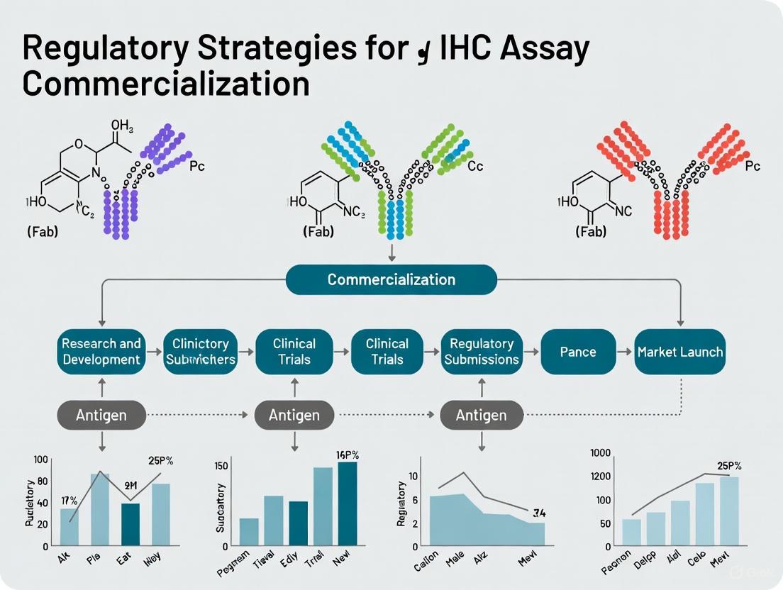
Regulatory Strategy for IHC Assay Commercialization
Navigating the regulatory landscape is critical for the commercialization of IHC assays, particularly when used for patient stratification or as companion diagnostics (CDx).
Risk Assessment and Regulatory Pathways
The regulatory strategy is fundamentally determined by the assay's intended use [9].
- Investigational Use: When an assay is not used to make treatment decisions, an Investigational Device Exemption (IDE) is generally not required [9].
- Clinical Decision-Making: If an assay is used for prospective patient stratification or to guide treatment, a Study Risk Determination (SRD) must be performed to evaluate if it represents a Significant Risk (SR) or Non-Significant Risk (NSR) device. This SRD can be assessed by the FDA via a Q-submission, by an Institutional Review Board (IRB), or included in a pre-IND briefing book [9]. An SR determination mandates the submission of an IDE to the FDA [9].
CLIA Validation vs. FDA Approval
A crucial distinction exists between laboratory validation and regulatory clearance:
- CLIA Validation: Compliance with the Clinical Laboratory Improvement Amendments (CLIA) is a baseline requirement for all clinical laboratories in the U.S. to ensure test reliability. However, CLIA does not specify how to satisfy each performance study [9].
- FDA Approval/Clearance: For commercialization as an In Vitro Diagnostic (IVD), especially a CDx, FDA approval is required. This process, via Premarket Approval (PMA) or 510(k), involves more extensive studies than CLIA, including multi-site reproducibility data for IVD kits [9]. The FDA recommends using Clinical Laboratory Standards Institute (CLSI) guidelines and often requires a pre-submission meeting to align on the analytical validation study design [9].
Global Commercialization: US vs. EU Framework
Commercializing an IHC assay globally requires navigating distinct regulatory frameworks, as summarized in Table 3.
Table 3: Comparison of US and EU Regulatory Pathways for IVD Assays [9]
| Aspect | United States (FDA) | European Union (IVDR) |
|---|---|---|
| Regulatory Authority | Food and Drug Administration (FDA) | Notified Bodies |
| CDx Classification | Class II or Class III | Uniformly Class C |
| Key Process | Modular PMA submission (review takes ~12-24 months) | Technical dossier review and QMS audit for CE marking (takes ~12-18 months) |
| Clinical Trial Requirement | SRD and potential IDE submission | Annex XIV submission to national competent authority in each EU country |
| Quality Standards | 21 CFR Part 820 (transitioning to integrate ISO 13485) | ISO 13485, ISO 14971 |
The following diagram outlines the key regulatory decision points and pathways for a clinical trial assay in the U.S.
The global IHC market, anchored in a $3.55 billion valuation in 2025, is poised for sustained growth, driven by the escalating cancer burden and the pivotal role of IHC in personalized medicine. Success in this evolving landscape extends beyond technological innovation. It necessitates a deep commitment to rigorous analytical validation and a strategically navigated regulatory pathway. For researchers and drug developers, integrating assay development with a clear regulatory strategy from the outset is not merely best practice—it is a fundamental requirement for translating promising IHC assays into commercially successful diagnostic tools that improve patient care.
This guide provides a comparative overview of the four key regulatory and standards frameworks that impact the commercialization of In Vitro Diagnostic (IVD) devices, including Immunohistochemistry (IHC) assays.
Navigating the regulatory landscape is a critical step in the successful commercialization of In Vitro Diagnostic (IVD) devices. For researchers and developers, understanding the distinct roles and requirements of the United States and European Union's regulatory systems is fundamental. The U.S. Food and Drug Administration (FDA) and the Clinical Laboratory Improvement Amendments (CLIA) provide the regulatory structure in the United States, while the European Union's In Vitro Diagnostic Regulation (IVDR) governs market access in Europe. Alongside these regulations, standards from the Clinical & Laboratory Standards Institute (CLSI) provide the foundational technical and quality practices that support compliance across all regimes [10] [11] [12].
Core Concepts and Comparative Analysis
Defining the Four Pillars
- CLIA (Clinical Laboratory Improvement Amendments): A U.S. federal regulation that establishes quality standards for all clinical laboratory testing [13]. CLIA regulates the laboratories themselves, ensuring the accuracy, reliability, and timeliness of test results regardless of where the test is performed [14].
- CLSI (Clinical & Laboratory Standards Institute): A global, nonprofit organization that develops voluntary consensus standards and guidelines for clinical laboratories [12]. CLSI documents are not regulations, but they provide the technical protocols and best practices for method evaluation, quality control, and verification that laboratories use to comply with CLIA and other regulatory requirements [15].
- FDA (U.S. Food and Drug Administration): The U.S. regulatory agency that oversees the safety and effectiveness of medical devices, including IVDs [10]. The FDA regulates the manufacturers of IVD products, controlling their entry into the market through premarket review and clearance/approval processes [16].
- IVDR (In Vitro Diagnostic Medical Devices Regulation): The European Union's regulation governing the market access and lifecycle of in vitro diagnostic devices [11]. It repeals the previous Directive and introduces a more stringent framework based on a risk classification system, with an emphasis on clinical evidence and post-market surveillance [17].
Comparative Analysis of Regulatory Frameworks
The following table summarizes the key characteristics of these frameworks for easy comparison.
Table 1: Comparative Overview of CLIA, CLSI, FDA, and IVDR
| Aspect | CLIA | CLSI | FDA (for IVDs) | EU IVDR |
|---|---|---|---|---|
| Primary Role | Regulates clinical testing laboratories [13] | Develops voluntary laboratory standards [12] | Regulates manufacturers of IVD products [10] | Regulates manufacturers of IVD products in the EU [11] |
| Geographic Scope | United States | Global (standards used internationally) | United States | European Union |
| Legal Status | Law (Federal Regulation) [13] | Voluntary guidance [12] | Law (Federal Regulation) [10] | Law (EU Regulation) [11] |
| Key Focus | Laboratory quality, accuracy, & proficiency testing [13] | Standardizing test methods & quality practices [12] | Premarket safety & effectiveness of the device [10] | Device safety, performance, & lifecycle oversight [11] |
| Basis for Regulation/Categorization | Test complexity (Waived, Moderate, High) [10] [14] | Technical best practices & consensus | Device risk classification (Class I, II, III) [10] | Device risk classification (Class A, B, C, D) [11] |
| Premarket Review | Not applicable (regulates labs, not devices) | Not applicable | Required (e.g., 510(k), PMA) [10] | Required (Conformity Assessment with Notified Body for most classes) [11] |
Experimental Protocols for Regulatory Compliance
Generating robust experimental data is a cornerstone of both FDA and IVDR submissions. The following protocols outline key studies required for IVD commercialization.
Protocol for Analytical Performance Validation
This protocol, which aligns with CLSI guidelines, is fundamental for demonstrating that an IHC assay performs as intended from an analytical perspective [12].
- Objective: To rigorously establish the analytical performance characteristics of a new IHC assay, including its sensitivity, specificity, precision, and reproducibility.
- Materials:
- Test Device: The IHC assay kit, including all reagents, antibodies, and controls.
- Specimens: A well-characterized panel of human tissue specimens, including both positive and negative cases for the analyte of interest. Tissues should be fixed and processed according to standard laboratory procedures.
- Instrumentation: Automated stainers, microscopes, and any other equipment used in the testing process.
- Comparator: A validated method, which could be a predicate IHC assay, a different analytical technique (e.g., flow cytometry, PCR), or a clinically confirmed diagnosis.
- Methods:
- Analytical Sensitivity (Limit of Detection): Perform a titration of the primary antibody or key reagent using known positive samples with low analyte expression. The limit of detection is the lowest concentration that can be consistently distinguished from a negative control.
- Analytical Specificity:
- Interference Testing: Spike samples with potentially interfering substances (e.g., hemoglobin, bilirubin) to assess impact on staining.
- Cross-reactivity: Test the assay against tissues or cell lines known to express structurally similar antigens to evaluate off-target binding.
- Precision:
- Repeatability: Have one operator run the assay multiple times on the same day using the same equipment and reagents.
- Reproducibility: Conduct the assay across multiple days, operators, and laboratories to assess inter-laboratory variability.
- Data Analysis: Calculate clinical sensitivity, specificity, and overall percent agreement with the comparator method. Statistical analysis, such as Cohen's Kappa for inter-rater agreement, is often employed.
Protocol for Clinical Validation (Clinical Utility)
This study is critical for both FDA and IVDR submissions to prove the assay's correlation with clinical outcomes [18].
- Objective: To evaluate the ability of the IHC assay to accurately identify a clinical condition or predict patient outcomes in a well-defined population.
- Study Design: A retrospective or prospective study using archived or prospectively collected patient samples.
- Materials:
- Patient Cohorts: Specimens from a clearly defined intended use population, along with associated clinical and outcome data.
- Reference Standard: The accepted "gold standard" for diagnosis (e.g., histopathology consensus, long-term clinical follow-up, outcome data).
- Methods:
- Blinded Testing: Perform the IHC assay on all samples in a blinded fashion, without knowledge of the reference standard result.
- Data Collection: Record the IHC assay results and correlate them with the reference standard results and clinical outcomes.
- Data Analysis: Determine clinical sensitivity (ability to correctly identify diseased patients) and clinical specificity (ability to correctly identify non-diseased patients). For predictive assays, statistical analyses such as hazard ratios and Kaplan-Meier survival curves may be required.
Essential Research Reagent Solutions
The table below lists key materials and their functions critical for conducting the experiments outlined above.
Table 2: Key Research Reagent Solutions for IHC Assay Development and Validation
| Item | Function in Regulatory Experiments |
|---|---|
| Analyte Specific Reagents (ASRs) | Antibodies or nucleic acid sequences for specific identification and quantification of an individual chemical substance; the core of the IHC test [10]. |
| Well-Characterized Tissue Microarrays (TMAs) | Provide multiple tissue specimens on a single slide; essential for efficient testing of sensitivity, specificity, and precision across many samples [18]. |
| Reference Standard Materials | Serve as the benchmark for comparison to demonstrate assay accuracy and validity during method validation studies [18]. |
| Quality Control Materials | Used to monitor the precision and consistency of the assay over time, a requirement for both CLIA compliance and regulatory submissions [10]. |
Regulatory Pathway and Workflow
The journey from assay development to market release involves parallel and interconnected processes with different regulatory bodies. The following diagram synthesizes the core workflow and relationships between the key frameworks.
Diagram: Integrated Regulatory Pathway for IHC Assays. This workflow shows how CLSI standards support premarket submissions to the FDA and EU IVDR, with CLIA governing the laboratory where the approved test is implemented.
A successful regulatory strategy for IHC assay commercialization requires a clear understanding of the distinct yet interconnected roles of CLIA, CLSI, FDA, and IVDR. CLSI provides the scientific and technical foundation for assay validation. The FDA and IVDR are the gatekeepers for the market, requiring rigorous data on safety and performance. Finally, CLIA ensures that the test is performed reliably in the clinical laboratory setting. By integrating the requirements of all four pillars from the earliest stages of development, researchers and drug development professionals can design more efficient studies, generate defensible data, and navigate the path to market with greater confidence and success.
The development and commercialization of immunohistochemistry (IHC) assays require precise definition of assay intent from the earliest stages. The classification of an assay as Research Use Only (RUO), In Vitro Diagnostics (IVD), or Companion Diagnostics (CDx) determines its regulatory pathway, clinical applicability, and commercial potential. Within the context of IHC assay commercialization, understanding these categories is fundamental to developing an effective regulatory strategy that aligns with the assay's intended purpose in patient care and drug development. This guide provides an objective comparison of these three critical classifications to inform researchers, scientists, and drug development professionals.
Classification and Regulatory Definitions
Table 1: Core Definitions and Regulatory Oversight of Assay Types
| Parameter | Research Use Only (RUO) | In Vitro Diagnostics (IVD) | Companion Diagnostic (CDx) |
|---|---|---|---|
| Definition | Tests for non-diagnostic research purposes | Medical devices used for disease diagnosis, monitoring, or prevention | Specialized tests providing essential information for safe and effective use of a specific therapeutic product [19] [20] |
| Regulatory Status | Not for diagnostic procedures | FDA cleared/approved for general diagnostic use | Requires regulatory approval as part of therapeutic product labeling [19] |
| Intended Use | Basic research, biomarker discovery, proof-of-concept studies | Diagnosis, monitoring, or risk assessment of diseases or conditions | Identifying patients who will benefit from specific treatments or have increased risk of serious side effects [20] |
| Regulatory Pathway | Laboratory development following internal QC | FDA 510(k), De Novo, or PMA submissions | Co-development and regulatory approval with pharmaceutical partner; FDA, CDRH, and/or EMA interactions [20] |
Performance Characteristics and Technical Standards
Table 2: Performance Validation and Technical Requirements
| Validation Parameter | RUO | IVD | CDx |
|---|---|---|---|
| Analytical Validation | Proof-of-concept studies; specimen selection and stability studies [20] | Full analytical validation per FDA/regulatory standards [20] | Stringent analytical and clinical validation aligned with therapeutic development timeline [20] |
| Clinical Validation | Not required | Clinical performance studies for intended use population | Biomarker discovery and validation linked to therapeutic response [20] |
| Quality Standards | Custom assay development; may follow CLSI guidelines | CLIA, CAP, and FDA guideline compliance [20] | Globally standardized test validated for specific mutation detection [19] |
| Standards Compliance | Optional adherence to GCLP, ISO | Mandatory compliance with region-specific regulations (CLIA, ISO) [21] [22] | Adherence to CLSI, GCLP, ISO, and specific regional regulatory requirements [21] [22] |
Experimental Data and Performance Comparison
Diagnostic Performance in Clinical Applications
Table 3: Experimental Performance Data Across Methodologies
| Assay Application | Methodology | Sensitivity (%) | Specificity (%) | PPV/NPV (%) | Agreement (Kappa) |
|---|---|---|---|---|---|
| MSI Status in EC [23] | IHC vs. PCR (Gold Standard) | 89.3 | 87.3 | PPV: 78.1, NPV: 94.1 | 0.74 (Substantial) |
| p53 Status in EC [23] | IHC vs. NGS (Gold Standard) | 92.3 | 77.1 | PPV: 60.0, NPV: 96.4 | 0.59 (Moderate) |
| Universal IHC Analyzer [24] | AI (PH-LUB) vs. Pathologist | N/A | N/A | N/A | 0.578 (Substantial) |
| Conventional IHC Model [24] | AI (H-B SC-model) vs. Pathologist | N/A | N/A | N/A | 0.509 (Moderate) |
Deep Learning Model Performance
Table 4: AI-Based IHC Prediction Model Performance for Gastrointestinal Cancers [25]
| IHC Biomarker | AUC | Accuracy (%) | Clinical Application |
|---|---|---|---|
| P40 | 0.90-0.96 | 83.04-90.81 | Squamous differentiation |
| Pan-CK | 0.90-0.96 | 83.04-90.81 | Epithelial origin confirmation |
| Desmin | 0.90-0.96 | 83.04-90.81 | Submucosal invasion assessment |
| P53 | 0.90-0.96 | 83.04-90.81 | Mutation-associated overexpression |
| Ki-67 | 0.90-0.96 | 83.04-90.81 | Proliferation index quantification |
Detailed Experimental Protocols
Protocol 1: IHC Validation for Clinical Use
For IVD and CDx assays, validation follows standardized protocols:
- Sample Preparation: Formalin-fixed paraffin-embedded (FFPE) tissues sectioned at 4-5μm [23] [24]
- Staining Method: Antibody-based detection with DAB chromogen and hematoxylin counterstain [26]
- Image Analysis: Whole slide imaging (WSI) using automated scanners (KF-PRO-020 or Pannoramic 250 Flash Scanner) [25]
- Interpretation: Pathologist evaluation with standardized scoring criteria (e.g., HER2 IHC 3+ requires strong complete, basolateral, or lateral membrane staining in ≥10% of tumor cells for surgical specimens) [27]
- Quality Control: Inclusion of positive and negative controls with each batch
Protocol 2: Deep Learning-Based IHC Analysis
The Universal IHC (UIHC) analyzer development protocol:
- Training Data Collection: 134 WSIs including H&E and IHC pairs with 415,463 automatically extracted tiles [25]
- Image Registration: H&E to IHC alignment using HEMnet neural network combining affine transformation and B-spline-based non-rigid registration [25]
- Model Architecture: Mean Teacher semi-supervised learning framework with ResNet-50 backbone pretrained on ImageNet [25]
- Preprocessing: Stain normalization using Vahadane method with iterative luminosity standardization [25]
- Performance Validation: Multi-reader multi-case (MRMC) study with pathologists reading both AI-IHC and conventional IHC with washout period [25]
Regulatory Pathway Diagram
Technology Development Workflow
The Scientist's Toolkit: Essential Research Reagent Solutions
Table 5: Key Materials and Technologies for IHC Assay Development
| Tool/Technology | Function | Application Context |
|---|---|---|
| Whole Slide Scanners (KF-PRO-020, Pannoramic 250) [25] | Digital conversion of glass slides for analysis | All phases: RUO through CDx |
| HEMnet Neural Network [25] | H&E to IHC alignment and label transfer | AI-based assay development |
| VGG Image Annotator (VIA) [25] | Pathologist verification of automated annotations | Model training and validation |
| Mean Teacher Framework [25] | Semi-supervised learning for biomarker prediction | AI-IHC model development |
| Universal IHC (UIHC) Analyzer [24] | DL-based tool quantifying protein expression across cancers and IHC types | Cross-platform assay validation |
| Color Deconvolution Algorithms [26] | Separation of DAB and hematoxylin channels for quantification | Automated H-score calculation |
| Vahadane Stain Normalization [25] | Inter-slide color variability minimization | Preprocessing for computational analysis |
The distinction between RUO, IVD, and CDx assays represents a continuum of increasing regulatory scrutiny and clinical application. RUO assays serve vital functions in basic research and early development but lack the validation required for diagnostic use. IVD assays undergo rigorous validation for general diagnostic purposes but are not linked to specific therapeutics. CDx assays represent the most stringent category, requiring co-development with pharmaceutical products and demonstrating clinical utility for specific treatment decisions. The emerging field of AI-powered IHC analysis shows substantial agreement with conventional IHC (kappa scores 0.578 for MC-models vs. 0.509 for SC-models) [24] and offers promising approaches for standardizing quantification across assay types. Understanding these categories enables researchers to strategically navigate the regulatory landscape and advance IHC assays toward appropriate clinical applications.
Within the strategic commercialization of immunohistochemistry (IHC) assays, the classification of an investigational device as presenting a Significant Risk (SR) or Nonsignificant Risk (NSR) constitutes a pivotal regulatory determination. This assessment directly dictates the development pathway, regulatory burden, and timeline for bringing a novel IHC from research to clinical use [9]. For researchers and drug development professionals, understanding this dichotomy is not merely an administrative exercise but a fundamental component of efficient experimental design and strategic planning. The U.S. Food and Drug Administration (FDA) defines these categories based on the potential for serious risk to the health, safety, or welfare of a subject, with profound implications for the required regulatory approvals [28] [29]. As IHC assays continue to serve as critical companion diagnostics in targeted therapies, mastering these risk assessment fundamentals becomes indispensable for navigating the complex transition from biomarker discovery to commercially viable clinical trial assay.
Defining Significant Risk and Nonsignificant Risk
The FDA provides a definitive framework for categorizing medical device studies under 21 CFR 812.3(m) [28]. A Significant Risk (SR) study involves an investigational device that meets one or more of the following criteria:
- Is intended as an implant and presents a potential for serious risk to the health, safety, or welfare of a research subject.
- Is used to support or sustain human life and presents a potential for serious risk to the health, safety, or welfare of a subject.
- Plays an important role in diagnosing, curing, mitigating, or treating disease, or otherwise preventing impairment of human health and presents a potential for serious risk to the health, safety, or welfare of a subject.
- Otherwise presents a potential for serious risk to the health, safety, or welfare of a subject [28] [29].
Conversely, a Nonsignificant Risk (NSR) device study is one that does not meet the SR definition above [28]. It is critical to recognize that this SR/NSR determination is unique to device regulations and is separate from the "minimal risk" assessment used for certain institutional review board (IRB) reviews [28]. An NSR determination does not automatically equate to minimal risk; it is possible for an NSR study to be considered greater than minimal risk while still not meeting the threshold for "significant risk" [28].
Table 1: Core Definitions and Regulatory Implications
| Aspect | Significant Risk (SR) | Nonsignificant Risk (NSR) |
|---|---|---|
| Regulatory Definition | Presents potential for serious risk to health, safety, or welfare; meets specific implant, life-support, or diagnostic/treatment criteria [29] | Does not meet the definition of Significant Risk [28] |
| Primary Regulatory Oversight | FDA + IRB | IRB (acting as FDA surrogate) |
| IDE Requirement | Required - must submit IDE application and receive FDA approval [28] | Not required - study may proceed with IRB approval alone [28] |
| Key Regulatory Consideration | Risk determination is based on how the device is used in the study, not solely on the device itself [28] | IRB makes final risk determination if FDA has not previously ruled on the device |
The Risk Determination Process and Regulatory Pathways
The process for determining device risk classification follows a structured pathway with clearly defined responsibilities. The study sponsor is responsible for proposing the initial risk determination based on regulatory criteria, which is then submitted to the IRB for consideration [28]. In some cases, the FDA may have already made a risk determination before the study reaches the IRB, in which case the FDA's determination is final [28]. If the FDA has not previously ruled on the device, the IRB must decide whether it concurs with the sponsor's assessment, considering factors such as the basis for the risk determination, the type of harm resulting from device use, and any additional procedures subjects may undergo as part of the study [28].
For IHC assays specifically, risk evaluation is fundamentally based on how the device is used in the investigational therapeutic study [9]. When an IHC assay is not used to make treatment determinations, an Investigational Device Exemption (IDE) is generally not required—unless the sample is obtained through a high-risk procedure [9]. However, when an assay is used for prospective stratification or clinical decision-making, it is necessary to perform a Study Risk Determination (SRD) to evaluate if an IDE is required [9]. Manufacturers have the option of submitting an SRD Q-submission to the FDA for a formal agency determination, having the IRB assess risk as an FDA surrogate, including a risk assessment in the pre-Investigational New Drug (IND) briefing book, or simply assuming significant risk and submitting an IDE [9].
The following diagram illustrates the decision pathway for IHC assay regulatory submission strategy:
Experimental Data and Concordance Studies in IHC Validation
Robust experimental validation is fundamental to the risk assessment process for IHC assays. Analytical validation studies provide the critical evidence needed for regulatory submissions and inform the risk classification by demonstrating assay reliability. Recent studies highlight both the challenges and solutions in IHC standardization.
A comprehensive analytical comparison of commonly used laboratory-developed Ki-67 IHC tests revealed significant interlaboratory heterogeneity [30]. When compared against the reference Ki-67 IHC MIB-1 pharmDx assay at a 20% cutoff, none of the laboratory-developed tests achieved high overall agreement (predetermined as ≥85%). The clones MIB-1 on Dako Autostainer Link 48 and K2 on Leica BOND-III showed high specificity (99.5% and 100% respectively) but poor sensitivity (24.8% and 25.1%), while clone 30-9 on Ventana BenchMark ULTRA showed high sensitivity (99.3%) but markedly reduced specificity (53.6%) [30]. This variability underscores the importance of rigorous validation, particularly for assays used in treatment decisions where false positives or negatives could directly impact patient care.
In HER-2 testing for breast cancer, a prospective study demonstrated an 82.0% total concordance between IHC and fluorescence in situ hybridization (FISH), with a Kappa coefficient of 0.640 (P < 0.001) [31]. However, significant discordance rates were observed across IHC scores: 19.2% in IHC 0 and 1+, 30.0% in IHC 2+, and 7.1% in IHC 3+ [31]. These findings support the strategy of using IHC as an initial screening tool with FISH confirmation for equivocal cases, reflecting how performance characteristics directly influence clinical implementation and risk classification.
Table 2: Experimental Performance Data for IHC Assays
| Assay Type | Performance Metric | Results | Clinical/Risk Implications |
|---|---|---|---|
| Ki-67 IHC (LDT vs Reference) [30] | Sensitivity/Specificity at 20% cutoff | MIB-1: 24.8% sens, 99.5% specK2: 25.1% sens, 100% spec30-9: 99.3% sens, 53.6% spec | High variability between platforms affects reliability for clinical decision-making |
| HER-2 IHC vs FISH [31] | Overall Concordance | 82.0% (Kappa = 0.640, P < 0.001) | Supports IHC as initial screen with FISH confirmation for equivocal cases (IHC 2+) |
| HER-2 Discordance by IHC Score [31] | Discordance Rate | IHC 0/1+: 19.2%IHC 2+: 30.0%IHC 3+: 7.1% | Informs reflexive testing protocols and risk mitigation strategies |
| Universal IHC AI Analyzer [24] | Cohen's Kappa (vs pathologists) | Multi-cohort model: 0.578Single-cohort model: 0.509 | AI standardization may reduce inter-observer variability and improve reproducibility |
Advanced computational approaches are now addressing these validation challenges. A novel Universal IHC (UIHC) analyzer, utilizing deep learning to quantify protein expression across different cancers and IHC types, has demonstrated superior performance compared to conventional single-cohort models, achieving a Cohen's kappa score of 0.578 versus up to 0.509 for analyzing unseen IHC images [24]. This multi-cohort trained model showed consistent performance across varying positive staining cutoff values, representing a significant advancement in quantitative IHC analysis that could potentially streamline the validation process for novel assays [24].
The Scientist's Toolkit: Essential Research Reagent Solutions
Successful IHC assay development and validation requires meticulous attention to reagent selection and experimental conditions. The following toolkit outlines critical components and their functions based on current protocols and methodologies:
Table 3: Essential Research Reagent Solutions for IHC Assay Development
| Reagent/Component | Function | Key Considerations |
|---|---|---|
| Primary Antibodies [6] | Specific binding to target antigen | Monoclonal (batch consistency) vs. polyclonal (sensitivity); species selection to minimize cross-reactivity |
| Antigen Retrieval Solutions [32] [6] | Unmask epitopes cross-linked during fixation | Acidic or basic buffers for HIER; enzymatic retrieval for limited antigens (e.g., cytokeratins) |
| Protein Blocking Agents [32] | Reduce nonspecific background staining | Normal serum, BSA, gelatin, or commercial synthetic peptide mixes; critical for Fc receptor-rich tissues |
| Detection Systems [32] | Visualize antibody-antigen complexes | Peroxidase- or alkaline phosphatase-based; require endogenous enzyme blocking with H₂O₂ or levamisol |
| Control Tissues [6] | Validate assay performance | Positive controls with known low/intermediate expression; negative controls; tissue microarrays for higher throughput |
| Fixation Media [32] | Preserve tissue architecture and antigenicity | 10% neutral buffered formalin (24 hours, room temperature); tissue to fixative ratio 1:1 to 1:20 critical |
| Automated IHC Platforms [6] | Standardize staining process | Dako, Leica, and Ventana systems; choice based on client preference and ultimate assay purpose |
The validation process for IHC assays requires systematic optimization of multiple parameters. Precision for Medicine follows a standard approach when developing or optimizing a new IHC assay, typically evaluating two to three antibodies (from different vendors or species) at three different concentrations with two different antigen retrieval times [6]. Depending on initial performance, they may further vary incubation times or alter temperatures for antigen retrieval to achieve the desired sensitivity and specificity [6].
The classification of an IHC assay as presenting Significant or Nonsignificant Risk fundamentally shapes its developmental trajectory from research tool to commercialized product. This determination directly influences regulatory strategy, validation requirements, and ultimately, the pathway to clinical implementation. As the field advances with sophisticated computational approaches like universal AI analyzers [24] and more standardized protocols [32] [6], the precision of risk assessments will continue to improve. For researchers and drug development professionals, embedding these risk assessment fundamentals throughout the assay development process—from initial antibody selection through clinical validation—is essential for efficient navigation of the regulatory landscape. This integrated approach ensures that IHC assays not only provide robust scientific insights but also comply with the appropriate regulatory standards for their intended use, ultimately supporting their successful commercialization and clinical adoption in precision medicine.
Building a Compliant IHC Assay: From CAP 2024 Validation to Strategic Regulatory Pathways
The College of American Pathologists (CAP) released a significant update to the "Principles of Analytic Validation of Immunohistochemical Assays" in February 2024, marking the first major revision since the original 2014 publication [8]. This guideline update aims to address evolving practices in immunohistochemistry (IHC) and reduce variation in laboratory procedures to ensure assay accuracy and reliability [8]. For researchers and drug development professionals, understanding these changes is crucial for developing robust regulatory strategies for IHC assay commercialization.
The update was necessitated by significant evolution in the field of clinical immunohistochemistry since the original guideline publication [8]. Through a systematic review of medical literature, the CAP panel created two strong recommendations, one conditional recommendation, and 12 good practice statements using rigorous development principles [8]. These guidelines particularly impact assays that guide therapeutic decision-making for cancer treatment, making them essential for researchers developing companion diagnostics [33].
Key Changes in the 2024 Update
Harmonized Requirements for Predictive Markers
The updated guideline harmonizes validation requirements for all predictive markers, replacing the previous approach that outlined distinct requirements for HER2, ER, and PR predictive markers [8]. This standardization creates a more uniform framework for assay validation regardless of the specific predictive marker being tested.
Expanded Scope for Cytology Specimens
A significant advancement in the 2024 update addresses the validation of IHC assays performed on cytology specimens that are not fixed identically to tissues used for initial assay validation [8]. This change responds to frequent laboratory requests for more definitive validation guidelines in this area [8]. The literature has shown variable sensitivity of IHC assays performed on specimens collected in fixatives often used in cytology laboratories compared with formalin-fixed, paraffin-embedded (FFPE) tissues [8].
Standardized Concordance Threshold
The updated guideline establishes a uniform 90% concordance requirement for all IHC assays, replacing the varying concordance requirements previously recommended for estrogen receptor, progesterone receptor, and HER2 IHC performed on breast carcinomas [8]. This standardization simplifies validation target setting while maintaining rigorous performance standards.
Explicit Verification for FDA-Approved Assays
The update provides more explicit verification requirements for unmodified United States Food and Drug Administration (FDA) approved/cleared assays [8]. This clarification helps laboratories better navigate the regulatory landscape when implementing commercially available assays.
Detailed Validation Requirements
Specimen-Specific Validation Protocols
Specimen Processing Pathways in IHC Validation
The updated guidelines establish distinct validation pathways based on specimen type and processing methods. For cytology specimens fixed differently from standard FFPE tissues used in initial validation, separate validations are now required [8]. This includes alcohol-fixed smears, liquid-based cytology preparations, and specimens collected in alternative fixative media [34].
Quantitative Validation Requirements
Table 1: Case Requirements for Initial Analytic Validation
| Assay Type | Minimum Positive Cases | Minimum Negative Cases | Key Considerations |
|---|---|---|---|
| Nonpredictive LDTs | 10 | 10 | Include high and low expressors; span expected range of clinical results [34] |
| All Predictive Markers | 20 | 20 | Include high and low expressors; span expected range of clinical results [34] |
| Cytology Specimens | 10 | 10 | Required for each new fixation method; increase for predictive markers [8] [34] |
| Rare Antigens | Director-determined | Director-documented | Rationale for reduced case numbers must be documented [34] |
Table 2: Revalidation Requirements for Assay Modifications
| Type of Change | Validation Requirement | Documentation Needed |
|---|---|---|
| New Antibody Lot | 1 known positive + 1 known negative tissue | Control tissue with known positive/negative cells sufficient [34] |
| Antibody Dilution/Vendor/Incubation Times | 2 known positive + 2 known negative tissues | Verification of performance [34] |
| Fixative Type, Antigen Retrieval, Detection System | Sufficient tissues to ensure consistent results | Laboratory director determines number of cases [34] |
| Antibody Clone Change | Full revalidation | Equivalent to initial analytic validation [34] |
Assay-Scoring System Combinations
For predictive IHC assays with distinct scoring systems like HER2 and PD-L1, the updated guideline stipulates that laboratories must separately validate/verify each assay-scoring system combination [8] [33]. This requirement acknowledges that scoring systems may vary by tumor site and clinical indication, potentially affecting assay performance and interpretation [8].
Experimental Protocols for Validation
Validation Study Design Options
The CAP guideline provides a range of study design options for validation, ordered from most to least stringent [8]:
- Comparison to Protein Calibrators: Comparing new assay results to IHC results from cell lines containing known amounts of protein [8]
- Non-IHC Method Comparison: Comparing with results from flow cytometry or FISH [8]
- Inter-laboratory Comparison: Testing same tissues in another laboratory using validated assay [8]
- Intra-laboratory Comparison: Comparing with prior testing of same tissues with validated assay in same laboratory [8]
- Clinical Trial Laboratory Comparison: Comparing with results from laboratories that performed testing for clinical trials [8]
- Antigen Localization Verification: Comparing with expected architectural and subcellular localization of antigen [8]
- Published Data Comparison: Comparing against percent positive rates in published clinical trials [8]
- Proficiency Testing Challenges: Comparing with previously graded tissue challenges from formal PT programs [8]
Recommended Research Reagent Solutions
Table 3: Essential Research Reagents for IHC Validation
| Reagent Category | Specific Examples | Research Application |
|---|---|---|
| Reference Standards | Cell lines with known protein content, Calibrators | Serve as quantitative standards for assay comparison [8] |
| Control Tissues | FFPE tissues with known antigen status, Cytology specimens with alternative fixatives | Provide positive/negative controls for validation studies [34] |
| Antibody Clones | FDA-approved/cleared clones, Laboratory-developed clones | Determine specificity; clone changes require full revalidation [34] |
| Detection Systems | Various detection platforms | Changes require performance verification [34] |
| Fixation Media | Formalin, Alcohol-based fixatives, Alternative fixative media | Impact antigen preservation; require separate validations [8] |
Regulatory Strategy Integration
Navigating Multiple Regulatory Frameworks
Integrated Regulatory Framework for IHC Assays
Implementing the updated CAP guidelines requires integration with broader regulatory strategies. For clinical laboratories, compliance with Clinical Laboratory Improvement Amendments (CLIA) regulations remains fundamental, as CLIA applies to all US facilities testing human specimens for health assessment or disease diagnosis [9]. However, CAP emphasizes that CLIA validation alone may be insufficient for assays intended for commercial development [9].
For companion diagnostic commercialization in the US, the FDA typically requires studies that exceed CLIA requirements through a modular pre-market approval (PMA) process [9]. The European Union follows a different pathway under the In Vitro Diagnostic Regulation (IVDR), where companion diagnostics are uniformly classified as Class C devices [9]. Successfully commercializing IHC assays globally requires parallel validation strategies that address both US and EU requirements from the outset [9].
Proficiency Testing Considerations
Laboratories must also prepare for updated proficiency testing (PT) requirements under CLIA, with significant changes implemented in January 2025 [35]. These include revised grading criteria for acceptable performance and additional regulated analytes [35]. While not all laboratories can perform full organism identification, CAP recommends performing at least a Gram stain as best practice to help clinical teams determine if growth indicates infection versus colonization [35].
Impact on Laboratory Practice
Previous data demonstrates that evidence-based guideline implementation significantly improves laboratory validation practices. Following the 2014 CAP guideline publication, validation rates for predictive marker assays increased from 74.9% to 99% [36]. Laboratories with written validation procedures for predictive markers increased from 45.9% to 73.8% during the same period [36].
The 2024 CAP Laboratory Accreditation Program checklists have been updated to integrate these revised validation requirements, along with other CLIA final rule changes [37]. This alignment between evidence-based guidelines and accreditation standards ensures laboratories can practically implement the updated recommendations while maintaining compliance.
While these updated CAP recommendations represent best practices, they are not currently mandated by the Laboratory Accreditation Program or any regulatory/accrediting agency [8]. Laboratories are encouraged to adopt these evidence-based recommendations to increase the quality and safety of clinically important IHC assays [8].
For researchers and drug development professionals commercializing immunohistochemistry (IHC) assays, designing robust validation studies is a critical step in the regulatory pathway. The intended use of an assay—whether for research, patient enrollment in clinical trials, or as a companion diagnostic (CDx)—directly determines the stringency of validation requirements and the regulatory strategy [9]. A well-designed validation study must demonstrate that an assay is reliable, reproducible, and fit for its specific clinical purpose. This guide objectively compares different validation approaches and parameters by examining current methodologies and data from recent studies, providing a framework for selecting appropriate sample sizes, concordance targets, and comparators.
Core Parameters for IHC Assay Validation
The design of a validation study rests on three foundational parameters: sample size, concordance rates, and the selection of an appropriate comparator. The table below summarizes typical targets and considerations for each.
Table 1: Key Parameters for IHC Assay Validation Study Design
| Parameter | Typical Targets & Considerations | Application Examples |
|---|---|---|
| Sample Size | Guided by CLSI standards and regulatory feedback; must include a range of expression levels and sample types [9]. | A recent PD-L1 assay study used 136 NSCLC samples to ensure a mix of adenocarcinomas, squamous cell carcinomas, and borderline cases [38]. |
| Concordance Rates | Overall Percent Agreement (OPA) ≥85% is a common minimum target for non-inferiority [38]. Positive/Negative Percentage Agreement (PPA/NPA) are also critical [39]. | A HER2 assay ring study reported a PPA of 84.8% (for HER2-low) and an NPA of 69.2% (for HER2 IHC 0) [39]. |
| Appropriate Comparators | FDA-approved companion diagnostics are the standard for CDx claims [40]. For LDTs, assays with established clinical utility are used. | The novel PD-L1 CAL10 assay was compared to the FDA-approved VENTANA PD-L1 (SP263) Assay [38]. |
Experimental Protocols for Key Validation Studies
The following section details the methodologies from recent, relevant experiments that successfully generated data for regulatory submissions.
Protocol 1: Analytical Concordance Study for a Novel PD-L1 Assay
A 2025 feasibility study aimed to demonstrate the concordance of a novel PD-L1 CAL10 assay (Leica Biosystems) with a validated comparator in Non-Small Cell Lung Cancer (NSCLC) [38].
- Objective: To determine the concordance between the test assay and the comparator at Tumor Proportion Score (TPS) cutoffs of ≥1% and ≥50% [38].
- Sample Selection and Size: The study utilized 136 FFPE NSCLC tissue samples. The cohort was designed to include a mix of resection and biopsy cases, with 60-70% adenocarcinomas and 30-40% squamous cell carcinomas. Crucially, it included 21 borderline cases (TPS 40-60%) to rigorously challenge the assay's performance around the critical clinical cutoff [38].
- Experimental Workflow:
- Pre-screening: Cases were pre-characterized for PD-L1 expression using a different clone to ensure a full dynamic range (TPS 0-100%) [38].
- Staining: Samples were stained with the test (CAL10) and comparator (SP263) assays on their respective automated staining platforms (BOND-III and Benchmark Ultra) [38].
- Blinded Reading: Randomized, anonymized slides were independently scored by two pathologists for TPS [38].
- Digital Analysis (Informational): The CAL10 slides were scanned, and pathologists re-scored the digital whole-slide images after a washout period [38].
- Statistical Analysis: A one-sided, exact non-inferiority test for a single proportion was applied. The pre-defined success criterion was a lower bound of the 95% confidence interval (CI) for the OPA of at least 85% [38].
Protocol 2: Global Ring Study for HER2-Low Concordance
A 2025 global ring study assessed the real-world concordance of different HER2 assays in identifying the challenging HER2-low category [39].
- Objective: To assess the concordance between the Ventana PATHWAY 4B5 CDx assay and various comparator assays used in clinical laboratories for identifying HER2-low breast cancer [39].
- Sample Selection and Size: The study used 50 breast cancer samples centrally scored using the PATHWAY 4B5 assay. These samples represented the full spectrum of HER2 expression: IHC 0, 1+, 2+, and 3+ [39].
- Experimental Workflow:
- Central Characterization: Samples were stained and scored at a central lab using the CDx assay [39].
- Distribution: Unstained samples were sent to 68 laboratories across North and South America, Europe, and Asia-Pacific [39].
- Local Testing: Participating labs stained and scored the samples using their own routine HER2 IHC assays and protocols [39].
- Virtual Alignment: Pathologists underwent virtual training on HER2 IHC scoring guidelines before re-scoring the samples ("postalignment" scores) [39].
- Statistical Analysis: The primary endpoints were Positive Percentage Agreement (PPA) and Negative Percentage Agreement (NPA) for HER2-low vs. HER2 IHC 0, calculated from the postalignment scores [39].
The following diagram illustrates the general workflow common to rigorous assay validation studies, integrating elements from both protocols described above.
Comparative Performance Data
The outcomes of the featured studies provide concrete data on achievable performance benchmarks.
Table 2: Comparative Performance Data from Recent IHC Validation Studies
| Study & Assay Focus | Concordance Metric | Reported Rate (95% CI) | Key Takeaway |
|---|---|---|---|
| PD-L1 CAL10 Assay (NSCLC) [38] | OPA at ≥50% TPS | >86.2% (Lower bound of 95% CI) | Met pre-specified non-inferiority target, supporting regulatory submission. |
| OPA at ≥1% TPS | >94.0% (Lower bound of 95% CI) | Higher concordance at a lower, more inclusive clinical cutoff. | |
| HER2 4B5 Global Ring (Breast Cancer) [39] | PPA (HER2-low) | 84.8% (83.6%-86.0%) | Demonstrates moderate agreement in identifying HER2-low disease. |
| NPA (HER2 IHC 0) | 69.2% (67.0%-71.2%) | Highlights significant challenge in differentiating HER2 0 from HER2-low. | |
| MI Cancer Seek (NGS Assay) [40] | PPA & NPA for CDx | >97% (vs. FDA-approved tests) | Benchmarks for comprehensive genomic assays used as CDx. |
The Scientist's Toolkit: Essential Research Reagent Solutions
Successful validation requires not only a sound design but also high-quality, well-characterized reagents and platforms.
Table 3: Key Research Reagent Solutions for IHC Validation
| Reagent / Platform | Function in Validation | Example in Use |
|---|---|---|
| Automated IHC Stainers | Ensure standardized, reproducible staining runs; critical for minimizing technical variability. | BOND-III [38], Benchmark Ultra [38], and Ventana platforms [39] are industry standards. |
| FDA-Approved Companion Diagnostic Assays | Serve as the gold-standard comparator for non-inferiority studies for CDx claims. | VENTANA PD-L1 (SP263) assay used as a comparator for a novel PD-L1 test [38]. |
| Validated Primary Antibodies | Specifically bind the target biomarker; clone specificity is critical for performance. | The Ventana PATHWAY anti-HER2/neu (4B5) [39] and PD-L1 CAL10 [38] clones. |
| FFPE Tissue Microarrays (TMAs) | Contain multiple tissue samples on a single slide, enabling efficient staining optimization and initial reproducibility assessments. | While not explicitly mentioned in results, TMAs are a ubiquitous tool in IHC assay development. |
| Multitissue Control Blocks | Used as positive and process controls on every run to ensure staining protocol is working. | A block containing tonsil and placenta tissue was used as a positive control in the PD-L1 study [38]. |
Designing a validation study for an IHC assay demands a strategic approach aligned with the final regulatory goal. As evidenced by recent studies, successful designs incorporate statistically justified sample sizes that include challenging borderline cases, target concordance rates with OPA lower bounds exceeding 85%, and employ appropriate FDA-approved comparator assays. The experimental data shows that while high concordance is achievable, specific clinical contexts like HER2-low classification present significant challenges, lowering expected agreement rates. A deep understanding of these parameters, combined with robust experimental protocols and high-quality reagents, provides the foundation for generating the compelling data required for successful IHC assay commercialization in both the US and EU markets.
The commercialization of Immunohistochemistry (IHC) assays, particularly Companion Diagnostics (CDx), requires navigating complex regulatory frameworks that vary significantly across regions. These regulatory pathways ensure that medical devices are safe and effective for their intended use, especially when they play a critical role in therapeutic decision-making. In the United States, the Food and Drug Administration (FDA) employs a risk-based classification system with three primary marketing pathways for in vitro diagnostic (IVD) devices: Premarket Notification (510[k]), De Novo classification, and Premarket Approval (PMA) [41] [10]. CDx devices, which provide essential information for the safe and effective use of corresponding therapeutic products, are typically classified as high-risk Class III devices and thus require the rigorous PMA pathway [42] [43].
Conversely, the European Union's In Vitro Diagnostic Regulation (IVDR) establishes a different framework where CDx devices are uniformly classified as Class C devices [9]. Understanding these distinct pathways is crucial for researchers, scientists, and drug development professionals seeking to successfully commercialize IHC assays globally. This guide provides a comprehensive comparison of these regulatory strategies, supported by experimental validation data and structured protocols essential for navigating the approval process.
US Regulatory Pathways: A Comparative Analysis
Key Pathway Definitions and Applicability
Premarket Notification (510(k)): A pathway for devices demonstrating substantial equivalence to a legally marketed predicate device [10] [44]. Most Class II and some Class I devices use this route, which typically does not require clinical trials but does need laboratory performance testing [45] [46].
De Novo Classification: A risk-based pathway for novel devices of low to moderate risk without a predicate [41] [10]. Upon successful review, the FDA creates a new classification and special controls, establishing a potential predicate for future 510(k) submissions [41] [44].
Premarket Approval (PMA): The most stringent pathway for Class III devices that support or sustain human life, are of substantial importance in preventing impairment of health, or present potential unreasonable risk [41] [46]. PMA requires extensive scientific evidence, including comprehensive clinical data, to demonstrate safety and effectiveness [41] [44].
Comparative Analysis of US Regulatory Pathways
Table 1: Comparison of Key Features of US Regulatory Pathways for Medical Devices
| Feature | 510(k) | De Novo | PMA |
|---|---|---|---|
| Device Risk Level | Low to Moderate (Class I, II) | Low to Moderate (Class I, II) | High (Class III) |
| Predicate Device | Required | Not required (creates new classification) | Not required |
| Clinical Data Requirements | Sometimes (for certain modifications) | Usually required (~80% of submissions) | Always required |
| FDA Review Timeline | 30-90 days [44] | 150 days (user fee goal) [41] | 180 days [44] |
| User Fees (FY2025) | $13,260 [41] | $162,235 [41] | $540,783 [41] |
| Post-Market Changes | 510(k) guidance [41] | 510(k) modification standard [41] | PMA supplements required [41] |
Table 2: FDA Application Volume for Fiscal Year 2024
| Regulatory Pathway | Applications Received by CDRH |
|---|---|
| 510(k) | 3,643 |
| De Novo | 78 |
| Premarket Approval (PMA) | 69 |
| Humanitarian Device Exemption (HDE) | 2 |
Data extracted from FDA MDUFA V Performance Report [41]
Companion Diagnostics and the PMA Pathway
Companion diagnostics are defined as IVD devices that provide essential information for the safe and effective use of corresponding therapeutic products [42] [47]. The FDA considers CDx to be high-risk devices that typically require PMA approval [43]. The modular PMA is the preferred submission format for CDx devices, consisting of separate modules for Quality Systems, Non-Clinical (Analytical) Performance, Clinical Performance, and Labeling [43]. This modular approach allows for staged submission and review, potentially streamlining the overall process.
For CDx commercialization, the FDA favors a modular PMA process where "each module is reviewed independently and must be approved before submitting the next module" [9]. The overall timeline for review is approximately 12 to 24 months, and compliance with 21 CFR Part 820 and a Bioresearch Monitoring (BIMO) audit of the facility are required prior to approval [9].
CDx PMA Submission Pathways
EU IVDR Class C Framework for CDx
Understanding the IVDR Classification System
The European Union's In Vitro Diagnostic Regulation (IVDR) establishes a risk-based classification system with classes A (lowest risk) through D (highest risk) [9]. Under this framework, companion diagnostics are uniformly classified as Class C devices [9]. This represents a significant shift from the previous directive and imposes more stringent requirements on CDx manufacturers.
The regulatory authority in the EU is the Notified Body, which differs from the centralized FDA approach in the United States [9]. The approval process for a CDx under IVDR requires a technical dossier including both analytical and clinical data, consultation with a competent authority or the EMA, and an audit of the Quality Management System (QMS) by a Notified Body [9]. The estimated timeline for CE marking under IVDR is approximately 12 to 18 months [9].
Key Requirements for IVDR Class C Devices
For CDx devices under IVDR Class C, manufacturers must address several critical requirements:
Technical Documentation: Comprehensive documentation demonstrating conformity with the general safety and performance requirements outlined in Annex I of the IVDR [9]
Performance Evaluation: Including scientific validity, analytical performance, and clinical performance data [9]
Quality Management System: Implementation of a QMS in accordance with Article 10(9) of the IVDR [9]
Post-Market Surveillance: Establishment of a post-market surveillance system according to Chapter VII of the IVDR [9]
Clinical Evidence: Compilation of clinical evidence based on performance evaluation data [9]
Experimental Validation Protocols for Regulatory Submissions
Analytical Validation of IHC Assays
Robust analytical validation is fundamental for all regulatory pathways, with stringency increasing with device risk classification. The College of American Pathologists (CAP) provides evidence-based guidelines for analytical validation of IHC assays, which were updated in 2024 to ensure accuracy and reduce variation in laboratory practices [8].
Table 3: Key Analytical Validation Requirements for IHC Assays
| Validation Parameter | Protocol Requirements | Acceptance Criteria |
|---|---|---|
| Accuracy | Comparison to a validated method or known positive/negative tissue samples | ≥90% concordance for predictive markers [8] |
| Precision | Intra-run, inter-run, inter-operator, and inter-lot reproducibility testing | CV <15% for quantitative assays |
| Analytical Specificity | Cross-reactivity with similar antigens and interfering substances | <5% cross-reactivity |
| Analytical Sensitivity | Limit of detection studies with serially diluted biomarkers | Detection at clinically relevant levels |
| Robustness | Deliberate variations in protocol parameters (e.g., incubation times, temperatures) | Maintained performance within specifications |
Clinical Validation and Bridging Studies
For CDx devices requiring PMA, clinical validation must demonstrate that the assay accurately identifies patients who will respond to the corresponding therapeutic. When different assays are used during clinical development versus the final CDx, bridging studies are required [43].
Bridging Study Protocol:
- Sample Selection: Bank both biomarker-positive and biomarker-negative samples from all screened subjects in the registrational trial [43]
- Testing: Test banked samples using the final validated CDx assay [43]
- Concordance Analysis: Demonstrate high concordance between the clinical trial assay and the final CDx [43]
- Clinical Correlation: Show that clinical efficacy observed with the clinical trial assay is maintained with the final CDx [43]
Critical considerations for bridging studies include biomarker prevalence, harmonization of biomarker rules, potential for missing samples, and missing clinical outcome data in the negative population [43].
Bridging Study Workflow for CDx Development
Strategic Regulatory Planning for Global Commercialization
US vs EU Regulatory Comparison
Table 4: Comparison of US and EU Regulatory Requirements for CDx
| Parameter | US FDA (PMA Pathway) | EU (IVDR Class C) |
|---|---|---|
| Classification | Class III (High Risk) | Class C |
| Regulatory Authority | FDA Center for Devices and Radiological Health (CDRH) | Notified Body |
| Review Timeline | 12-24 months [9] | 12-18 months [9] |
| Clinical Evidence | Extensive clinical data from registrational trials | Clinical performance data from performance evaluation |
| Quality System | 21 CFR Part 820 (Transitioning to QMSR incorporating ISO 13485) [9] | ISO 13485 required |
| Post-Market Surveillance | PMA Periodic Reports, Adverse Event Reporting | Post-Market Performance Follow-up (PMPF) |
The Scientist's Toolkit: Essential Reagents and Materials
Table 5: Key Research Reagent Solutions for IHC Assay Development
| Reagent/Material | Function | Regulatory Considerations |
|---|---|---|
| Primary Antibodies | Specific binding to target antigen | Specificity validation, lot-to-lot consistency |
| Detection Systems | Signal amplification and visualization | Sensitivity optimization, background reduction |
| Control Materials | Assay performance monitoring | Positive/Negative controls, reference standards |
| Tissue Sections | Analytical validation substrate | FFPE tissue microarrays with known biomarker status |
| Antigen Retrieval Solutions | Epitope exposure | Optimization for specific antibody-epitope pairs |
| Blocking Reagents | Reduction of non-specific binding | Species-specific blocking for antibody validation |
Pre-Submission Strategy and Meeting Planning
The pre-submission process is a critical strategic tool for navigating complex regulatory pathways, particularly for novel devices or those with uncertain classification [10]. The FDA encourages pre-submission meetings when devices involve new technology, new intended use, or when assistance is needed in defining possible regulatory pathways [10].
Key benefits of pre-submission meetings include [10]:
- Beginning a dialogue with the FDA and promoting greater understanding
- Reducing the cost of research studies by focusing on important information needed for approval
- Facilitating the review process since the FDA will already be familiar with the device
For CDx devices specifically, it is recommended to "align with the FDA on PMA Shell Content prior to submitting the first module via Q-submission" [9]. This alignment is crucial for streamlining the modular PMA process.
Successfully navigating the complex regulatory pathways for IHC assay commercialization requires a strategic approach that accounts for the specific requirements of each jurisdiction. The PMA pathway in the US demands the most rigorous evidence for CDx devices, while the EU's IVDR Class C framework presents its own distinct challenges under the Notified Body system.
Key differentiators between pathways include the type and amount of clinical data required, review timelines, associated costs, and post-market obligations. For companion diagnostics, which are uniformly considered high-risk, the PMA pathway in the US and Class C requirements under IVDR in the EU necessitate comprehensive analytical and clinical validation.
A harmonized global validation strategy that incorporates requirements from multiple regions from the outset can significantly streamline the commercialization process. By understanding these regulatory frameworks and implementing robust experimental validation protocols, researchers and drug development professionals can more effectively navigate the path to market for their IHC-based companion diagnostics.
For researchers and developers commercializing In Vitro Diagnostic (IVD) assays, particularly immunohistochemistry (IHC) tests, navigating the U.S. Food and Drug Administration (FDA) regulatory pathway presents significant challenges. The analytical validation phase—which demonstrates that an test accurately and reliably detects the target analyte—requires substantial investment in time and resources. A misstep in validation study design can lead to costly delays or failed submissions. Pre-submission meetings, formally known as Q-Submission Program interactions, provide a critical mechanism for sponsors to align with the FDA on analytical validation strategies before committing to extensive laboratory studies [48]. These structured interactions allow developers to present proposed validation plans and receive agency feedback, thereby de-risking the development process and increasing the likelihood of successful regulatory review.
The importance of this alignment is particularly acute for IHC-based companion diagnostics (CDx), where technological complexities and subjective interpretation elements create unique validation challenges. As noted by Precision for Medicine, "Since it is not always clear how to apply the CLSI guidelines to every assay and scientific methodology, the FDA suggests a pre-submission meeting to align on the appropriate designs for analytical validation study prior to conducting them" [9]. This article examines the function, process, and strategic value of pre-submission meetings within the context of IHC assay commercialization, providing researchers and drug development professionals with evidence-based guidance for optimizing their regulatory strategy.
The Q-Submission Program: Framework for FDA Interaction
Program Scope and Submission Types
The Q-Submission Program provides formal mechanisms for device sponsors to request interactions with the FDA regarding medical device submissions [48]. This program covers a comprehensive range of submission types, including:
- Pre-submission (Pre-Sub) meetings: The primary mechanism for obtaining FDA feedback on proposed analytical validation studies
- Informal meetings: Suitable for less complex issues
- Submission issue requests: For resolving specific problems with pending applications
- Study risk determinations: Important for assays used in clinical investigations [48] [9]
The FDA's guidance document "Requests for Feedback and Meetings for Medical Device Submissions: The Q-Submission Program" outlines the general framework for these interactions, though specific recommendations for IHC analytical validation often emerge through the meeting process itself rather than being detailed in written guidance [48].
The Strategic Value of Pre-Submission Meetings
Pre-submission meetings offer several strategic advantages for assay developers:
- Risk Mitigation: Alignment with FDA prior to conducting validation studies prevents potentially costly missteps in study design [9]
- Efficiency Optimization: Understanding agency expectations helps focus resources on the most critical validation elements
- Relationship Building: Establishing ongoing dialogue with FDA reviewers creates collaborative regulatory partnerships
- Timeline Management: Early identification and resolution of potential issues accelerates the overall development pathway
As evidenced by Foundation Medicine's experience with their liquid biopsy assay, thoughtful engagement with regulatory requirements through appropriate channels can lead to more efficient validation strategies, such as their "tumor-agnostic" approach to analytical validation [49].
Analytical Validation Fundamentals for IHC Assays
Core Validation Parameters
Analytical validation for IHC assays must demonstrate that the test consistently performs as intended across key performance parameters. The College of American Pathologists (CAP) and Clinical Laboratory Standards Institute (CLSI) provide foundational guidance, though specific requirements vary based on intended use and regulatory classification [9].
Table 1: Core Analytical Validation Parameters for IHC Assays
| Validation Parameter | Definition | Typical Study Approach |
|---|---|---|
| Accuracy | Agreement with a reference method | Testing against validated comparator or clinical outcome |
| Precision | Consistency of repeated measurements | Intra-run, inter-run, inter-operator, inter-site reproducibility |
| Analytical Sensitivity | Ability to detect low analyte levels | Limit of detection studies with serial dilutions |
| Analytical Specificity | Ability to detect only target analyte | Interference, cross-reactivity, and sample stability testing |
| Reportable Range | Range of reliable results | Testing samples with known values across expected range |
For IHC assays specifically, precision becomes particularly critical due to the technical and interpretative variables involved. The 2025 study by Chan et al. highlights how validation approaches are evolving for quantitative IHC assays, with their high-sensitivity HER2 assay demonstrating a coefficient of variation below 10% through rigorous validation [50].
Evolving Standards for Quantitative IHC
Traditional IHC validation has followed subjective, pathologist-read approaches with recommendations for 20-40 case validations depending on predictive status [50]. However, as quantitative digital pathology advances, validation standards are evolving toward more rigorous, metrically-driven approaches. As noted in the HS-HER2 assay validation, "for this first pass as validation of a truly analytic assay to measure protein on histopathology slides, we have combined criteria for IHC with criteria for Ligand Binding Assays to produce a rigorous approach" [50].
This hybrid approach acknowledges that quantitative IHC assays function more like traditional analytical chemistry methods while maintaining the histological context of traditional IHC. The resulting validation framework demands more stringent statistical analysis and objective performance criteria than traditional IHC validation.
Designing Robust Analytical Validation Studies
Sample Selection and Sizing Strategies
Appropriate sample selection is fundamental to successful analytical validation. Key considerations include:
- Sample Types: Use of well-characterized clinical samples or cell line models that represent intended use population
- Sample Size: Justification through statistical power calculations rather than arbitrary numbers
- Positive/Negative Distribution: Inclusion of samples spanning the dynamic range of detection
- Pre-analytical Variables: Representation of expected variations in sample collection, fixation, and processing
The FoundationOneLiquid CDx validation approach demonstrated how extensive sample sets (31,826 samples across 335 disease ontologies) can support novel validation strategies, such as their "DNA-is-DNA" tumor-agnostic approach [49]. While not all validations require this scale, the principle of comprehensive sample representation remains critical.
Statistical Approaches and Acceptance Criteria
Defining statistically justified acceptance criteria prior to study initiation is essential for objective validation assessment. The FDA's Q2(R2) guidance, though developed for pharmaceuticals, provides a useful framework for considering validation statistics [51]. Common approaches include:
- Precision Studies: Calculation of percent agreement, Cohen's kappa, or intraclass correlation coefficients with predefined confidence intervals
- Accuracy Studies: Sensitivity/specificity calculations with minimum performance thresholds
- Linearity/Quantitation: Regression analysis with R² values and slope constraints
For AI-based IHC scoring systems, as examined in the meta-analysis by Oliveira et al., performance metrics become particularly important, with pooled sensitivity of 0.97 and specificity of 0.82 for distinguishing HER2-positive from negative cases [52]. Establishing these performance benchmarks through pre-submission alignment ensures they will be acceptable during formal review.
Regulatory Strategy: Navigating US and EU Pathways
Risk Classification and Submission Planning
IVD assays used for treatment selection typically require significant risk determination, which directly impacts the regulatory pathway. In the U.S., manufacturers must perform a study risk determination (SRD) to establish whether an Investigational Device Exemption (IDE) is required [9]. The algorithm below illustrates the decision process for IVD regulatory strategy:
Diagram 1: IVD Regulatory Submission Strategy Algorithm. SRD can be submitted via Q-Submission or determined by IRB. FDA is ultimate arbiter of significant risk [9].
Comparative Regulatory Frameworks
Understanding differences between U.S. and European Union (EU) regulatory requirements is essential for global assay commercialization. The table below highlights key distinctions:
Table 2: Comparison of US and EU Regulatory Requirements for IVD Assays
| Regulatory Aspect | United States (FDA) | European Union (IVDR) |
|---|---|---|
| Classification of CDx | Class II or Class III | Uniformly Class C |
| Regulatory Authority | FDA Center for Devices and Radiological Health (CDRH) | Notified Bodies |
| Key Standards | CLIA, CLSI, 21 CFR Part 820 | ISO 13485, ISO 14971, ISO 15189 |
| Pre-submission Process | Q-Submission Program | Country-specific consultations with Competent Authorities |
| Clinical Evidence | Modular PMA process (12-24 months) | Technical dossier with clinical data (12-18 months) |
Manufacturers can develop parallel validation strategies that satisfy both U.S. and EU requirements by building comprehensive validation packages from the outset, incorporating both CLSI standards and ISO requirements [9].
Case Studies: Successful Validation Strategies
FoundationOneLiquid CDx: Tumor-Agnostic Validation
The analytical validation of FoundationOneLiquid CDx provides a compelling case study in leveraging pre-submission meetings to advance novel validation approaches. By demonstrating through 31,247 clinical samples and 579 validation samples that "analytical assessment of precision and concordance and coverage are comparable among tumor types," the sponsor successfully argued for a tumor-agnostic validation approach [49]. This strategy challenged conventional requirements for tumor-specific validation and illustrates how robust data presented during pre-submission interactions can support more efficient regulatory pathways.
HS-HER2 Assay: Hybrid Validation Model
The high-sensitivity HER2 (HS-HER2) quantitative assay validation exemplifies the evolving nature of IHC validation [50]. By combining traditional IHC validation elements with principles from ligand binding assays, the developers created a hybrid approach suitable for quantitative measurement of HER2 in attomoles/mm². This methodology included:
- Construction of a calibrator cell line microarray with LC-MS/MS quantification
- Precision studies showing coefficient of variation below 10%
- Definition of a reportable range and limits of detection/quantification
- Prospective testing on 316 core biopsy specimens
This case demonstrates how engaging with FDA through appropriate channels can facilitate validation of novel technologies that don't fit traditional regulatory paradigms.
The Scientist's Toolkit: Essential Research Reagents and Materials
Successful analytical validation requires carefully selected reagents and materials. The following table details essential components for IHC assay validation:
Table 3: Research Reagent Solutions for IHC Assay Validation
| Reagent/Material | Function | Validation Considerations |
|---|---|---|
| Primary Antibodies | Target antigen detection | Specificity, lot-to-lot consistency, optimal dilution |
| Cell Line Microarrays | Assay calibration and standardization | Quantitative target measurement, expression range |
| Control Tissues | Process monitoring and quality control | Positive, negative, and borderline expression levels |
| Detection Systems | Signal amplification and visualization | Sensitivity, background, linear range |
| Antigen Retrieval Solutions | Epitope exposure | Consistency, optimization for specific targets |
| Whole Slide Imaging Systems | Digital pathology and quantification | Resolution, linearity, imaging conditions |
For the HS-HER2 assay, researchers constructed a calibrator cell line microarray using nine cell lines with different HER2 expression levels quantitatively measured by LC-MS/MS [50]. This approach provided an objective standard for assay calibration, demonstrating how traditional reagents can be adapted for modern quantitative applications.
Pre-submission meetings represent a strategic tool for efficient IHC assay commercialization when properly integrated into the overall development timeline. The optimal regulatory strategy engages the FDA early and often, beginning with Pre-Sub meetings to align on analytical validation designs, followed by additional Q-Subs as needed for study risk determinations or submission issue resolutions [48] [9]. The workflow below illustrates this iterative engagement process:
Diagram 2: Pro Forma Timeline for IHC Clinical Trial Assay Development. Early FDA engagement through Q-Subs de-risks later development stages [9].
For researchers and drug development professionals, the evidence is clear: strategic use of pre-submission meetings to align on analytical validation study designs significantly enhances regulatory success. As regulatory standards evolve toward more quantitative approaches and global harmonization, these structured interactions with regulatory agencies will become increasingly vital for efficient IHC assay commercialization.
Overcoming Commercialization Hurdles: Standardization, LDTs, AI Integration, and Audit Preparedness
Immunohistochemistry (IHC) serves as a critical technique in both diagnostic pathology and research, enabling the visualization of specific protein targets within tissue architecture. However, the path to commercializing robust IHC assays is fraught with standardization challenges that can compromise result reliability and regulatory approval. Protocol variability introduced through differing sample handling and analytical methods, inter-observer concordance in result interpretation, and reagent lot-to-lot validation requirements constitute significant hurdles that directly impact assay precision and clinical utility. The College of American Pathologists (CAP) emphasizes that validation ensures accuracy and reduces variation in IHC laboratory practices, forming the foundation for reliable clinical and research applications [8]. Within the framework of regulatory strategies for commercialization, addressing these challenges is not merely methodological but fundamental to demonstrating analytical validity and achieving successful market entry for IHC-based tests.
Protocol variability in IHC arises from multiple pre-analytical and analytical factors, each introducing potential deviations in assay performance. Recognizing and controlling these sources is the first step toward robust standardization.
Sample Fixation and Processing: The type of fixative and fixation time significantly impact antigen preservation. Formalin-fixed, paraffin-embedded (FFPE) tissue represents the gold standard, but fixation duration must be optimized to avoid under-fixation or over-fixation, which can mask target epitopes through protein cross-linking [53] [54]. Furthermore, the 2024 CAP guideline update specifies that IHC assays performed on cytology specimens fixed in alternative fixatives require separate validation with a minimum of 10 positive and 10 negative cases due to variable sensitivity compared to FFPE tissues [8].
Antigen Retrieval Methods: The process of antigen retrieval reverses formaldehyde-induced cross-links to expose epitopes. Methods vary considerably, including heat-induced epitope retrieval (HIER) using microwave ovens, pressure cookers, or water baths, and enzyme-induced retrieval. Heat-induced methods are most common, but the specific buffer, pH, temperature, and incubation time must be optimized and consistently applied for each antibody [53] [55].
Primary Antibody Incubation: Both antibody concentration and incubation conditions (time, temperature) require precise optimization. Monoclonal antibodies offer higher specificity, while polyclonal antibodies may provide greater sensitivity. The CAP guidelines indicate that validation should demonstrate a minimum of 90% overall concordance with expected results for all IHC assays, establishing a performance benchmark [33].
Experimental Validation of Protocol Consistency
A standardized experimental approach is essential for quantifying and controlling protocol variability. The following protocol provides a framework for assessing procedural robustness:
- Sample Selection and Preparation: Select a set of 5-10 well-characterized FFPE tissue blocks with known antigen expression levels, encompassing expected expression ranges. Section each block consecutively to ensure identical sample material across testing.
- Variable Introduction: Systematically alter one protocol parameter at a time (e.g., fixation duration, antigen retrieval pH and time, antibody dilution) while keeping all other conditions constant.
- Staining and Visualization: Perform IHC staining using a standardized detection system, such as a polymer-based method for enhanced sensitivity, followed by chromogenic development with DAB [53] [55].
- Quantitative Analysis: Score staining intensity and percentage of positive cells using image analysis software for objective quantification. Calculate the coefficient of variation (CV) for staining intensity across the different protocol conditions.
- Acceptance Criteria: Establish pre-defined acceptance criteria, such as a CV of less than 15% for staining intensity across minor, justified protocol variations, ensuring results remain within clinically or analytically acceptable bounds.
Table 1: Key Validation Requirements from CAP Guidelines (2024 Update)
| Validation Aspect | Requirement | Applicable Scope |
|---|---|---|
| Overall Concordance | ≥90% | All IHC assays [33] |
| Cytology Specimens | 10 positive and 10 negative cases | Specimens fixed in alternative fixatives [8] |
| Predictive Markers | Separate validation for each antibody-scoring system combination | Assays with distinct scoring systems (e.g., PD-L1, HER2) [8] [33] |
| Precision | Assess repeatability and reproducibility | All clinical assays [56] |
Figure 1: Experimental Workflow for Validating Protocol Robustness. CV: Coefficient of Variation.
Inter-Observer Concordance: Quantifying and Improving Scoring Reproducibility
The subjective interpretation of IHC staining, particularly for biomarkers with complex scoring systems, introduces significant inter-observer variability, potentially impacting patient management in clinical settings.
Establishing Concordance Metrics
Quantifying inter-observer concordance is a cornerstone of analytical validation, especially for predictive markers like PD-L1 and HER2, which employ distinct scoring systems [8]. The CAP validation framework recommends specific study designs to assess this variability, breaking it down into intra-observer (repeatability) and inter-observer (reproducibility) components [56]. The primary statistical endpoint for such studies is often Fleiss' kappa (κ), a robust measure of agreement between multiple raters for categorical data. A kappa value exceeding 0.9 indicates almost perfect agreement and is a typical CAP-recommended threshold for high-precision assays [56]. Percent agreement serves as a valuable secondary endpoint.
A 2023 multi-center study on a 7-biomarker IHC prognostic assay for melanoma provides a exemplary model for inter-observer validation. The study design included five distinct phases to isolate different sources of variability [56]:
- Intra-observer: A single observer scored the same assay run on three different days, separated by two-week washout periods.
- Inter-observer: Three independent observers scored a single assay run.
This rigorous approach demonstrated nearly perfect concordance, with Fleiss' kappa ranging from 0.864 to 1.000 and overall percent agreement between 95% and 100% [56].
Experimental Protocol for Concordance Assessment
A standardized protocol for determining inter-observer concordance is critical for assay validation and regulatory submissions.
- Sample Set Curation: Assemble a validation set of 20-30 patient samples that represent the entire spectrum of staining results (negative, weak, moderate, strong) and relevant biological subtypes [56].
- Rater Selection and Blinding: Engage 3-5 qualified pathologists or scientists as observers. Each observer must be blinded to the others' scores, their own previous scores, and all clinical and pathological data to prevent assessment bias.
- Standardized Scoring: Provide all raters with the same validated scoring algorithm or guideline. For assays with binary outcomes (e.g., high-risk/low-risk), this is straightforward. For more complex systems (e.g., Combined Positive Score for PD-L1), detailed training is essential.
- Statistical Analysis: Calculate Fleiss' kappa to assess agreement between all raters for the entire sample set. Determine the overall percent agreement and positive/negative percent agreements.
Table 2: Inter-Observer Concordance Data from a 7-Marker IHC Assay Validation Study
| Validation Phase | Fleiss' Kappa | Percent Agreement | Sample Size (N) | Interpretation |
|---|---|---|---|---|
| Intra-Observer | 0.895 - 0.947 | 95% - 100% | 20 | Almost perfect repeatability [56] |
| Inter-Observer | 0.864 - 1.000 | 95% - 100% | 20 | Almost perfect reproducibility [56] |
| Inter-Run | 0.901 | 95% | 20 | High reproducibility across runs [56] |
Figure 2: Workflow for Inter-Observer Concordance Validation.
Reagent Lot-to-Lot Validation: Ensuring Assay Consistency Over Time
The performance of IHC assays can be significantly influenced by variations between different lots of critical reagents, particularly primary antibodies. A robust lot-to-lot validation strategy is therefore non-negotiable for maintaining assay consistency throughout the product lifecycle and is a key focus during regulatory evaluations for commercialization [9].
Core Validation Strategy
The fundamental principle of lot-to-lot validation is to demonstrate analytical equivalence between the current (qualified) reagent lot and a new (incoming) lot. This process should be guided by a pre-defined statistical plan with clear acceptance criteria. The strategy involves testing both lots in parallel using the same set of characterized tissue samples to control for other variables. The CAP guidelines, through their focus on precision, implicitly require that assays demonstrate minimal performance shift due to reagent changes, supporting the need for such validation [8]. For in vitro diagnostic (IVD) kits, this forms a core part of the technical dossier for the EU's In Vitro Diagnostic Regulation (IVDR) and FDA submissions [9].
Experimental Protocol for Lot-to-Lot Validation
A systematic experimental approach ensures that new reagent lots do not compromise the assay's diagnostic or analytical performance.
- Bridging Study Design: Select a minimum of 10-20 well-characterized tissue samples that cover the assay's dynamic range, including low-positive and negative cases that challenge the assay's cutoff. The sample number should provide sufficient statistical power.
- Parallel Staining: Stain the selected sample set with both the current and new reagent lots in the same run, or in a balanced design across multiple runs, to eliminate run-to-run variability as a confounding factor.
- Quantitative Comparison: Use image analysis to obtain continuous data (e.g., H-score, percentage of positive cells, staining intensity). For binary assays, manual scoring by multiple observers may be used.
- Statistical Analysis and Acceptance Criteria: Perform a concordance analysis. For continuous data, use a correlation coefficient (e.g., Pearson's r > 0.95) and a paired t-test or equivalence test to show no significant difference (p > 0.05). For categorical data, calculate the overall percent agreement, which should meet or exceed the laboratory's standard validation threshold (e.g., ≥90%) [8] [33].
Table 3: Key Reagent Solutions for IHC Standardization Experiments
| Research Reagent | Function in IHC Protocol | Standardization Consideration |
|---|---|---|
| Primary Antibody | Binds specifically to the target antigen | Source, clone, concentration, and lot-to-lot variability are critical; requires precise optimization [53] [55]. |
| Detection System | Amplifies and visualizes the signal (e.g., HRP polymer) | Polymer-based systems are preferred for high sensitivity and low background [53]. |
| Antigen Retrieval Buffer | Reverses cross-links from fixation to expose epitopes | Buffer type (e.g., citrate, EDTA) and pH must be optimized and kept consistent for each target [53] [55]. |
| Chromogen (e.g., DAB) | Produces an insoluble colored precipitate at the antigen site | Must be freshly prepared and monitored; development time is critical to avoid background [55]. |
| Blocking Serum | Reduces non-specific antibody binding | Should be from the same species as the secondary antibody or use a universal blocker [53] [55]. |
Addressing the trifecta of protocol variability, inter-observer concordance, and reagent lot-to-lot validation is not an isolated technical exercise but a strategic imperative integrated throughout the IHC assay commercialization pipeline. The updated 2024 CAP guidelines provide a clear framework for analytic validation, harmonizing requirements across predictive markers and specifying stringent criteria for novel specimen types [8] [33]. As demonstrated by successful validation studies, a rigorous, data-driven approach—incorporating statistical measures like Fleiss' kappa and percent agreement—is essential for demonstrating assay robustness to regulatory bodies such as the FDA and EU notified bodies [9] [56]. For researchers and drug development professionals, proactively embedding these standardized validation protocols into development workflows is the most effective strategy to de-risk the path to market, ensure reliable patient results, and ultimately, support the commercialization of impactful IHC-based diagnostic and therapeutic products.
Table 1: Key Definitions in LDT Validation and Regulation
| Term | Definition | Relevance to LDTs |
|---|---|---|
| Laboratory Developed Test (LDT) | An in vitro diagnostic test that is developed, validated, and performed within a single clinical laboratory [57]. | The subject of ongoing regulatory debate and validation requirements. |
| Indirect Clinical Validation (ICV) | A process for validating LDTs that provides evidence of clinical relevance without conducting new clinical trials [58]. | Essential methodology when clinical trial validation is not feasible for laboratories. |
| Analytical Validity | The accuracy and reliability of a test in detecting the analyte it is designed to measure [59]. | Fundamental requirement for all LDTs under CLIA regulations. |
| Clinical Validity | The accuracy of a test in identifying or predicting a clinical condition or status [59]. | Required for tests used in clinical decision-making; focus of ICV. |
| FDA Enforcement Discretion | The FDA's policy of generally not enforcing applicable device requirements for most LDTs [57]. | Recently reinstated after the vacating of the 2024 Final Rule [60] [61]. |
The regulatory framework for Laboratory Developed Tests (LDTs) is in a state of significant flux. In 2024, the U.S. Food and Drug Administration (FDA) issued a final rule aiming to explicitly regulate LDTs as medical devices, phasing out its long-standing enforcement discretion approach over four years [57]. However, on March 31, 2025, a federal district court vacated this rule, asserting the FDA had exceeded its statutory authority [62]. Subsequently, in September 2025, the FDA issued a new final rule reverting its regulations to the pre-2024 status quo [60]. Despite this legal shift, the scientific and clinical imperatives for robust validation of LDTs remain critical for patient safety and test reliability. This guide provides strategies for implementing Indirect Clinical Validation (ICV)—a methodological cornerstone for LDTs—within this dynamic regulatory environment.
The Regulatory Context: A Timeline of Recent FDA LDT Oversight
The debate over LDT regulation centers on balancing innovation and patient safety. While historically the FDA exercised enforcement discretion, the agency argued that modern LDTs—used for critical care decisions, often with high-tech instrumentation and software, and performed in large volumes—present greater risks than earlier, simpler tests [57]. Concerns over potentially inaccurate, unsafe, or ineffective LDTs that could lead to patient harm drove the push for increased oversight [57].
Opponents of the FDA's rule, including the American Hospital Association (AHA), argued that applying medical device regulations to hospital and health system LDTs would stifle innovation, reduce patient access, and impose undue regulatory burdens [61]. The federal court's decision to vacate the rule effectively maintained the primary oversight role of the Centers for Medicare & Medicaid Services (CMS) under the Clinical Laboratory Improvement Amendments (CLIA), which focuses on analytical validity and laboratory quality but does not specifically require demonstration of clinical validity [63] [64].
Figure 1: The Recent Shifting Regulatory Landscape for LDTs. The period from 2024 to 2025 saw rapid changes in the proposed federal oversight of LDTs, culminating in a return to the pre-2024 regulatory framework.
Foundational Validation Concepts: Analytical vs. Clinical Validity
For any clinical test, particularly LDTs, demonstrating two key types of validity is paramount [59]:
- Analytical Validity refers to the test's performance in detecting the analyte. This includes metrics like accuracy, precision, analytical sensitivity, and specificity. CLIA regulations primarily focus on this sphere of validation.
- Clinical Validity establishes the test's ability to accurately identify or predict the clinical condition or status of interest. It is measured by clinical sensitivity, specificity, and predictive values. This is the primary focus of Indirect Clinical Validation.
A third concept, Clinical Utility, which refers to whether the use of the test leads to improved patient outcomes, is also a consideration but is distinct from validity [59].
Core Strategy: Implementing Indirect Clinical Validation (ICV)
For predictive and prognostic biomarkers in oncology and other fields, direct clinical validation through new clinical trials is not feasible for individual clinical laboratories [58]. Indirect Clinical Validation (ICV) serves as a fit-for-purpose methodology to bridge this gap. The specific approach to ICV is dictated by the biological and clinical characteristics of the biomarker, leading to a grouped framework [58].
Table 2: Groups for Indirect Clinical Validation (ICV) of Predictive Biomarkers
| ICV Group | Biomarker Characteristics | Examples | Purpose of ICV |
|---|---|---|---|
| Group 1 | Detects a specific biological event triggering a tumor driver; minimal tumor heterogeneity. | Gene fusions (e.g., ALK, NTRK), gene amplification (e.g., HER2) [58]. | Provide evidence that the LDT is highly accurate in detecting the specific biological event itself. |
| Group 2 | Detects molecular events informative for therapy response; often characterized by tumor heterogeneity and use of specific cut-offs. | Tumor Mutational Burden (TMB), Microsatellite Instability (MSI), PD-L1 expression [58]. | Provide evidence of diagnostic equivalence to a gold-standard assay (usually a CDx) by showing the LDT stratifies patients into "positive" and "negative" categories identically. |
| Group 3 | Technical screening assays used to exclude patients unlikely to benefit from definitive testing. | ROS1 IHC, pan-TRK IHC as screens for subsequent molecular testing [58]. | Demonstrate high accuracy compared to a definitive biomarker assay (requires diagnostic validation similar to Group 2). |
Figure 2: Decision Workflow for Indirect Clinical Validation (ICV) Strategy. The appropriate ICV methodology is determined by the biological and clinical characteristics of the biomarker.
Experimental Protocols for Indirect Clinical Validation
Protocol for ICV Group 1 (e.g., for an NTRK Fusion LDT using IHC and FISH)
- Objective: To validate that the LDT accurately identifies the presence of the specific gene fusion (the biological event) known to be predictive of response to therapy.
- Sample Selection: Obtain a minimum of 50 residual clinical specimens with a known truth status, determined by an orthogonal, validated method (e.g., RNA-based NGS). The sample set should include both positive and negative cases.
- Testing: Perform the LDT (e.g., pan-TRK IHC) on all specimens in a blinded fashion.
- Data Analysis:
- Calculate the diagnostic sensitivity and specificity of the LDT against the truth standard.
- Determine the positive and negative predictive values.
- Assess inter-observer concordance if the readout involves pathologist interpretation.
Protocol for ICV Group 2 (e.g., for a PD-L1 LDT using IHC)
- Objective: To demonstrate that the LDT stratifies patients into "positive" and "negative" categories equivalently to the FDA-approved Companion Diagnostic (CDx) assay.
- Sample Selection: Obtain a minimum of 60 residual clinical specimens representing the full spectrum of expression (negative, low, high) for the biomarker. The sample set should be enriched around the clinical cut-off (e.g., TPS ≥1% or ≥50%).
- Testing: Perform both the LDT and the CDx assay on serial sections from the same tissue block for all specimens. The testing and interpretation should be performed blinded.
- Data Analysis:
- Calculate the percent agreement between the LDT and the CDx, including overall, positive, and negative agreement.
- Perform Cohen's kappa statistic to assess agreement beyond chance.
- Use Passing-Bablok regression and Bland-Altman plots for continuous scores to assess systematic bias.
The Scientist's Toolkit: Essential Research Reagent Solutions for LDT Validation
Table 3: Key Research Reagent Solutions for LDT Development and Validation
| Reagent / Solution | Critical Function in LDT Development | Considerations for Use |
|---|---|---|
| Analyte Specific Reagents (ASRs) | Active ingredients (e.g., antibodies, nucleic acid probes) used in LDTs to detect a specific analyte [63]. | FDA regulates ASRs as Class I, II, or III devices. Their use in an LDT is subject to specific labeling and promotion restrictions [63]. |
| Research Use Only (RUO) Reagents | Reagents labeled and sold for use in basic research, not for diagnostic procedures [63] [65]. | Laboratories must perform a full LDT validation if using RUO reagents for clinical testing. The FDA monitors for misuse (e.g., "RUO creep") [64] [65]. |
| Investigational Use Only (IUO) Reagents | Reagents intended for use in test systems where the results are not reported for clinical decision-making [65]. | Permissible for use in clinical trials only under a specific FDA Investigational Device Exemption (IDE) [65]. |
| Reference Materials & Controls | Well-characterized specimens (commercial or in-house) used to establish and verify test performance characteristics [58]. | Essential for both initial validation and ongoing quality control. Must cover positive, negative, and cut-off levels where applicable. |
| Digital Image Analysis / AI Algorithms | Software tools used to assist or automate the readout of in situ assays like IHC [58] [65]. | If not part of an approved CDx, the algorithm itself requires validation to ensure it provides readouts equivalent to the clinically validated method. |
Navigating Future Regulatory Uncertainty
Despite the court's ruling, the FDA may still seek to influence the LDT landscape through alternative pathways. Potential future actions could include [64]:
- Regulating Components: Exercising authority over physical components used in LDTs (e.g., instruments, reagents, software) that are defined as devices under the FD&C Act.
- Targeting "RUO Creep": Increasing enforcement actions against manufacturers whose RUO-labeled products are being widely used for clinical diagnostics without proper validation [64].
- Supporting Legislative Action: Encouraging Congress to pass explicit statutory authority for FDA oversight of LDTs, such as the previously proposed Verifying Accurate Leading-edge IVCT Development (VALID) Act [62].
Furthermore, laboratories must remain aware of state-level oversight, such as the New York State Department of Health Clinical Laboratory Evaluation Program (NYS CLEP), which requires pre-approval of LDTs used on specimens from New York and includes review of clinical validity [57] [65]. Adherence to such state programs may become a de facto standard for test quality.
In the current post-ruling environment, a proactive and strategic approach to LDT development is essential. The following recommendations are critical:
- Prioritize Rigorous Validation: Adhere to the highest standards of analytical validation under CLIA and implement fit-for-purpose Indirect Clinical Validation following the group-based framework, meticulously documenting all processes and results.
- Adopt a "Quality by Design" Mindset: Integrate robust quality control and assurance practices throughout the test lifecycle, from development and validation to routine clinical use and post-market monitoring.
- Maintain Regulatory Vigilance: Stay informed of potential new federal legislation, FDA guidance, and state-level regulatory changes that could impact LDT operations.
- Engage with Stakeholders: Participate in professional societies and advocacy groups that contribute to the development of consensus standards and rational, risk-based regulatory policies for LDTs.
By implementing these strategies, the diagnostic and research communities can ensure the continued availability of reliable, innovative LDTs that meet critical patient care needs while operating within a clear and scientifically sound framework of evidence generation.
In the field of diagnostic pathology and biomarker research, immunohistochemistry (IHC) is a cornerstone technique for identifying specific cellular antigens within tissue samples, playing a vital role in diagnosing diseases, particularly cancers, and informing treatment decisions [9] [66]. However, traditional IHC scoring is inherently limited by subjectivity and inter-observer variability, leading to inconsistent results across different pathologists and laboratories [66]. This manual quantification is especially challenging in borderline cases, such as distinguishing HER2-low from HER2-ultralow expression in breast cancer, where subtle differences can significantly impact patient eligibility for targeted therapies [67] [68].
The integration of Artificial Intelligence (AI) and automation presents a transformative solution to these challenges. AI-powered image analysis enhances diagnostic precision by providing quantitative, reproducible scoring, thereby reducing human error and variability [67]. Furthermore, the automation of IHC workflows through digital pathology and AI tools streamlines processes, increases throughput, and ensures consistent application of scoring criteria [69]. This technological evolution is critical for advancing the reproducibility and reliability of IHC assays, which is a fundamental requirement for their successful commercialization and regulatory approval in clinical trials and companion diagnostics [9].
Comparative Analysis of AI Solutions for IHC Scoring
The market offers a diverse ecosystem of AI-powered solutions designed to assist with IHC scoring and analysis. These platforms vary in their specific applications, from focused companion diagnostic support to flexible, developer-centric toolkits. The table below provides a structured comparison of leading alternatives based on recent developments and validation studies.
Table 1: Comparison of AI-Powered Digital Pathology Solutions for IHC Optimization
| Solution/Company | Primary Application in IHC | Key Features & Technology | Reported Performance/Validation Data |
|---|---|---|---|
| Mindpeak HER2 AI [67] [69] | HER2-low & ultralow scoring in breast cancer | AI-based image analysis for digital HER2 IHC assessment; integrates into routine workflows. | In a 6-center study, AI boosted pathologist agreement to 86.4% (from 73.5%) for HER2-low and to 80.6% (from 65.6%) for HER2-ultralow. HER2-null misclassification decreased by 65%. |
| AstraZeneca QCS [67] | TROP2 scoring for patient selection in NSCLC trials | Quantitative Continuous Scoring (QCS) computational pathology; FDA Breakthrough Device Designation as a companion diagnostic. | In TROPION-Lung02 trial (NSCLC), QCS-positive patients showed a trend toward prolonged PFS. Used for patient stratification in ongoing pivotal trials (TROPION-Lung07/08). |
| Aiforia [69] | Customizable IHC analysis across disease areas | Cloud-based deep learning AI models for quantifying and analyzing complex medical images. | Aims to increase the speed, accuracy, and consistency of IHC analysis in clinical, pre-clinical, and academic labs. |
| Indica Labs HALO AP [69] | Companion diagnostic reporting of IHC biomarkers | Suite of AI software deployed in a clinical digital pathology workflow (HALO AP platform). | Offers AI modules for Breast IHC (e.g., ER/PR, HER2) and Lung PD-L1, designed to bring efficiency and quality gains to clinical pathology laboratories. |
| Johnson & Johnson MIA:BLC-FGFR [67] | Predicting FGFR alterations in bladder cancer | Foundation model (Vision Transformer) trained on >58,000 WSIs to predict molecular status from H&E slides. | Achieved 80-86% AUC for predicting FGFR+ status directly from H&E-stained slides of NMIBC patients, concordant with traditional testing. |
| Artera Multimodal AI (MMAI) [67] | Prognostication in prostate cancer | Multimodal AI combining H&E image analysis with clinical data (e.g., PSA levels, Gleason grade). | In 640 post-radical prostatectomy patients, the MMAI score independently predicted metastasis. High-risk patients had an 18% 10-year risk of metastasis vs. 3% for low-risk. |
| PathAI AIM-TumorCellularity [69] | Tumor cellularity assessment for molecular testing | AI-powered tool for tumor detection and cellularity estimation from H&E slides across multiple tumor types. | Validation studies at University Hospital Zurich showed strong correlation with genomic tumor purity estimates, outperforming manual assessment. |
Experimental Protocols & Validation Methodologies
Robust experimental design and multi-institutional validation are prerequisites for the clinical acceptance and regulatory approval of AI tools for IHC. Below are detailed methodologies from key studies that demonstrate this rigor.
Protocol: Development of a Deep Learning-Based IHC Biomarker Prediction Model
A 2025 study detailed an automated pipeline for constructing deep learning models that generate virtual IHC staining (AI-IHC) from H&E whole-slide images (WSIs) for gastrointestinal cancers [25].
- Sample Preparation: 134 WSIs (H&E and paired IHC stains) were retrospectively collected from 73 patients with GI cancers. The study targeted five IHC biomarkers: P40, Pan-CK, Desmin, P53, and Ki-67.
- Automatic Annotation: The HEMnet neural network was used to align IHC and H&E WSIs, transferring molecular labels from the IHC slide to its corresponding H&E slide. This process combined rigid (affine transformation) and non-rigid (B-spline-based) registration techniques to correct for global and local tissue deformations.
- Pathologist Verification: The automatically generated annotations on H&E slides were reviewed and corrected by a pathologist using the VGG Image Annotator (VIA) platform, ensuring label accuracy for model training.
- Model Training and Architecture: A total of 415,463 tiles were extracted from the H&E slides. A Mean Teacher semi-supervised learning framework with a ResNet-50 backbone was used for training. The model was optimized using a combined loss function (supervised binary cross-entropy and consistency loss). All H&E image tiles were stain-normalized using the Vahadane method to minimize color variability.
- Validation (MRMC Study): An additional 150 WSIs from 30 patients were used in a multi-reader, multi-case study. Three pathologists interpreted each case twice—once using AI-IHC and once using conventional IHC—with a minimum 2-week washout period between readings to prevent recall bias.
Table 2: Performance of Deep Learning-Based IHC Prediction Models [25]
| IHC Biomarker | Area Under Curve (AUC) | Accuracy | Pathologist Concordance (AI-IHC vs. Conventional IHC) |
|---|---|---|---|
| P40 | 0.96 | 90.81% | 96.67% - 100% |
| Pan-CK | 0.90 | 83.04% | 96.67% - 100% |
| Desmin | 0.93 | 86.96% | 96.67% - 100% |
| P53 | 0.93 | 86.96% | ~70.00% |
| Ki-67 | 0.92 | 85.87% | ICC = 0.415 (P=0.015); Variability: 17.35% ±16.2% |
Protocol: AI Microscope for HER2 IHC Scoring
A 2025 study developed an AI microscope to accurately interpret HER2 IHC scores of 0 and 1+ in breast cancer, a critical distinction for HER2-low treatment eligibility [68].
- Data Collection and Gold Standard: 698 HER2 IHC slides were used to develop the AI models. A separate test set of 501 HER2 IHC 0 and 1+ slides was interpreted by one junior and three senior pathologists to establish a gold standard.
- Model Development - Two-Stage AI:
- Invasive Breast Cancer (IBC) Region Segmentation (Model I): A bilateral segmentation network (BiSeNet v2) was trained to segment the IBC region on the slide, achieving a mean intersection over union (MIoU) score of 0.879.
- Nuclei Detection (Model II): A fully convolutional network (FCN) was used to detect and segment individual cell nuclei, achieving an F1-score of 0.866.
- Threshold Optimization: The optimal thresholds for membrane staining percentage (th1) and staining intensity (th2) were determined by searching all possible combinations against the pathologist-derived gold standard.
- Performance Assessment: The AI microscope's performance was compared to the interpretations of the junior and senior pathologists.
Table 3: Performance of AI Microscope for HER2 IHC 0 vs. 1+ Scoring [68]
| Metric | AI Microscope at 20x | AI Microscope at 40x | Junior Pathologist |
|---|---|---|---|
| F1 Score | 0.878 | 0.906 | 0.871 |
| Accuracy | 0.856 | 0.890 | 0.848 |
| Consistency with Senior Pathologists (Kappa) | 0.703 | 0.774 | N/A |
The Scientist's Toolkit: Essential Research Reagents & Materials
Successful development and deployment of AI for IHC relies on a foundation of specific reagents, platforms, and software.
Table 4: Essential Research Reagents and Solutions for AI-IHC Integration
| Item | Function & Relevance to AI Integration |
|---|---|
| Ventana anti-HER2/neu (4B5) Antibody [68] | A standardized, clinically validated rabbit monoclonal primary antibody for HER2 IHC staining. Consistent reagent quality is critical for generating reliable and reproducible training data for AI models. |
| Whole Slide Image (WSI) Scanners (e.g., KFBIO, 3DHISTECH Pannoramic) [25] | High-throughput scanners are the gateway to digital pathology. They convert glass slides into high-resolution digital images that are the primary input for all subsequent AI analysis. |
| Stain Normalization Algorithms (e.g., Vahadane method) [25] | Computational methods that minimize inter-slide color variability in H&E and IHC images. This is an essential pre-processing step to ensure AI models are robust to staining differences across labs and batches. |
| Annotation Software (e.g., VGG Image Annotator (VIA), Labelme) [25] [68] | Software tools that allow pathologists to manually delineate regions of interest (e.g., tumor areas, positive cells) on digital slides. These "ground truth" annotations are required for supervised training of AI models. |
| Foundation Models (e.g., Vision Transformer) [67] | Large AI models pre-trained on vast datasets of whole slide images. Researchers can fine-tune these models for specific tasks (e.g., predicting FGFR status), reducing the need for massive, task-specific datasets and accelerating development. |
| Digital Pathology Platforms (e.g., Proscia Concentriq, HALO AP) [67] [69] | Enterprise software that manages the entire digital pathology workflow, from image storage and viewing to the integration and deployment of AI algorithms into the diagnostic or research pathway. |
Workflow and Regulatory Pathway for AI-IHC Commercialization
Integrating AI into IHC is not just a technical challenge but also a regulatory one. A clear strategy is required to navigate the path from research to commercialized product. The following diagram illustrates the key stages in the development and regulatory validation of an AI-powered IHC assay.
AI-IHC Assay Development and Regulatory Pathway
This workflow highlights several critical success factors. First, regulatory strategy must be developed early, as requirements differ significantly between the US (e.g., FDA PMA, CLIA) and EU (IVDR, Class C under In Vitro Diagnostic Regulation) [9]. Second, engagement with regulators via a pre-submission meeting is highly recommended to align on validation study designs before they are conducted [9] [21]. Finally, the entire process—from multi-center data collection for robust AI training to rigorous analytical and clinical validation—must be meticulously documented to build the evidence base required for regulatory approval and successful commercialization [9] [66].
The integration of AI and automation into IHC scoring is no longer a futuristic concept but a present-day solution to the long-standing challenges of subjectivity, variability, and inefficiency. As demonstrated by robust validation studies, AI tools consistently enhance diagnostic agreement among pathologists, enable the precise detection of subtle biomarker expressions like HER2-low, and unlock prognostic insights from standard H&E slides [67] [25] [68]. For researchers and drug developers, leveraging these technologies is imperative for developing robust, reproducible, and commercially viable IHC assays. Success in this endeavor requires a holistic strategy that couples state-of-the-art technical development with a proactive and informed regulatory approach, ensuring that these powerful tools can be translated reliably from the research bench to the clinical bedside.
The regulatory environment for healthcare reimbursement is undergoing a significant transformation, marked by intensified audit activities and stricter compliance requirements. For researchers and drug development professionals commercializing Immunohistochemistry (IHC) assays, understanding this landscape is crucial not only for regulatory success but also for ensuring financial viability. The Centers for Medicare & Medicaid Services (CMS) has announced a substantial expansion of its audit programs, particularly for Medicare Advantage plans, leveraging advanced technology and increased audit volume to recoup billions in suspected overpayments [70] [71]. This heightened scrutiny directly impacts laboratories and developers, as payer audits are accelerating with a reported 30% increase in total at-risk amounts and a troubling rise in denials [72]. Within this context, a proactive approach to financial and compliance audits is no longer optional but essential for successful IHC assay commercialization.
This guide provides a strategic framework for mitigating reimbursement risks, comparing traditional reactive methods with modern proactive strategies. It synthesizes the latest 2025 data and regulatory trends to equip research scientists with the knowledge to navigate the complex intersection of assay validation, regulatory strategy, and reimbursement compliance. By integrating financial audit preparedness into the early stages of assay development, organizations can protect their revenue streams and accelerate the path to market.
The Changing Audit Environment: A 2025 Snapshot
Key Regulatory Shifts and Their Implications
The audit landscape in 2025 is characterized by expanded scope, technological advancement, and stricter enforcement. A cornerstone of this change is CMS's plan to initiate annual Risk Adjustment Data Validation (RADV) audits for all Medicare Advantage plans, a dramatic increase from auditing approximately 60 to 550 plans annually [70] [71]. This initiative, backed by a planned increase of medical coders from 40 to 2,000 by September 2025, aims to address a backlog from 2018-2024 and ensure diagnosis codes submitted for risk adjustment payments are supported by medical records [71].
Concurrently, laboratories are facing intense pressure. CMS and commercial payers are increasing improper payment audits, with a particular focus on high-complexity services. Key targets include specific CPT panels, G-codes in toxicology, high-volume genetic test claims, and modifier usage [73]. The financial stakes are immense; one analysis flagged over $1.6 billion in improper lab payments in 2024, with the Office of Inspector General (OIG) targeting high-volume pathology and molecular labs for "patterned overbilling" [73]. For IHC assays, which often fall into high-cost pathology categories, this represents a significant compliance challenge. The table below summarizes the core components of these expanded audit programs.
Table 1: Key Components of Expanded CMS and Payer Audit Programs in 2025
| Program Component | Previous Scope | 2025 Expanded Scope | Primary Impact |
|---|---|---|---|
| RADV Audit Volume | ~60 MA Plans/year [71] | ~550 MA Plans/year [70] [71] | MA Plans & their Provider Networks |
| Records Audited/Plan | ~35 records/plan [71] | 35-200 records/plan (size-dependent) [70] | Increased scrutiny & extrapolation risk |
| CMS Coder Workforce | 40 medical coders [71] | 2,000 medical coders (by 9/1/25) [71] | Faster, more thorough record review |
| Payer Audit Focus | General compliance | 30% increase in at-risk amount/cases; 45% driven by commercial payers [72] | All provider types, especially hospitals/labs |
| Key Lab Audit Targets | Broad reviews | CPT panels, G-codes, modifier misuse, CLIA/NPI mismatches [73] | Clinical Labs, especially high-complexity |
Quantifying the Impact: Rising Denials and Financial Risks
The practical consequences of this audit expansion are visible in key performance indicators. Denial rates are climbing, with the average denied amount for hospitals rising by 14% for outpatient services and 12% for inpatient services [72]. Particularly alarming for diagnostic development is the nearly fivefold increase in Request for Information and medical necessity denials for Medicare Advantage plans, coupled with an 84% surge in telehealth-related denials [72]. These denials often stem from issues with documentation, claim submission errors, and non-covered charges.
For laboratories, coding-related denials have worsened, with outpatient coding denials rising 26% in 2025 after a 126% spike in 2024 [72]. This trend underscores a critical vulnerability in the revenue cycle. The financial impact is direct and severe, with one report noting that the average at-risk amount for a payer audit in a hospital setting is approximately $17,000, creating substantial financial liability for organizations [72]. The following diagram illustrates the typical workflow and major risk points in the RADV audit process that ultimately drive these denials.
Diagram 1: RADV Audit Workflow and Key Risk Points (2025)
Strategic Approach: Proactive vs. Reactive Audit Preparedness
A comparison of strategic approaches reveals a stark contrast in outcomes between organizations that are proactive versus those that remain reactive. The traditional model of addressing compliance after denials occur is proving to be costly and unsustainable [72]. The following table objectively compares these two methodologies based on 2025 benchmarking data.
Table 2: Comparative Analysis of Proactive vs. Reactive Audit Strategies
| Strategy Component | Reactive/Traditional Approach | Proactive/Modern Approach | Performance Impact (2025 Data) |
|---|---|---|---|
| Compliance Focus | Address denials post-occurrence [72] | Pre-bill risk mitigation & continuous monitoring [72] [74] | 30% lower denial rates with pre-bill audits [72] |
| Technology Utilization | Manual coding, basic claim scrubbing | AI-driven analytics, automated rules, risk-based audits [75] [72] | 25% more risk-based audits; 30% faster response [72] [74] |
| Documentation Process | Scattered, post-audit compilation | Centralized, version-controlled, time-stamped artifacts [74] | Enables 3-day ICAR response vs. missed deadlines [74] |
| Financial Outcome | Recoupments, penalties, 14% higher denials [72] | Revenue protection, 98% claim approval rates [75] | $521K revenue recovery in case study [73] |
| Staff Preparation | Annual audit training | Quarterly mock audits, role-based dashboards [74] | Identifies root causes before CMS notification [74] |
Experimental Protocol for Validating a Proactive Strategy
To objectively compare the efficacy of a proactive strategy, the following experimental protocol can be implemented and measured over a 120-day period, as demonstrated in a case study of a pathology lab [73].
Objective: To determine if a proactive audit readiness strategy significantly reduces denial rates and financial recoupment compared to a reactive approach.
Methodology:
- Baseline Analysis (Week 1): Pull 12 months of historical denial data categorized by CARC (Claim Adjustment Reason Code). Identify the top 20 CPT codes by volume and revenue, and audit them for documentation gaps, prior authorization compliance, and CLIA/NPI mapping accuracy [73].
- Intervention (Weeks 2-3):
- Implement automated billing rules for modifier usage and CLIA loops specific to each payer.
- Integrate a prior authorization portal directly into the test ordering workflow.
- Update compliance policies based on the latest MAC (Medicare Administrative Contractor) audit alerts [73].
- Training & Workflow Adjustment (Week 4): Conduct targeted training for coding staff on 2025 payer policy updates. Establish a continuous risk monitoring workflow using a dedicated platform [72] [76].
- Measurement & Analysis (Post-Implementation): Track denial rates, audit success rates, and net revenue recovery over 120 days. Compare these metrics against the pre-intervention baseline.
Expected Outcomes: Based on a real-world case study, this protocol can yield a denial rate reduction from 18.3% to 10.7%, an audit success rate of 93%, and significant net revenue recovery [73].
The Laboratory Connection: Integrating Compliance with IHC Assay Validation
For researchers commercializing IHC assays, financial audit risks are inextricably linked to the technical and regulatory validation process. The Clinical Laboratory Improvement Amendments (CLIA) set the baseline for laboratory testing, but assays intended for patient treatment decisions often require more robust validation, sometimes exceeding CLIA requirements [9]. The regulatory strategy, informed by the assay's intended use and patient risk, directly informs the laboratory strategy for development and validation [9].
A critical connection exists between analytical validation and reimbursement. Payers are increasingly denying claims for lack of prior authorization or missing medical necessity justification, particularly for high-cost pathology interpretations like IHC panels [73]. Furthermore, CLIA compliance and cross-state testing complications present a significant audit risk. Laboratories must ensure tests are performed within the authorized CLIA scope and that claims accurately reflect the performing laboratory's CLIA number and NPI [73]. Mismatches have led to massive claim denials, such as an Illinois-based molecular lab facing $420,000 in denials due to CLIA certificate and test complexity mismatches [73]. The diagram below outlines an integrated framework connecting assay validation to ongoing financial compliance.
Diagram 2: Integrated Framework Connecting Assay Validation to Reimbursement Compliance
The Scientist's Toolkit: Essential Research Reagent Solutions for Compliant Assay Development
Successfully navigating the commercialization pathway requires more than scientific reagents; it demands "reagents" for regulatory and financial compliance. The following toolkit outlines essential resources for building a compliant and reimbursable IHC assay.
Table 3: Research Reagent Solutions for Compliant Assay Commercialization
| Tool/Solution | Function | Role in Mitigating Audit Risk |
|---|---|---|
| Risk-Managed QC Software | Shifts quality control from statistical metrics to managing patient risk of medically incorrect results (MIR) [77]. | Prevents erroneous results that lead to incorrect billing and audit flags; aligns with ISO 15189:2022 [77]. |
| CLSI Guideline Documents | Provides standardized recommendations for study designs, requirements, and acceptance criteria for assay validation [9]. | Creates a defensible, standards-based validation package that satisfies regulatory and payer requirements for analytical validity [9]. |
| Pre-Submission Meeting (FDA) | A formal process to align with the FDA on analytical validation study designs prior to execution [9]. | Prevents costly missteps in validation design that could later invalidate claims data or regulatory submissions. |
| Automated Payer Policy Rules | AI or rules-based software that checks ICD-10-to-modifier pairing and medical necessity before claim submission [73]. | Directly reduces denials from modifier misuse (e.g., 91, 59) and lack of medical necessity, which are key 2025 audit targets [73]. |
| Continuous Risk Monitoring Platforms | Cloud-based platforms that provide real-time monitoring of billing, coding, and payment trends [72] [76]. | Enables proactive identification of audit risks before claims are submitted, transforming revenue integrity from defensive to proactive [72]. |
The 2025 audit climate demands a fundamental shift from reactive compliance to proactive revenue integrity. For researchers and developers in the IHC assay space, this means integrating reimbursement strategy and audit preparedness into the earliest stages of assay development. The experimental data and comparative analysis presented confirm that organizations leveraging technology, automation, and a continuous monitoring posture are achieving measurable improvements in denial reduction, audit success, and revenue protection.
The margin for error has shrunk. Successful navigation of this complex environment requires treating compliance not as a back-end headache, but as a frontline revenue strategy [73]. By building a culture of continuous readiness and connecting assay validation to billing integrity, organizations can mitigate financial risks, ensure sustainable reimbursement, and bring innovative diagnostics to market with confidence.
CDx vs. LDT vs. Global Markets: A Comparative Analysis of Validation and Commercialization Strategies
Companion Diagnostics (CDx) and Laboratory Developed Tests (LDTs) represent two distinct pathways for biomarker testing in modern precision medicine, each with a unique profile of validation requirements and regulatory burdens. A CDx is an in vitro diagnostic test that provides information essential for the safe and effective use of a corresponding therapeutic product, typically undergoing rigorous regulatory review and approval [78] [79]. In contrast, LDTs are diagnostic tests developed, validated, and used within a single laboratory without regulatory approval from bodies like the FDA [80] [81]. The selection between these pathways significantly impacts drug development timelines, clinical implementation strategies, and patient access to targeted therapies, making a systematic comparison vital for researchers and drug development professionals navigating IHC assay commercialization.
The regulatory landscape for these tests has undergone significant evolution. Recently, a U.S. district court vacated the FDA's Final Rule that would have subjected LDTs to greater FDA oversight, reverting their primary regulation to the Clinical Laboratory Improvement Amendments (CLIA) framework administered by the Centers for Medicare & Medicaid Services (CMS) [82] [83]. Conversely, the European Union has implemented the In Vitro Diagnostic Regulation (IVDR), which has increased regulatory requirements for both CDx and LDTs [84] [79]. Understanding these dynamic regulatory frameworks is crucial for developing effective commercialization strategies for IHC assays.
Comparative Analysis: Validation Requirements
Analytical and Clinical Validation
The validation pathways for CDx and LDTs differ substantially in both scope and regulatory scrutiny. CDx tests must undergo comprehensive validation that includes analytical validation, clinical validation, and rigorous quality system controls throughout the manufacturing process [78] [43]. This process requires demonstrating acceptable analytical performance and clinical validity to regulatory agencies before marketing approval.
For LDTs, the primary focus is on analytical validation under CLIA regulations, which ensures the test successfully detects the intended biomarker but does not require demonstration of clinical validity [83] [81]. However, laboratories using LDTs for predictive biomarkers must consider indirect clinical validation to establish clinical relevance, especially when CDx tests are unavailable or when modifying existing CDx assays [85].
Table 1: Comparative Validation Requirements for CDx versus LDTs
| Validation Component | Companion Diagnostic (CDx) | Laboratory Developed Test (LDT) |
|---|---|---|
| Analytical Validation | Required as part of premarket approval; must follow CLSI guidelines and quality system regulations [43] | Required under CLIA; focuses on accuracy, precision, analytical sensitivity, and specificity [81] [85] |
| Clinical Validation | Mandatory; must demonstrate clinical utility in the intended population through clinical trials [78] [86] | Not formally required under CLIA; indirect clinical validation recommended for predictive biomarkers [85] |
| Quality Systems | Must comply with Quality System Regulation (QSR) including design controls, process validation, and change control [43] [83] | Follows CLIA quality requirements; less comprehensive than QSR [82] [81] |
| Reagent Controls | Strict controls on reagent qualification, supply chain, and manufacturing consistency [43] | More flexibility in reagent sourcing; can switch suppliers without regulatory approval [81] |
| Post-Market Surveillance | Required; includes adverse event reporting and post-approval studies [83] | Not formally required under CLIA; laboratories may monitor performance as part of quality assurance [82] |
Indirect Clinical Validation for LDTs
For predictive biomarkers in oncology, clinical validation is typically established through clinical trials, an approach not feasible for individual clinical laboratories developing LDTs. The International Quality Network for Pathology (IQN Path) recommends a structured approach to indirect clinical validation based on biomarker categories [85]:
- ICV Group 1: Biomarkers detecting specific biological events (e.g., fusion events, gene amplification) require demonstration of high accuracy in detecting the specific biological event using validated reference materials and comparator assays.
- ICV Group 2: Biomarkers with clinical cutoffs (e.g., PD-L1, TMB) require evidence of diagnostic equivalence to the gold standard CDx assay, showing identical patient stratification.
- ICV Group 3: Technical screening assays require diagnostic validation against a definitive biomarker assay.
This framework provides laboratories with a practical pathway to establish clinical relevance for LDTs when full clinical validation is not feasible.
Comparative Analysis: Regulatory Burdens
United States Regulatory Landscape
The regulatory pathways for CDx and LDTs in the United States have recently undergone significant changes, particularly following the March 2025 court ruling that vacated the FDA's Final Rule on LDTs [82]. This decision reaffirmed that LDTs are regulated under CLIA by CMS rather than as medical devices by the FDA [82] [83].
Table 2: Comparative Regulatory Burdens in the United States
| Regulatory Aspect | Companion Diagnostic (CDx) | Laboratory Developed Test (LDT) |
|---|---|---|
| Premarket Review | Required via PMA or 510(k) pathway; extensive data submission [43] | No FDA premarket review required; laboratory validation under CLIA suffices [82] |
| Registration & Listing | Must register establishment and list devices with FDA [83] | No FDA registration or listing requirements [82] [81] |
| Labeling Requirements | Strict FDA labeling regulations including intended use, limitations [83] | No specific FDA labeling requirements; must follow CLIA laboratory reporting standards [81] |
| Quality System Regulation | Must comply with FDA Quality System Regulation [43] [83] | Quality systems under CLIA requirements; less comprehensive than QSR [82] |
| Adverse Event Reporting | Mandatory medical device reporting (MDR) to FDA [83] | No FDA reporting requirements; handled through laboratory quality assurance [82] |
| Post-Market Surveillance | Required; may include post-approval studies [43] | No FDA requirements; internal quality control under CLIA [81] |
For CDx development, the FDA considers these high-risk devices that typically require Premarket Approval (PMA) [43]. The preferred submission pathway is a modular PMA, which includes four modules covering quality systems, software, analytical validation, and clinical validation [43]. This process requires extensive interaction with the FDA through pre-submission meetings to align on requirements and timelines.
The reversal of the FDA's Final Rule on LDTs has preserved the flexibility and innovation associated with these tests, preventing what many industry experts warned would create compliance complications, slowed innovation, raised costs, and restricted patient access to critical diagnostics [82].
European Union Regulatory Landscape
The European Union has implemented the In Vitro Diagnostic Regulation (IVDR), which has significantly altered the regulatory landscape for both CDx and LDTs [84] [79]. Under IVDR, CDx tests are now classified as moderate-to-high-risk devices requiring comprehensive review and approval processes [79]. For LDTs (referred to as "in-house tests" in the EU), the IVDR states that labs can only use them if no equivalent CE-marked IVD is available on the market [81].
The EU's definition of CDx differs slightly from the US approach. While both regions consider assays used to identify patients who may benefit from treatment as CDx, the EU does not classify devices used to monitor treatment concentration within the therapeutic window as CDx [84]. This distinction can impact regulatory strategy for sponsors seeking approval in both markets.
Impact on Drug Development and Commercialization
The regulatory burden associated with CDx development has significant implications for drug development timelines and strategies. The ideal co-development pathway for a targeted drug and CDx involves parallel development with use of the final CDx assay in Phase 3 trials to maximize the likelihood of contemporaneous approval [43]. However, this ideal pathway is often challenging to achieve in practice.
When LDTs are used for patient enrollment in registrational studies, a bridging study is typically required to demonstrate that the clinical efficacy observed with the LDT is maintained with the final CDx assay [43]. These studies can create significant delays in regulatory submission and approval, particularly if they cannot begin until the Phase 3 study is complete.
The one-drug/one-test model inherent in current CDx regulations has created unintended barriers to diagnostic innovation [86]. In some cases, multiple pharmaceutical companies have partnered separately with diagnostic companies to co-develop different CDx tests for the same biomarker, leading to clinical confusion and inefficient testing practices [86]. For example, in PD-L1 testing for immunotherapies, four different tests have been approved with different expression cut-offs or scoring algorithms, requiring laboratories to potentially perform multiple tests for the same biomarker [86].
Performance Comparison: Experimental Data
Diagnostic Accuracy and Clinical Utility
Comparative studies have demonstrated significant differences in performance between CDx and LDTs. A 2022 study on PD-L1 testing in non-small cell lung cancer (NSCLC) found that CDx tests showed 93% accuracy compared to 73% accuracy for LDTs, indicating that LDTs could lead to a 20% greater chance of misdiagnosis [80]. This accuracy differential translated to substantial clinical impact, with approximately 1 in 4 patients potentially receiving incorrect treatment based on LDT results [80].
The same study demonstrated that IVD testing provided a 19% increase in successful diagnosis and treatment despite adding only 0.4% to overall diagnostic costs [80]. This cost-effectiveness analysis using the German healthcare system as a model showed that improved diagnostic accuracy with CDx tests led to reduction in overall healthcare costs associated with disease progression, management of adverse events, and end-of-life care [80].
Implementation in Clinical Trials
The choice between CDx and LDTs has significant implications for clinical trial design and execution. From 1998 to 2024, among 217 new molecular entities (NMEs) approved for oncological and hematological malignancies, 78 (36%) were linked to one or more CDx tests [78]. For 67% of these NMEs, both the drug and CDx received approval simultaneously, while in the remaining 33%, CDx approval followed through a supplemental process [78].
For tissue-agnostic drugs, CDx approval delays have been particularly challenging. Among nine tissue-agnostic drugs approved by the FDA, eight experienced significant delays in CDx approval compared to the drug approval date, with a mean delay of 707 days (ranging from 0 to 1,732 days) [78]. These delays create challenges in implementing precision medicine approaches in clinical practice.
Regulatory Pathway Visualization
Regulatory Pathways for CDx and LDTs - This diagram illustrates the distinct regulatory pathways for Companion Diagnostics (CDx) and Laboratory Developed Tests (LDTs), including the potential need for bridging studies when LDTs are used in registrational trials.
The Scientist's Toolkit: Research Reagent Solutions
Table 3: Essential Research Reagents for IHC Assay Development
| Reagent/Resource | Function in Assay Development | Key Considerations |
|---|---|---|
| Primary Antibodies | Target protein detection; determines assay specificity | Specificity, sensitivity, lot-to-lot consistency, validation data [81] [85] |
| Detection Systems | Signal amplification and visualization; impacts sensitivity | Compatibility with primary antibody, signal-to-noise ratio, background [81] |
| Reference Materials | Analytical validation and quality control; ensures accuracy | Well-characterized, appropriate positive/negative controls [85] |
| Quality Control Reagents | Monitoring assay performance over time | Stability, reproducibility, clinically relevant thresholds [43] [85] |
| Oligonucleotides & Probes | Molecular assay components (for complementary assays) | Specificity, quenching efficiency, manufacturing quality [81] |
When developing IHC assays for either CDx or LDT applications, selection of appropriate research reagents is critical. For CDx development, reagent traceability and manufacturing consistency are paramount due to Quality System Regulation requirements [43] [83]. For LDTs, laboratories have greater flexibility in reagent selection but must still establish performance characteristics through rigorous validation [81] [85].
For molecular assays, probe chemistry selection (e.g., BHQ, BHQplus, MGB, LNA probes) significantly impacts assay performance, with dual-quenched probes generally providing better signal-to-noise ratios [81]. Suppliers with vertical integration in manufacturing (producing their own CPG and phosphoramidites) typically offer more reliable supply chains, a crucial consideration for clinical implementations [81].
The choice between CDx and LDT pathways involves navigating a complex landscape of validation requirements and regulatory burdens with significant implications for IHC assay commercialization. CDx tests offer regulatory certainty and established clinical validity but require substantial investment in preclinical testing, regulatory submissions, and quality systems. LDTs provide flexibility and rapid implementation but may face questions about clinical validity and have uncertain regulatory futures despite recent court victories.
For researchers and drug development professionals, the decision framework should consider:
- Target population size - CDx may be preferable for large markets, LDTs for niche applications
- Available resources - CDx development requires significant regulatory expertise and financial investment
- Timeline constraints - LDTs typically reach patients faster than CDx tests
- Clinical evidence requirements - CDx provides definitive clinical validation, while LDTs rely on indirect validation
- Commercial strategy - CDx offers broader commercialization potential, while LDTs remain restricted to individual laboratories
The evolving regulatory landscape necessitates ongoing vigilance, as current frameworks may change through legislation, additional rulemaking, or further legal challenges. By understanding the comparative validation requirements and regulatory burdens outlined in this guide, researchers can make informed strategic decisions that align with their commercial objectives and patient care goals.
The evolution of precision medicine in oncology has elevated the importance of predictive biomarkers, making their accurate detection crucial for patient stratification and treatment selection. For clinical laboratories, the development and implementation of Laboratory Developed Tests (LDTs) present unique regulatory and methodological challenges, particularly when Companion Diagnostic (CDx) assays are unavailable or unsuitable for a laboratory's specific context. Clinical validation, which establishes the relationship between the biomarker and clinical outcomes, is typically only feasible within the controlled environment of clinical trials [58]. This creates a significant gap for laboratories implementing LDTs, as they cannot conduct new clinical trials for validation purposes.
To bridge this gap, the concept of indirect clinical validation (ICV) has emerged as a critical methodological framework. This approach provides a pathway for laboratories to demonstrate that their LDTs deliver clinically relevant results comparable to assays validated in clinical trials [58] [87]. The International Quality Network for Pathology (IQN Path) has developed expert consensus guidance that forms the basis for this framework, emphasizing that laboratories must perform and document their assessment for the need for ICV and execute it according to established guidelines when required [58].
This framework is particularly relevant in the current regulatory landscape, where the FDA's Final Rule on LDTs (published May 6, 2024) has expanded the agency's oversight, making robust validation protocols essential for compliance [63]. The framework addresses the fundamental distinction that while clinical laboratories routinely perform technical/analytical validation, this alone may be insufficient to provide evidence of an LDT's clinical relevance for predictive biomarkers in oncology [58].
Biomarker Classification and ICV Framework
Categorizing Biomarkers for Validation
The IQN Path guidance establishes a classification system that categorizes predictive biomarkers into three distinct groups based on their biological characteristics and clinical application. This classification is fundamental to determining the appropriate validation approach, as each group requires different evidence to establish clinical validity [58].
Table 1: Biomarker Classification for Indirect Clinical Validation
| ICV Group | Biomarker Characteristics | Representative Examples | Primary ICV Objective |
|---|---|---|---|
| Group 1 | Detects specific biological events triggering tumor driver presence/overexpression; minimal tumor heterogeneity | ALK, NTRK, and HER2 gene fusions or amplifications | Demonstrate high accuracy in detecting the specific biological event |
| Group 2 | Detects molecular events informative about immunological responses; often exhibits tumor heterogeneity; uses clinically validated cutoffs | PD-L1, TMB, MSI, c-MET protein overexpression | Provide evidence of diagnostic equivalence to gold standard assay in stratifying patients as "positive" or "negative" using established cutoffs |
| Group 3 | Technical screening assays to reduce testing cost and turnaround time for excluding negative patients | ROS1 and pan-TRK IHC assays as screens for definitive testing | Demonstrate diagnostic accuracy compared to a definitive biomarker assay |
Conceptual Workflow for Indirect Clinical Validation
The following diagram illustrates the decision pathway and methodological approach for implementing indirect clinical validation based on biomarker classification:
Experimental Protocols and Methodologies
Sample Selection and Preparation
Robust experimental design begins with appropriate sample selection that represents the intended use population. For ICV studies, the College of American Pathologists (CAP) recommends using a minimum of 10 positive and 10 negative cases for validation of IHC assays on cytology specimens fixed in alternative fixatives [8]. For assays with separate scoring systems employed depending on tumor site or clinical indication, laboratories should separately validate each assay-scoring system combination [8].
The sample set should encompass the spectrum of expression levels expected in clinical practice, including cases near critical clinical decision points. For biomarkers with established cutoffs, it is essential to include cases representing all scoring categories (e.g., for HER2: 0, 1+, 2+, and 3+) with emphasis on cases near the clinically relevant thresholds [52]. This approach ensures that the validation reflects real-world scenarios where diagnostic accuracy is most challenging.
Comparison Methodologies and Reference Standards
The selection of an appropriate comparator is fundamental to ICV study design. CAP guidelines provide a hierarchy of comparators, ordered from most to least stringent [8]:
- Comparison to IHC results from cell lines containing known amounts of protein ("calibrators")
- Comparison with a non-immunohistochemical method (e.g., flow cytometry or FISH)
- Comparison with results of testing the same tissues in another laboratory using a validated/verified assay
- Comparison with prior testing of the same tissues with a validated/verified assay in the same laboratory
For ICV Group 2 biomarkers, the comparison must demonstrate that the LDT stratifies patients into "positive" and "negative" categories equivalently to the comparator CDx assay using established clinical cutoffs [58]. This requires rigorous statistical analysis of concordance rates at the relevant decision boundaries.
Pathologist Training and Scoring Standardization
For assays requiring manual interpretation, pathologist training on standardized scoring algorithms is essential. Recent studies on Ki-67 LDTs found that training on reference scoring algorithms did not substantively alter within-assay or within-pathologist agreement, suggesting that some assay variability may be inherent to technical rather than interpretive factors [88] [30].
The integration of artificial intelligence tools for scoring standardization shows significant promise. In HER2 IHC assessment, AI demonstrated high accuracy in predicting eligibility for trastuzumab-deruxtecan (T-DXd), with a pooled sensitivity of 0.97 and specificity of 0.82 across multiple studies [52]. The performance improved with higher HER2 scores, achieving near-perfect performance for score 3+.
Research Reagent Solutions and Essential Materials
Table 2: Essential Research Reagents and Materials for IHC LDT Validation
| Category | Specific Examples | Function in Validation | Considerations |
|---|---|---|---|
| Primary Antibodies | Clones MIB-1, K2, 30-9 for Ki-67; 22C3 for PD-L1 | Target antigen detection | Clone selection significantly impacts staining intensity and specificity; documented evidence required for analytical validation |
| Detection Systems | Polymer-based detection, avidin-biotin systems | Signal amplification and visualization | Must be optimized for each antibody-platform combination; impacts sensitivity and background |
| Staining Platforms | Dako Autostainer Link 48, Leica BOND-III, Ventana BenchMark ULTRA | Automated staining processing | Platform affects staining results even with same antibody clone; requires separate validation |
| Reference Materials | Cell lines with known protein expression, formalin-fixed paraffin-embedded tissue controls | Assay calibration and quality control | Essential for maintaining day-to-day consistency; should represent various expression levels |
| Image Analysis Tools | Mindpeak Breast Ki-67, other AI-assisted scoring algorithms | Quantitative assessment and standardization | Reduces inter-observer variability; requires validation of algorithm performance |
Comparative Performance Data Across Biomarker Types
Quantitative Comparison of LDT Performance
Table 3: Experimental Performance Data of LDTs vs. Reference Assays
| Biomarker | LDT Platform | Reference Assay | Sensitivity (95% CI) | Specificity (95% CI) | Overall Agreement | Key Findings |
|---|---|---|---|---|---|---|
| Ki-67 (20% cutoff) | MIB-1 on Dako Autostainer Link 48 | Ki-67 IHC MIB-1 pharmDx (Dako Omnis) | 24.8% (20.2-29.9) | 99.5% (98.6-99.8) | Not provided | Specificity comparable to reference but sensitivity substantially lower |
| Ki-67 (20% cutoff) | K2 on Leica BOND-III | Ki-67 IHC MIB-1 pharmDx (Dako Omnis) | 25.1% (20.5-30.3) | 100% (99.4-100) | Not provided | Specificity comparable to reference but sensitivity substantially lower |
| Ki-67 (20% cutoff) | 30-9 on Ventana BenchMark ULTRA | Ki-67 IHC MIB-1 pharmDx (Dako Omnis) | 99.3% (97.6-99.8) | 53.6% (49.6-57.5) | Not provided | Sensitivity comparable but specificity substantially different from reference |
| HER2 (AI vs. pathologist) | Various AI algorithms | Pathologist visual scoring | 0.97 (0.96-0.98) for 1+/2+/3+ vs 0 | 0.82 (0.73-0.88) for 1+/2+/3+ vs 0 | 88-97% across scores | Performance improved with higher HER2 scores; highest agreement at score 3+ (97%) |
Analysis of Variability Factors
The data reveal significant variability in LDT performance across different platforms, even when detecting the same biomarker. For Ki-67 assessment, none of the commonly used LDTs achieved high overall agreement (≥85%) with the FDA-approved benchmark at the 20% cutoff [88] [30]. This illustrates the profound impact of technical variables including antibody clone selection and staining platform.
The inter-observer consistency in biomarker interpretation, as measured by intraclass correlation coefficient (ICC), ranged from 0.6 to 0.8 across pathologists in the Ki-67 study, indicating good but not perfect consistency [88]. AI-assisted analysis demonstrated comparable consistency (ICC=0.7), suggesting its potential role in standardization [88].
For HER2 classification, AI performance varied substantially based on methodology. Meta-regression analysis revealed better performance with deep learning approaches and patch-based analysis, while performance declined in externally validated studies and those utilizing commercially available algorithms [52].
Implementation Framework and Regulatory Strategy
Integrated Validation Workflow
Implementing a successful ICV strategy requires a systematic approach that integrates technical validation with regulatory planning. The following workflow outlines the key stages in developing and validating LDTs for predictive biomarkers:
Regulatory Considerations Across Jurisdictions
The implementation of LDTs must account for an evolving regulatory landscape. In the United States, the FDA's Final Rule on LDTs (May 2024) has expanded the agency's oversight, though with targeted enforcement discretion for "1976-type" LDTs and tests addressing unmet needs [63]. Laboratories must be aware that:
- CLIA validation alone may be insufficient for predictive biomarkers used in clinical decision-making [9]
- Risk assessment is critical when assays are used for prospective stratification or treatment decisions [9]
- The FDA favors a modular PMA process for CDx commercialization, with a typical review timeline of 12-24 months [9]
For global implementation, significant differences exist between US and EU regulatory frameworks. While the US classifies CDx as either Class II or III devices, the EU uniformly classifies them as Class C devices under IVDR [9]. The EU requires Annex XIV submission to national competent authorities for assays with medical purpose in clinical trials, adding complexity due to country-specific requirements [9].
Quality Management and Ongoing Verification
Successful LDT implementation requires robust quality management systems. The updated CAP guidelines harmonize concordance requirements for all predictive IHC assays to 90% [8]. Laboratories should implement:
- Proficiency testing programs with regular participation
- Quality control procedures including daily controls and monitoring of key reagents
- Revalidation protocols for changes in reagents, equipment, or procedures
- Documentation systems that track lot-to-lot variability and assay performance over time
The integration of AI-assisted tools requires particular attention to algorithm validation and monitoring for drift in performance. Studies comparing AI with pathologist assessment for HER2 scoring have shown that while AI can achieve high performance, this varies significantly based on the algorithm and validation approach [52].
For researchers and drug development professionals commercializing immunohistochemistry (IHC) assays, navigating the divergent regulatory pathways of the United States (US) and European Union (EU) has become increasingly complex. The global MedTech regulatory landscape shows a sharp and growing split between these major markets, creating different strategic needs for innovators [89]. Where the US maintains a pro-innovation stance with more predictable pathways, the EU under the In Vitro Diagnostic Regulation (IVDR) presents a more cautious, complex environment with significant systemic challenges [89]. This guide provides a detailed, evidence-based comparison of these frameworks, specifically contextualized for IHC assay commercialization, to inform strategic regulatory planning and resource allocation for scientific teams operating in the global precision medicine landscape.
The analysis reveals a clear regulatory divergence with direct consequences for IHC assay development strategy. The US framework, centered on FDA oversight with well-established Class I-III risk classifications and pre-submission mechanisms, offers greater predictability and engagement. Conversely, the EU's IVDR has dramatically expanded oversight, requiring Notified Body involvement for 80-90% of IVDs compared to only 10-20% under the previous directive [90]. This shift, compounded by a constrained Notified Body ecosystem with only 19 designated IVDR bodies [91] and application processing times of 13-18 months [89], has solidified a "US-First" launch model for many MedTech innovators [89]. Since IVDR implementation, choice of the EU as a first launch market has dropped by approximately 40% for large IVD manufacturers [89], fundamentally altering global commercialization strategies.
Comparative Analysis of US and EU Regulatory Frameworks
Philosophical Foundations and Strategic Posture
The US and EU approaches stem from fundamentally different regulatory philosophies that directly impact IHC assay development timelines and strategies.
US Framework: Pro-Innovation Stance: The US Food and Drug Administration (FDA) operates as an active partner in technological progress through a stable, well-understood regulatory system [89]. This pro-innovation attitude is codified in policy, exemplified by the FDA's final guidance on Predetermined Change Control Plans (PCCPs) for AI-enabled devices, which provides a structured way to manage the evolution of adaptive technologies without requiring new submissions for each change [89]. This approach supports faster development cycles crucial for software and AI-driven IHC assays.
EU Framework: Precautionary Principle: Europe's system under IVDR follows a precautionary approach with significantly increased complexity [89]. The requirement for "exact equivalence" presents a higher barrier for novel assays compared to the US "substantial equivalence" standard [89]. This philosophical divergence has measurable market impacts: the US now holds approximately 46.4% of the global MedTech market, while Europe represents 26.4% [89].
Side-by-Side Comparison of Regulatory Requirements
The table below provides a detailed, quantitative comparison of specific regulatory requirements for IHC assays in the US and EU markets.
Table 1: Comprehensive Comparison of US FDA and EU IVDR Requirements for IHC Assays
| Regulatory Aspect | US FDA Requirements | EU IVDR Requirements |
|---|---|---|
| Regulatory Authority | US Food and Drug Administration (FDA) [10] | Notified Bodies (NBs) overseen by national competent authorities [9] |
| Governing Regulations | FD&C Act; 21 CFR Parts 807, 809, 810, 820; CLIA '88 [10] | Regulation (EU) 2017/746 (IVDR) [90] |
| Risk Classification | Class I (low risk), II (moderate risk), III (high risk) [90] | Class A (lowest risk), B, C, D (highest risk) [90] |
| Classification Basis | Risk-based system [90] | Rules-based system using 7 classification rules [90] |
| Premarket Pathway | 510(k), De Novo, or PMA [10] | Conformity assessment based on risk class [90] |
| Notified Body Involvement | Not applicable | Required for ~80-90% of IVDs [90] |
| Clinical Evidence Requirements | Required for Class III; often for Class II [90] | Performance Evaluation Report (PER) required for all classes [90] |
| Post-Market Surveillance | Reactive system focusing on adverse event reporting [90] | Proactive system with Periodic Safety Update Reports (PSURs) for Class C&D and Post-Market Performance Follow-up (PMPF) [90] |
| Quality System Requirements | 21 CFR Part 820 (QSR) [10] | Quality Management System per Annexes IX-XI of IVDR [90] |
| Unique Device Identification | UDI required [90] | UDI required with basic UDI-DI format [90] |
Notified Body Capacity and Certification Timelines
A critical strategic consideration for EU market entry is the constrained Notified Body ecosystem. As of early 2025, only 19 Notified Bodies are designated under IVDR [91] to handle the enormous volume of applications from approximately 38,000 MedTech companies in Europe [91]. With over 28,489 MDR applications already filed and only 12,177 certificates issued (a 43% issuance rate) [89], the system faces significant bottlenecks. The average certification process takes 13-18 months from application to final certificate [89] [91], with incomplete manufacturer submissions contributing to approximately 58% of total processing time [89]. This creates substantial timeline uncertainty for IHC assay commercialization in the EU.
Experimental Protocols and Validation Methodologies
IHC Assay Validation Requirements
For IHC assays to meet regulatory standards in either market, comprehensive validation following established experimental protocols is essential. The intended use of the assay directly determines the level of validation required, with assays informing patient treatment decisions requiring the most robust validation [9]. The following diagram illustrates the core validation workflow for IHC assays destined for regulatory submission.
Key Analytical Validation Experiments
For IHC assays, analytical validation requires specific experimental protocols to demonstrate reliability. The table below details essential experiments and their regulatory functions.
Table 2: Essential Experimental Protocols for IHC Assay Validation
| Experiment Type | Protocol Overview | Key Performance Metrics | Regulatory Function |
|---|---|---|---|
| Analytical Specificity | Cross-reactivity studies; interference testing | % Cross-reactivity; interference limits | Demonstrates assay selectively detects target analyte [9] |
| Analytical Sensitivity | Limit of detection (LOD) studies; serial dilution | LOD value; dilution factor | Establishes lowest detectable analyte level [9] |
| Precision/Reproducibility | Inter-site, inter-operator, inter-lot testing | % Coefficient of variation (CV) | Quantifies assay consistency across conditions [9] |
| Robustness | Deliberate variation in assay conditions | Success rate under varied conditions | Evaluates method resilience to procedural changes [9] |
The Scientist's Toolkit: Essential Research Reagent Solutions
Successful IHC assay validation requires specific, high-quality reagents and materials. The following table details essential components of the regulatory validation toolkit.
Table 3: Essential Research Reagent Solutions for IHC Assay Validation
| Reagent/Material | Function in Validation | Key Quality Requirements |
|---|---|---|
| Validated Primary Antibodies | Specific biomarker detection | Specificity, sensitivity, lot-to-lot consistency [9] |
| Reference Standard Materials | Assay calibration and standardization | Well-characterized, traceable source [9] |
| Control Cell Lines/Tissues | Assay performance monitoring | Known positive/negative expression status [9] |
| Tissue Microarray (TMA) Sets | Validation across multiple tissue types | Diverse tissue representation,病理 confirmed [9] |
Companion Diagnostic Commercialization Pathways
Strategic Considerations for Companion Diagnostics
Companion diagnostics (CDx) represent a particularly complex regulatory challenge with significant strategic implications. In the US, CDx may be classified as either Class II or III devices, while in the EU they are uniformly classified as Class C devices under IVDR [9]. The regulatory authority differs substantially—in the US, the FDA maintains direct oversight, while in the EU, Notified Bodies serve as the primary evaluators with consultation from competent authorities or the European Medicines Agency (EMA) [9]. For pharmaceutical companies developing targeted therapies, these divergent pathways create substantial complexity. The "one-drug/one-test" model, while ensuring consistency in clinical trials, has created unintended barriers to diagnostic innovation and patient access [86]. In practice, this has led to situations where multiple companion diagnostics are approved for the same biomarker, creating confusion and implementation challenges for laboratories and clinicians [86].
Commercialization Timelines and Evidence Requirements
Commercialization pathways and timelines differ significantly between regions. In the US, the FDA favors a modular PMA process for companion diagnostics, with each module reviewed independently in a process spanning 12-24 months [9]. This requires compliance with 21 CFR Part 820 and successful Bioresearch Monitoring (BIMO) audit prior to approval [9]. In the EU, the CE marking process under IVDR typically requires 12-18 months, involving a technical dossier with analytical and clinical data, consultation with competent authorities, and audit of the quality management system by a Notified Body [9]. A critical strategic consideration is that data from FDA 510(k) submissions can often be leveraged for EU regulatory submissions, potentially reducing duplication of effort [90]. Similarly, validation studies performed in US-based laboratories can be designed to meet both CLIA and Clinical Laboratory Standards Institute (CLSI) standards while simultaneously supporting EU submissions under ISO 13485 and Good Clinical Laboratory Practice (GCLP) guidelines [9].
Emerging Technologies: AI Integration and Cybersecurity
Regulatory Approaches to AI-Enabled IVDs
The regulatory divide is particularly pronounced for AI-enabled IHC assays and digital pathology systems. The US and EU have established fundamentally different frameworks for governing artificial intelligence and machine learning in medical devices.
US AI Framework: The FDA has established a voluntary and flexible approach centered on the National Institute of Standards and Technology (NIST) AI Risk Management Framework (AI RMF) [89]. This framework utilizes four core steps—Govern, Map, Measure, and Manage—providing a flexible process to identify, assess, and reduce AI risks with a strong focus on building a culture of risk management [89]. The FDA's Predetermined Change Control Plan (PCCP) allows for iterative improvements to AI algorithms without requiring new submissions for each change [89].
EU AI Framework: The EU AI Act establishes a mandatory and integrated approach where almost all AI medical devices are classified as "high-risk" [89]. These systems must follow strict requirements for risk control, data governance, human oversight, and transparency, with risk management as a continuous process throughout the device lifecycle [89]. Manufacturers must comply with both IVDR and AI Act requirements, creating a dual regulatory burden [89].
Cybersecurity Requirements for Connected Systems
As IHC assays increasingly incorporate digital imaging, connectivity, and software components, cybersecurity has become a critical regulatory requirement in both markets.
US Cybersecurity Framework: In June 2025, the FDA published updated guidance requiring a Secure Product Development Framework (SPDF) throughout the product lifecycle [89]. Premarket submissions must include a comprehensive Software Bill of Materials (SBOM), robust vulnerability management plans, and detailed security testing records [89].
EU Cybersecurity Framework: Europe maintains a multi-layered approach to cybersecurity with requirements distributed across multiple regulations [89]. The IVDR includes general safety requirements to prevent unauthorized access in Annex I, supplemented by the NIS 2 Directive for critical sectors, the EU AI Act for high-risk AI systems, and the Radio Equipment Directive (RED) for internet-connected radio devices [89].
The US and EU regulatory landscapes for IHC assays present fundamentally different challenges and opportunities. The US FDA's pro-innovation stance, predictable pathways, and efficient engagement processes support faster market entry, while the EU's IVDR offers comprehensive standardization but faces significant implementation challenges including Notified Body constraints and complex documentation requirements. For research scientists and drug development professionals, this analysis suggests several strategic imperatives: (1) adopt a "US-First" launch strategy to generate early revenue and real-world evidence; (2) engage with Notified Bodies early when pursuing EU market access, recognizing extended timelines; (3) design validation studies to simultaneously meet both FDA and IVDR requirements where possible; and (4) implement robust quality management systems aligned with both 21 CFR Part 820 and ISO 13485 standards. As regulatory frameworks continue to evolve—particularly for AI-enabled assays and companion diagnostics—maintaining agile development strategies and proactive regulatory intelligence will be essential for successful global IHC assay commercialization.
For researchers and drug development professionals commercializing immunohistochemistry (IHC) assays, navigating the divergent regulatory landscapes of the United States (US) and European Union (EU) presents a significant challenge. The traditional approach of sequential validation—completing one region's requirements before beginning another—often leads to duplicated efforts, extended timelines, and delayed market access. A parallel validation strategy, in which studies are designed from the outset to meet the requirements of multiple regulatory jurisdictions, offers a streamlined path to simultaneous commercialization [9] [92].
The complexity of IHC assays, evidenced by studies showing significant inter-laboratory heterogeneity even for common biomarkers like Ki-67, underscores the necessity of robust, globally-minded validation protocols [30]. Furthermore, the emergence of artificial intelligence (AI) tools for IHC scoring and biomarker prediction introduces additional validation considerations that must be addressed within modern regulatory frameworks [93] [25]. This guide provides a detailed comparison of US and EU requirements and outlines practical experimental protocols for developing a successful parallel validation strategy, framed within the broader context of regulatory strategy for IHC assay commercialization research.
US vs. EU Regulatory Frameworks: A Comparative Analysis
Understanding the distinct regulatory philosophies and requirements of the US and EU is the foundation of any parallel strategy. The US Food and Drug Administration (FDA) and the EU's In Vitro Diagnostic Regulation (IVDR) represent two different systems with unique classification schemes, review processes, and evidence expectations.
Table 1: Key Comparison of US and EU Regulatory Pathways for IHC Assays
| Aspect | United States (FDA) | European Union (IVDR) |
|---|---|---|
| Governing Regulation | Clinical Laboratory Improvement Amendments (CLIA), FDA regulations [9] | In Vitro Diagnostic Regulation (IVDR) [9] [92] |
| Typical Classification | Class II or III (for Companion Diagnostics) [9] | Class C (for Companion Diagnostics) [9] [92] |
| Regulatory Authority | FDA (Center for Devices and Radiological Health - CDRH) [9] | Notified Body [9] |
| Key Submission Types | Pre-market Approval (PMA), 510(k), De Novo [9] | Technical Documentation, Annex XIV (for clinical trials) [9] |
| Primary Quality Standard | 21 CFR Part 820 (Transitioning to integrated ISO 13485) [9] | ISO 13485 [9] |
| Clinical Evidence Focus | Prospective clinical trials often required for PMA [9] | Analytical/clinical performance, consultation with competent authority for CDx [9] |
A critical difference lies in the regulatory starting point. In the US, the intended use of the assay directly correlates with the level of validation required and the regulatory pathway [9]. Assays used for patient treatment decisions demand more robust validation than those for research. In the EU, the IVDR centers around the medical purpose and a risk-based classification system [9] [92]. For companion diagnostics (CDx), the EU mandates a consultation between the Notified Body and a national competent authority, a step absent in the US process [9].
The following workflow visualizes the core concept of a parallel validation strategy, highlighting simultaneous engagement with both regulatory systems.
Designing a Parallel Validation Strategy: Core Principles and Workflows
A parallel validation strategy is not merely conducting the same studies for two regions, but a fundamentally integrated approach to assay development.
Foundational Considerations
The strategy must account for several key variables from the outset:
- Intended Use and Risk Classification: The assay's purpose drives the validation strategy and regulatory pathway in both regions. A significant risk assessment in the US (e.g., when an assay is used for prospective patient stratification) may require an Investigational Device Exemption (IDE), whereas the EU's IVDR uses a classification system (Class C for CDx) to determine requirements [9].
- Assay Format: The validation data package for a single-site In Vitro Diagnostic (IVD) will be smaller than for an IVD kit due to the lack of a multi-site reproducibility requirement for the former [9].
- Standards and Guidelines: The strategy should be built upon recognized international standards. The Clinical Laboratory Standards Institute (CLSI) guidelines are widely recognized by the FDA and provide a foundation for study design, requirements, and acceptance criteria [9]. Concurrently, compliance with ISO 13485 for quality management is essential for the EU and is being integrated into US FDA regulations [9].
The Parallel Validation Workflow
The following diagram details the sequential stages of a parallel validation process, from initial planning to final submission.
Experimental Protocols for Global Validation
The experimental protocols for a parallel validation must be designed to satisfy the key analytical and clinical performance criteria demanded by both the FDA and EU Notified Bodies. The following provides detailed methodologies for critical validation experiments.
Protocol 1: Analytical Concordance Study
This experiment is fundamental for establishing assay accuracy, often by comparison to a validated method or gold standard.
Objective: To determine the positive, negative, and overall percent agreement of the IHC assay against a validated comparator method (e.g., FISH, sequencing, or another IHC assay) [94].
Materials and Reagents:
- Tissue Microarrays (TMAs) or Patient Samples: A well-characterized cohort including both positive and negative cases for the biomarker. The 2024 CAP guidelines recommend a minimum of 10 positive and 10 negative cases for initial validation on a specific specimen type (e.g., cytology) [8].
- Reference Standard: The validated method considered the gold standard (e.g., FISH for ALK rearrangements) [94].
- IHC Assay Components: Validated antibodies, detection system, and appropriate controls.
Methodology:
- Sample Selection: Select a cohort of cases with known status from the reference standard. The cohort should reflect the intended use population and include borderline cases to challenge the assay.
- Blinded Staining and Interpretation: Perform IHC staining on all samples in a blinded manner. Have pathologists score the IHC results independently, without knowledge of the reference standard results.
- Data Analysis: Calculate the sensitivity (positive percent agreement), specificity (negative percent agreement), and overall percent agreement against the reference standard. The updated CAP guidelines have harmonized the required concordance rate for predictive markers to 90% [8].
Protocol 2: Inter- and Intra-Site Precision (Reproducibility)
This protocol assesses the consistency of the assay across multiple runs, operators, days, and sites—a critical requirement for IVD kits and for demonstrating robust assay performance.
Objective: To validate that the IHC assay produces consistent results under varied conditions, meeting both CLSI and ISO standards [9].
Materials and Reagents:
- Characterized Tissue Samples: A set of 3-5 samples spanning the assay's dynamic range (negative, low-positive, high-positive).
- Standardized Reagent Lots: The same lots of antibodies and detection systems should be used across all testing sites.
- Multiple Platforms/Operators: If applicable, the same model of automated stainers should be used at different testing sites.
Methodology:
- Experimental Design: Design a study that includes intra-run, inter-run, inter-operator, inter-instrument, and inter-site comparisons. A minimum of 3 sites is typical for a robust reproducibility study for an IVD kit.
- Staining and Scoring: Each site stains the same set of characterized samples over multiple days (e.g., 5 days) with multiple runs per day. Scoring should be performed by multiple pathologists to also capture inter-observer variability.
- Data Analysis: Use statistical methods such as calculation of the intraclass correlation coefficient (ICC) for continuous data (e.g., Ki-67 index) or Cohen's kappa for categorical data. An ICC above 0.8 is generally considered to indicate excellent reproducibility [30] [25]. The variance components from the study inform the assay's precision.
Protocol 3: AI-Assisted IHC Scoring Validation
With the increasing integration of AI tools for IHC analysis, a separate validation protocol is required to ensure the algorithm's performance is equivalent to or better than manual pathologist scoring.
Objective: To clinically validate an AI-based IHC scoring algorithm against pathologist interpretation and/or clinical outcomes [93] [25].
Materials and Reagents:
- Whole Slide Images (WSIs): A retrospective cohort of H&E and IHC-stained WSIs with associated, validated biomarker status and, ideally, clinical outcome data.
- AI Algorithm: The trained model for biomarker prediction (e.g., DuoHistoNet for MSI/MMRd or PD-L1 prediction) [93].
- Computational Infrastructure: Adequate hardware (GPUs) and software for running the AI model.
Methodology:
- Model Training and Testing: Split the WSIs into independent training and validation sets. Train the model on the training set and evaluate its performance on the held-out validation set.
- Performance Metrics: Evaluate the model using Area Under the Receiver Operating Curve (AUROC), sensitivity, specificity, and accuracy. For example, a recent AI model for predicting MSI/MMRd in colorectal cancer achieved an AUROC > 0.97 [93].
- Clinical Correlation: The most critical step is to correlate the AI-predicted biomarker status with patient outcomes, such as time-on-treatment (TOT) or overall survival (OS) on a specific therapy, to demonstrate clinical utility [93].
Table 2: Key Experimental Data Requirements for US and EU Submissions
| Performance Characteristic | Experimental Protocol | Key Metrics | US FDA Expectation | EU IVDR Expectation |
|---|---|---|---|---|
| Analytical Accuracy | Concordance Study vs. Gold Standard | Sensitivity, Specificity, Overall Agreement (≥90%) [8] | Required | Required |
| Precision | Inter/Intra-site Reproducibility | ICC, Kappa, Variance Components | Required (expanded for kits) | Required |
| Limit of Detection | Titration with Low-Expression Cell Lines | Lowest detectable concentration | Required | Required |
| Robustness | Deliberate Variation in Protocol | Success rate under stress conditions | Expected | Expected (per ISO 14971) |
| Clinical Validity | Correlation with Clinical Outcomes | Hazard Ratio, Response Rate | Required for PMA | Required for Class C |
The Scientist's Toolkit: Essential Reagents and Materials
Successful parallel validation relies on carefully selected, well-characterized reagents and materials. The following table details key solutions for ensuring regulatory compliance and data integrity.
Table 3: Essential Research Reagent Solutions for IHC Assay Validation
| Reagent/Material | Function in Validation | Key Regulatory Considerations |
|---|---|---|
| Validated Antibody Clones | Specific detection of the target antigen. | Select clones with proven clinical utility (e.g., D5F3 for ALK). Performance varies significantly between clones [30] [94]. |
| Cell Line Controls | Serve as calibrated positive and negative controls for accuracy and precision studies. | Cell lines with known protein expression levels provide a stringent comparator [8]. |
| Characterized Tissue Microarrays (TMAs) | Provide a multi-tissue platform for efficient staining and scoring reproducibility studies. | Must be well-characterized with reference method results. Critical for inter-laboratory studies [30]. |
| cGMP-Grade Reagents | Ensure quality and consistency for the manufacturing of IVD kits. | Sourcing cGMP-compliant materials is essential for commercial products and is a common manufacturing hurdle [95]. |
| Standardized Staining Platforms | Automated platforms (e.g., Ventana, Dako, Leica) perform the IHC staining procedure. | Staining intensity and background can vary substantively between platforms and protocols; platform must be locked during validation [30] [94]. |
| AI Software for Biomarker Assessment | Provides quantitative, reproducible scoring of IHC slides, reducing pathologist workload and inter-observer variability. | Must be clinically validated against pathologist scores and patient outcomes. Performance is comparable to pathologists (ICC ~0.7) [30] [93]. |
Adopting a parallel validation strategy is no longer optional for organizations seeking efficient global commercialization of IHC assays. By integrating US FDA and EU IVDR requirements into a single, unified validation plan, developers can avoid costly duplication of effort, reduce time-to-market, and ensure that their assays meet the most rigorous international standards for analytical and clinical performance. This approach, built on a foundation of early regulatory engagement, well-designed experiments, and a deep understanding of comparative regulatory pathways, provides a streamlined and strategic framework for success in the complex landscape of IHC assay commercialization. As the field evolves with new technologies like AI, the principles of a parallel strategy will remain essential for navigating the convergence of diagnostic and therapeutic innovation.
Conclusion
Successful commercialization of IHC assays demands a proactive, integrated strategy that intertwines rigorous scientific validation with astute regulatory planning. The key takeaways are the non-negotiable need for robust analytic validation per the latest CAP guidelines, a clear understanding of the distinct and evolving pathways for CDx and LDTs, and the strategic importance of building a global validation package from the outset. Future success will be dictated by the field's ability to further standardize practices, fully harness AI for improved accuracy and efficiency, and navigate the increasing regulatory convergence while maintaining agility. For researchers and developers, mastering this complex landscape is not just a regulatory hurdle but a significant competitive advantage in the rapidly growing $5+ billion IHC market, ultimately accelerating the delivery of precise diagnostics to patients worldwide.
References
- 1. Immunohistochemistry Market worth $5.14 billion in 2030 [https://www.marketsandmarkets.com/PressReleases/immunohistochemistry.asp]
- 2. Immunohistochemistry Market Outlook 2030: Innovations, ... [https://www.marketsandmarkets.com/ResearchInsight/immunohistochemistry-regional-insights.asp]
- 3. Immunohistochemistry Market worth US$5.14 billion by ... [https://finance.yahoo.com/news/immunohistochemistry-market-worth-us-5-144500130.html]
- 4. Immunohistochemistry Market Size, Trends | Forecast 2025 ... [https://www.gminsights.com/industry-analysis/immunohistochemistry-market]
- 5. Global Immunohistochemistry (IHC) Market to Reach USD ... [https://www.linkedin.com/pulse/global-immunohistochemistry-ihc-market-reach-usd-ozklf]
- 6. From Biomarker to Clinical Trial Assay [https://www.precisionformedicine.com/blog/immunohistochemistry-precision-medicine-biomarker-clinical-trial-assay]
- 7. IHC Testing Aims to Expand Personalized Treatment in ... [https://www.curetoday.com/view/ihc-testing-aims-to-expand-personalized-treatment-in-lung-cancer]
- 8. Principles of Analytic Validation of Immunohistochemical ... [https://www.cap.org/protocols-and-guidelines/cap-guidelines/current-cap-guidelines/principles-of-analytic-validation-of-immunohistochemical-assays]
- 9. Optimizing Immunohistochemistry Validation and ... [https://www.precisionformedicine.com/blog/optimizing-ihc-validation-regulatory-strategies]
- 10. Overview of IVD Regulation [https://www.fda.gov/medical-devices/ivd-regulatory-assistance/overview-ivd-regulation]
- 11. New Regulations - Public Health - European Commission [https://health.ec.europa.eu/medical-devices-sector/new-regulations_en]
- 12. CLSI: Clinical & Laboratory Standards Institute [https://clsi.org/]
- 13. Clinical Laboratory Improvement Amendments (CLIA) [https://www.cdc.gov/clia/php/about/index.html]
- 14. Clinical Laboratory Improvement Amendments (CLIA) [https://www.fda.gov/medical-devices/ivd-regulatory-assistance/clinical-laboratory-improvement-amendments-clia]
- 15. CLSI's Clinical Chemistry Standards [https://clsi.org/resources/insights/clsi-s-chemistry-standards/]
- 16. Overview of Device Regulation [https://www.fda.gov/medical-devices/device-advice-comprehensive-regulatory-assistance/overview-device-regulation]
- 17. Regulation (EU) 2017/746 (EU IVDR) [https://euivdr.com/]
- 18. Clinical Study Management for FDA and EU Submissions [https://www.mdcassoc.com/clinical-study-management-for-fda-and-eu-submissions/]
- 19. Companion Diagnostics ( CDx ) - LeukoStrat CDx FLT3 Mutation Assays [https://invivoscribe.com/products/companion-diagnostics-cdx/]
- 20. Precision Companion Diagnostic Testing by Versiti | Laboratory... [https://versiticlinicaltrials.org/services/central-laboratory-services/companion-diagnostic-testing]
- 21. Right-sizing Your Validation and Regulatory Strategy [https://insidescientific.com/webinar/illuminating-immunohistochemistry-right-sizing-your-validation-and-regulatory-strategy/]
- 22. Right-sizing your Validation and Regulatory Strategy [https://www.precisionformedicine.com/illuminating-immunohistochemistry-validation-and-regulatory-strategy]
- 23. Diagnostic Performance of Immunohistochemistry ... [https://pmc.ncbi.nlm.nih.gov/articles/PMC10002995/]
- 24. A universal immunohistochemistry analyzer for ... [https://www.nature.com/articles/s41698-024-00770-z]
- 25. Development and clinical validation of deep learning-based ... [https://bmcgastroenterol.biomedcentral.com/articles/10.1186/s12876-025-04045-0]
- 26. Deep Learning–Based H-Score Quantification of ... [https://www.sciencedirect.com/science/article/pii/S0893395223003034]
- 27. HER2 IHC Testing Updates for mNSCLC [https://www.her2know.com/newsletter/her2-ihc-testing-mnsclc-jan-2025-update.html]
- 28. Medical Devices: “Significant Risk” Versus “Nonsignificant ... [https://www.advarra.com/blog/significant-risk-nonsignificant-risk-determinations-medical-devices/]
- 29. Significant Risk and Nonsignificant Risk Medical Device ... [https://www.fda.gov/regulatory-information/search-fda-guidance-documents/significant-risk-and-nonsignificant-risk-medical-device-studies]
- 30. Analytical Comparison of Commonly Used Laboratory ... [https://www.sciencedirect.com/science/article/pii/S0893395225001097]
- 31. Comparison of immunohistochemistry (IHC) and fluorescence ... [https://pmc.ncbi.nlm.nih.gov/articles/PMC2776594/]
- 32. Immunohistochemistry for Pathologists: Protocols, Pitfalls, ... [https://pmc.ncbi.nlm.nih.gov/articles/PMC5122731/]
- 33. CAP Updates Their 'Principles of Analytic Validation ... [https://www.bioanalysis-zone.com/cap-updates-their-guidelines-for-principles-of-analytic-validation-of-immunohistochemical-assays/]
- 34. Principles of Analytic Validation of Immunohistochemical ... [https://www.guidelinecentral.com/guideline/24377]
- 35. Get Ready for the PT CLIA Changes:… [https://www.cap.org/calendar/webinars/get-ready-for-the-pt-clia-changes-stay-ahead-stay-compliant]
- 36. Analytic Validation of Immunohistochemical Assays [https://pmc.ncbi.nlm.nih.gov/articles/PMC6938252/]
- 37. CAP Lab Accreditation Updates Integrate CLIA Final Rule ... [https://clpmag.com/lab-management/accreditation/cap-lab-accreditation-updates-integrate-clia-final-rule-changes/]
- 38. Development of a Novel IHC Assay for PD-L1 Detection ... [https://pmc.ncbi.nlm.nih.gov/articles/PMC12562258/]
- 39. Concordance Between Ventana PATHWAY Anti-HER2/neu ... [https://pubmed.ncbi.nlm.nih.gov/40789533/]
- 40. Clinical and analytical validation of MI Cancer Seek ® , a ... [https://www.oncotarget.com/article/28761/text/]
- 41. Guide to Premarket Approval (PMA) vs. De Novo [https://namsa.com/resources/blog/approval-pma-vs-de-novo/]
- 42. Companion Diagnostics (CDx) Based on Molecular Biology ... [https://pmc.ncbi.nlm.nih.gov/articles/PMC11595734/]
- 43. Companion Diagnostics: Key Considerations for ... [https://www.precisionformedicine.com/blog/companion-diagnostics-key-considerations-for-registrational-studies-and-effective-co-development-with-therapeutics]
- 44. PMA vs. 510(k): Everything You Need to Know (2024) [https://www.thefdagroup.com/blog/pma-vs-510k]
- 45. 510(k) vs De Novo vs PMA: How to Choose the Right ... [https://www.complizen.ai/post/510k-vs-denovo-vs-pma]
- 46. FDA Approval of Medical Devices: 510(k), De Novo & PMA ... [https://www.elexes.com/process-of-fda-approval-for-medical-devices/]
- 47. Companion Diagnostics Trends in Precision Medicine [https://www.leicabiosystems.com/us/educational-resources/articles/companion-diagnostics-precision-medicine-depth-review-and-future-trends/]
- 48. Requests for Feedback and Meetings for Medical Device ... [https://www.fda.gov/regulatory-information/search-fda-guidance-documents/requests-feedback-and-meetings-medical-device-submissions-q-submission-program]
- 49. Analytical validation and sequencing coverage studies ... [https://journals.plos.org/plosone/article?id=10.1371/journal.pone.0329392]
- 50. Validation and Prospective Testing of a High Sensitivity ... [https://www.medrxiv.org/content/10.1101/2025.05.06.25327097v1.full-text]
- 51. Q2(R2) Validation of Analytical Procedures March 2024 [https://www.fda.gov/regulatory-information/search-fda-guidance-documents/q2r2-validation-analytical-procedures]
- 52. Systematic review and meta-analysis of artificial ... [https://www.nature.com/articles/s41746-025-01483-8]
- 53. An Introduction to the Performance of Immunohistochemistry [https://pmc.ncbi.nlm.nih.gov/articles/PMC6749998/]
- 54. Immunohistochemistry (IHC): The Complete Guide [https://www.antibodies.com/applications/immunohistochemistry]
- 55. Immunohistochemistry (IHC): Experimental Protocols and ... [https://www.antibodysystem.com/archive/387.html]
- 56. Analytical Validation of an Immunohistochemical 7-Biomarker ... [https://pmc.ncbi.nlm.nih.gov/articles/PMC10572486/]
- 57. FDA Takes Action Aimed at Helping to Ensure the Safety ... [https://www.fda.gov/news-events/press-announcements/fda-takes-action-aimed-helping-ensure-safety-and-effectiveness-laboratory-developed-tests]
- 58. Indirect clinical validation for predictive biomarkers in ... [https://pmc.ncbi.nlm.nih.gov/articles/PMC12488805/]
- 59. Patient safety and healthcare quality of U.S. laboratory ... [https://pmc.ncbi.nlm.nih.gov/articles/PMC11334219/]
- 60. Laboratory Developed Tests [https://www.fda.gov/medical-devices/in-vitro-diagnostics/laboratory-developed-tests]
- 61. FDA vacates final rule regulating lab-developed tests as ... [https://www.aha.org/news/headline/2025-09-18-fda-vacates-final-rule-regulating-lab-developed-tests-medical-devices]
- 62. FDA's Laboratory-Developed Test Rule Struck Down in ... [https://www.sidley.com/en/insights/newsupdates/2025/04/fdas-laboratory-developed-tests-ldt-rule-struck-down-in-major-test-of-loper-bright]
- 63. Laboratory-developed tests and in vitro diagnostics [https://pmc.ncbi.nlm.nih.gov/articles/PMC12086060/]
- 64. Could the FDA take an indirect approach to regulate LDTs? [https://www.medtechdive.com/news/Lab-developed-tests-FDA-regulate-LDTs-approach/750795/]
- 65. Test Approval | New York State Department of Health, ... [https://www.wadsworth.org/regulatory/clep/clinical-labs/obtain-permit/test-approval]
- 66. Pathology in the artificial intelligence era: practical insights ... [https://www.sciencedirect.com/science/article/abs/pii/S1756231725000702]
- 67. Digital Pathology and AI Highlights from ASCO 2025 [https://proscia.com/digital-pathology-and-ai-highlights-from-asco-2025/]
- 68. AI microscope facilitates accurate interpretation of HER2 ... [https://pmc.ncbi.nlm.nih.gov/articles/PMC12339971/]
- 69. AI Innovation Pitch [https://www.ecdp2025.org/ai-innovation-pitch/]
- 70. CMS 2025 RADV audit expansion: How to prepare [https://medinsight.com/healthcare-data-analytics-resources/blog/cms-2025-radv-audit-expansion-key-changes-and-what-they-mean-to-ma-plans/]
- 71. CMS Ramps Up Medicare Advantage Plan Audits Aimed at ... [https://www.wachler.com/articles/2025-articles-by-wachler-associates-pc/cms-ramps-up-medicare-advantage-plan-audits-aimed-at-taking-back-billions-in-overpayment-dollars-heres-what-providers-can-expect/]
- 72. MDaudit's 2025 Benchmark Report Reveals Ongoing ... [https://www.morningstar.com/news/accesswire/1103228msn/mdaudits-2025-benchmark-report-reveals-ongoing-acceleration-of-payer-audits-troubling-rise-in-denials-and-outpatient-coding-issues]
- 73. 2025 Will Be the Year of Regulatory Pressure for Labs [https://www.adsc.com/blog/the-year-of-regulatory-pressure-for-labs]
- 74. The 2025-26 CMS Audit Calendar for Health Plans [https://www.inovaare.com/blog/cms-audit-calendar-2025-2026-health-plans/]
- 75. Medicare Auditing: Key Trends and Changes to Expect in ... [https://www.bristolhcs.com/blog/blog-detail/medicare-auditing-key-trends-and-changes-to-expect-in-2025]
- 76. Healthcare Provider Organizations are Reeling from an ... [https://mdaudit.com/blog/payer-audits-and-denials-in-2025/]
- 77. Manage Risk, Not Stats: A Wake-Up Call for QC [https://myadlm.org/science-and-research/scientific-shorts/2025/manage-risk-not-stats-a-wake-up-call-for-qc]
- 78. An analysis of FDA drug approvals for oncological ... [https://pmc.ncbi.nlm.nih.gov/articles/PMC12507643/]
- 79. The Evolution of Companion Diagnostic Testing and ... [https://odelletechnology.com/the-evolution-of-companion-diagnostic-testing-and-regulatory-landscape/]
- 80. IVD vs LDT - Which should you be using? | LabLeaders [https://diagnostics.roche.com/global/en/article-listing/lab-leaders/article/ivd-vs-ldt.html]
- 81. How lab-developed tests drive diagnostic innovations [https://blog.biosearchtech.com/lab-developed-tests-diagnostic-innovations]
- 82. March 2025 Court Ruling on FDA Regulation of LDTs [https://www.streamlinesci.com/blog/understanding-the-march-2025-court-ruling-regarding-ldt-regulation-by-the-fda]
- 83. FDA's Final Rule on Laboratory-Developed Tests [https://americanpharmacogenomicsassociation.com/education/fdas-final-rule-on-laboratory-developed-tests-implications-for-pharmacogenomics/]
- 84. Gene Therapy Assays: Regulatory Compliance with ... [https://www.precisionformedicine.com/blog/gene-therapy-assays-regulatory-compliance-unified-immunogenicity]
- 85. Indirect clinical validation for predictive biomarkers in ... [https://link.springer.com/article/10.1007/s00428-025-04169-4]
- 86. Challenges to Innovation Arising from Current Companion ... [https://pmc.ncbi.nlm.nih.gov/articles/PMC11873800/]
- 87. Indirect clinical validation for predictive biomarkers in ... [https://pubmed.ncbi.nlm.nih.gov/40670724/]
- 88. Analytical Comparison of Commonly Used Laboratory ... [https://pubmed.ncbi.nlm.nih.gov/40516608/]
- 89. The 2025 MedTech Regulatory Divide: A Strategic Analysis ... [https://htec.com/insights/a-strategic-analysis-of-the-2025-us-eu-medtech-regulatory-divide/]
- 90. A Comparison of IVDR to FDA IVD Regulatory Submission ... [https://www.rqmplus.com/blog/a-comparison-of-ivdr-to-fda-ivd-regulatory-submission-requirements/]
- 91. Notified Bodies in Europe: still too few to meet the MDR ... [https://abroadlink.com/blog/Notified-Bodies-MDR-IVDR]
- 92. Optimising Approval Flows in Regulated Design Environments [https://workflowmax.com/blog/optimising-approval-flows-in-regulated-design-environments]
- 93. Synergistic H&E and IHC image analysis by AI predicts ... [https://www.nature.com/articles/s43856-025-01045-9]
- 94. A Comparison of Immunohistochemical Assays and FISH ... [https://pmc.ncbi.nlm.nih.gov/articles/PMC4132045/]
- 95. Navigating the manufacturing, testing and regulatory ... [https://www.frontiersin.org/journals/immunology/articles/10.3389/fimmu.2025.1626085/full]