The Definitive Guide to Counterstaining in IHC/ICC: Choosing Between Hematoxylin and DAPI for Optimal Nuclear Visualization
This comprehensive guide explores the critical role of nuclear counterstaining in immunohistochemistry (IHC) and immunocytochemistry (ICC), focusing on hematoxylin and DAPI.
The Definitive Guide to Counterstaining in IHC/ICC: Choosing Between Hematoxylin and DAPI for Optimal Nuclear Visualization
Abstract
This comprehensive guide explores the critical role of nuclear counterstaining in immunohistochemistry (IHC) and immunocytochemistry (ICC), focusing on hematoxylin and DAPI. It provides researchers and drug development professionals with foundational knowledge on the chemistry and purpose of each stain, detailed step-by-step protocols for their application in multiplexed workflows, solutions to common challenges like signal masking and background, and a direct comparative analysis of their performance. The article synthesizes the advantages, limitations, and best-use cases for each counterstain, empowering scientists to select and implement the optimal strategy for validating targets and generating publication-quality images in biomedical research.
Hematoxylin vs. DAPI: Understanding the Core Chemistry and Biological Targets for Nuclear Staining
Nuclear counterstains, primarily hematoxylin in brightfield immunohistochemistry (IHC) and DAPI (4',6-diamidino-2-phenylindole) in immunofluorescence/immunocytochemistry (IF/ICC), are fundamental tools. They provide critical topological context by delineating nuclear architecture, allowing researchers to assess cellularity, morphology, and the subcellular localization of target antigens. This application note details their essential roles, validation protocols, and best practices within a research framework emphasizing rigorous experimental design.
The Dual Role of Nuclear Counterstains: Context and Architecture
Spatial Context & Morphological Assessment
Counterstains transform a "signal on a blank canvas" into biologically meaningful data. They enable:
- Cellular Identification: Distinguishing tumor cells from stromal or immune infiltrates.
- Tissue and Subcellular Architecture: Visualizing tissue layers, nuclear boundaries, and compartmentalization.
- Quantification Normalization: Serving as a reference for calculating indices like the Labeling Index (% positive cells) or H-Score.
Validation of Assay Integrity
A nuclear counterstain acts as an internal procedural control:
- Section/ Sample Quality: Confirms tissue or cell integrity, proper fixation, and absence of processing artifacts.
- Staining Success: Verifies that all staining steps (permeabilization, mounting) were performed correctly. Absent or poor nuclear staining indicates a failed assay.
- Imaging & Analysis Setup: Aids in focal plane adjustment and defines regions for segmentation in quantitative image analysis (QIA).
Quantitative Comparison: Hematoxylin vs. DAPI
Table 1: Comparative Analysis of Primary Nuclear Counterstains
| Feature | Hematoxylin (IHC) | DAPI (IF/ICC) |
|---|---|---|
| Detection Method | Brightfield, colorimetric | Fluorescence, fluorophore |
| Excitation/Emission (nm) | N/A (broad spectrum light) | ~358/461 |
| Binding Mode | Complex with DNA/RNA via metal ion mordant (e.g., Al³⁺) | Minor groove binding to AT-rich DNA sequences |
| Primary Use | Histopathology, diagnostic IHC, research IHC | Immunofluorescence, live/dead cell imaging, confocal microscopy |
| Compatibility | Compatible with enzyme substrates (DAB, Vector Red). Must be applied after DAB. | Compatible with multiple fluorophores (e.g., FITC, TRITC, Alexa Fluor dyes). |
| Permanence | Highly permanent, resin-mounted slides archive for decades | Fades upon repeated exposure; requires anti-fade mounting media |
| Quantitative Potential | Lower; intensity varies with differentiation. Qualitative/ semi-quantitative. | High; stoichiometric binding allows for DNA content analysis and nuclear segmentation for QIA. |
| Key Advantage | Provides exquisite morphological detail, standard in pathology. | Enables multiplexing, precise co-localization studies with other probes. |
Experimental Protocols
Protocol 1: Hematoxylin Counterstaining for IHC (Post-DAB Development)
Application: Standard brightfield IHC with chromogens like DAB or Vector Red. Key Reagent Solutions: See Table 2.
- After final wash following DAB development, rinse slides in deionized water for 2 minutes.
- Immerse slides in Harris Modified Hematoxylin (or equivalent) for 30-60 seconds. Note: Time is empirical and depends on hematoxylin age and desired intensity.
- Rinse slides thoroughly in running tap water for 1 minute to remove excess stain.
- Differentiate: Dip slides 3-5 times in Acid Alcohol (1% HCl in 70% ethanol). This step removes non-specific background staining. Rinse immediately in tap water.
- Bluing: Immerse slides in Ammonia Water (0.1% NH₄OH) or Scott's Tap Water Substitute for 30 seconds until nuclei turn blue-black. This stabilizes the stain.
- Rinse in tap water for 1 minute.
- Dehydrate through a graded ethanol series (70%, 95%, 100%), clear in xylene or xylene substitute, and mount with a permanent mounting medium (e.g., synthetic resin).
Protocol 2: DAPI Counterstaining for IF/ICC
Application: Immunofluorescence labeling of cultured cells or tissue sections. Key Reagent Solutions: See Table 2.
- After final wash following incubation with secondary antibody, prepare a DAPI working solution (e.g., 1 µg/mL in PBS or antibody diluent). Note: Concentration must be optimized for each system.
- Apply enough DAPI solution to cover the sample. Incubate at room temperature for 5-10 minutes protected from light.
- Wash the sample 3 x 5 minutes in PBS or wash buffer.
- Optional but Recommended: For fixed samples, rinse briefly in deionized water to remove salt crystals.
- Mount slides using a commercial antifade mounting medium (e.g., ProLong Gold, Vectashield). Carefully apply coverslip, avoiding bubbles.
- Seal edges with clear nail polish if required for long-term storage.
- Store slides flat at 4°C in the dark. Image as soon as possible.
Protocol 3: Validation of Counterstain Specificity & Optimization
Purpose: To ensure nuclear counterstain signal is specific, does not bleed into other detection channels, and is optimally intense for segmentation. Experimental Workflow:
Diagram 1: Counterstain Validation & Optimization Workflow
The Scientist's Toolkit: Research Reagent Solutions
Table 2: Essential Materials for Nuclear Counterstaining
| Item | Function & Key Characteristics |
|---|---|
| Harris Hematoxylin | A common, aluminum-based hematoxylin providing strong, clear nuclear definition. Often requires differentiation. |
| Mayer's Hematoxylin | A progressive, aluminum-based hematoxylin that does not require differentiation, offering more consistency. |
| Acid Alcohol (1% HCl/70%EtOH) | Differentiating solution removes excess hematoxylin from cytoplasm and connective tissue. |
| Scott's Tap Water Substitute | Alkaline solution (contains Mg²⁺) that accelerates the "bluing" of hematoxylin, stabilizing the final color. |
| DAPI Stock Solution (e.g., 5 mg/mL) | High-concentration stock in solvent (e.g., water, DMSO) used to prepare working dilutions. Stable at -20°C. |
| Antifade Mounting Medium (e.g., ProLong Gold) | Preserves fluorescence by reducing photobleaching. Some varieties contain DAPI or harden for permanent mounting. |
| Fluorescence-Compatible Coverslips (#1.5) | Coverslips of optimal thickness (0.17mm) for high-resolution microscopy objectives. |
| Nuclear Segmentation Software (e.g., CellProfiler, QuPath) | Open-source or commercial tools used to identify nuclei based on DAPI/hematoxylin signal for quantitative analysis. |
Signal Pathway & Artifact Avoidance
Diagram 2: Hematoxylin Staining Pathway & Common Artifacts
Within the context of a broader thesis on counterstaining in immunohistochemistry (IHC) and immunocytochemistry (ICC) research, hematoxylin remains the cornerstone nuclear stain against which fluorescent markers like DAPI are compared. Understanding its fundamental chemistry and permanent nature is critical for researchers and drug development professionals designing robust, reproducible assays. This application note details the metal-complex chemistry of hematoxylin, its binding mechanism to nuclear components, and provides protocols for its effective use as a permanent counterstain.
Part 1: Core Chemistry & Binding Mechanisms
The Chemistry of Hematein and Metal-Complex Formation
Hematoxylin itself is not the active stain. Upon oxidation (ripening or "aging") to hematein, it becomes a weakly anionic dye with limited affinity. The critical transformation occurs when hematein complexes with a polyvalent metal ion (commonly Al³⁺, Fe³⁺, or W⁴⁺), forming a strong cationic lake.
Table 1: Common Hematoxylin Formulations & Their Metal Complexes
| Formulation | Metal Ion (M⁺) | Complex Charge | Typical Use | Staining Time | Color |
|---|---|---|---|---|---|
| Harris Hematoxylin | Al³⁺ | Cationic | Routine histology, IHC | 1-8 minutes | Blue |
| Mayer's Hematoxylin | Al³⁺ (Ammonium alum) | Cationic | Delicate nuclear stain, ICC | 5-15 minutes | Pale blue |
| Gill's Hematoxylin | Al³⁺ (Aluminum sulfate) | Cationic | Standardized, consistent | 2-10 minutes | Blue |
| Weigert's Iron Hematoxylin | Fe³⁺ | Highly cationic | Differentiation-resistant, elastin | 5-10 minutes | Black/Blue-Black |
| Phosphotungstic Acid Hematoxylin (PTAH) | W⁴⁺ | Complex | Muscle striations, glial fibers | Overnight | Blue & Red |
Mechanism of Nuclear Binding: Histones vs. DNA
The cationic hematein-metal complex binds electrostatically to anionic (phosphate) groups on the DNA backbone. However, its primary and stronger affinity is for the basic amino acids (lysine and arginine) in histone proteins associated with DNA. This dual-binding mechanism confers permanence.
Key Quantitative Binding Data:
- Binding Constant (Kd): Estimated in the low micromolar (µM) range for histone-hematein-Al³⁺ complexes, significantly stronger than for DNA alone.
- Stoichiometry: Multiple hematein molecules can bind per histone octamer, with the exact ratio dependent on pH and metal ion.
Part 2: Application Notes & Protocols
Protocol: Hematoxylin Counterstaining for IHC (DAB-based, Permanent Mounting)
Objective: To provide a permanent, high-contrast nuclear counterstain for chromogenic IHC (e.g., DAB - brown).
Research Reagent Solutions & Materials:
| Item | Function/Brief Explanation |
|---|---|
| Mayer's or Gill's Hematoxylin (Alum-based) | Provides standardized, clear nuclear staining. |
| 0.1% Acid Alcohol (1% HCl in 70% EtOH) | Differentiator - removes non-specific background stain. |
| Scott's Tap Water Substitute (or 0.1% NaHCO₃) | "Bluing" agent - converts stain color to stable blue by adjusting pH to ~8. |
| Xylene or Xylene Substitutes | Clearing agent for dehydration prior to permanent mounting. |
| Permanent Mounting Medium (e.g., DPX, Entellan) | Seals coverslip for long-term preservation. |
| Dehydrated Ethanol Series (70%, 95%, 100%) | Removes water from tissue/section for clearing. |
Methodology:
- Post-IHC Staining: Following DAB development and water rinse, place slides in hematoxylin bath.
- Stain: Immerse for 30 seconds to 2 minutes (optimize for specific batch). Over-staining leads to high background.
- Rinse: Rinse slides thoroughly in running tap water for 1 minute.
- Differentiate: Dip slides 5-10 times in 0.1% Acid Alcohol. Immediately rinse in tap water. Nuclei should be sharp; cytoplasm clear.
- Bluing: Immerse slides in Scott's Tap Water or 0.1% ammonium hydroxide for 1 minute. Rinse. Nuclei should appear blue.
- Dehydrate, Clear, Mount: Pass slides through graded alcohols (70%, 95%, 100% - 1 min each), clear in xylene (2 changes, 2 mins each), and mount with a resinous medium under a coverslip.
Protocol: Sequential Hematoxylin & DAPI Counterstaining for ICC (Fluorescent Imaging)
Objective: To compare permanent chemical staining (hematoxylin) with fluorescent intercalation (DAPI) in the same sample, useful for assay validation.
Research Reagent Solutions & Materials:
| Item | Function/Brief Explanation |
|---|---|
| Methanol-free, mild formaldehyde fixative (e.g., 4% PFA) | Preserves morphology and antigenicity without autofluorescence. |
| Triton X-100 (0.1-0.5%) | Permeabilizes cell membranes for antibody and stain access. |
| Hematoxylin (Mayer's, diluted 1:2 in PBS) | Provides permanent nuclear label. |
| Antifade Mounting Medium with DAPI (e.g., ProLong Gold) | Contains DAPI for fluorescent labeling and reduces photobleaching. |
| Phosphate-Buffered Saline (PBS), pH 7.4 | Isotonic washing buffer. |
Methodology:
- Fixation & Permeabilization: Fix cells with 4% PFA for 15 min, permeabilize with 0.25% Triton X-100 for 10 min. Wash with PBS.
- Primary/Secondary Antibody Incubation: Perform standard ICC protocol. Complete final wash in PBS.
- Hematoxylin Staining (Pre-Mount): Incubate with diluted Mayer's hematoxylin for 2 minutes. Rinse in PBS.
- Bluing: Incubate in alkaline PBS (pH 8.5) for 1 minute to blue the stain. Rinse in standard PBS.
- Mounting with DAPI: Apply a small drop of commercial antifade mounting medium containing DAPI directly to the cells. Gently lower a coverslip, avoiding bubbles.
- Imaging & Analysis:
- First, image DAPI channel (ex ~358 nm, em ~461 nm) using fluorescence microscope.
- Note cell locations.
- Switch to brightfield illumination to image the same fields for hematoxylin (blue-purple nuclei).
- Co-localization analysis confirms nuclear integrity and allows comparison of staining sensitivity.
Part 3: Visualizing Relationships & Workflows
Title: Hematoxylin Activation and Binding Pathway
Title: Post-IHC/ICC Hematoxylin Counterstain Workflow
Within the context of immunohistochemistry (IHC) and immunocytochemistry (ICC) research, counterstaining serves the critical function of providing cellular and architectural context to localized antigen signals. While hematoxylin stains nuclei in a broad histological context, the fluorescent counterstain 4',6-diamidino-2-phenylindole (DAPI) is indispensable for fluorescence-based techniques. This application note details the biophysical properties of DAPI, its binding mechanism, and provides optimized protocols for its use in conjunction with hematoxylin in multiplexed assays, supporting a broader thesis on advanced counterstaining methodologies.
Fluorophore Properties & Quantitative Data
DAPI is a blue-fluorescing, cell-permeant minor groove-binding dye. Its spectral properties shift upon binding to DNA, significantly enhancing fluorescence quantum yield.
Table 1: Spectral Properties of DAPI
| Property | Free DAPI (in aqueous buffer) | DAPI bound to dsDNA (AT-rich sites) |
|---|---|---|
| Primary Excitation (λ_max) | ~345 nm | ~358 nm |
| Primary Emission (λ_max) | ~465 nm | ~461 nm |
| Molar Extinction Coefficient | ~27,000 M⁻¹cm⁻¹ | ~24,000 M⁻¹cm⁻¹ |
| Quantum Yield | ~0.04 | ~0.92 |
| Fluorescence Enhancement | Baseline | >20-fold |
Table 2: Binding Characteristics of DAPI
| Characteristic | Value / Description |
|---|---|
| Binding Mode | Non-intercalative, minor groove binding |
| Sequence Preference | AT-rich clusters (≥3 consecutive A-T base pairs) |
| Binding Site Size | ~3 base pairs |
| Association Constant (K_a) | ~10⁵ - 10⁷ M⁻¹ (sequence-dependent) |
| Stoichiometry | ~1 dye molecule per 4-5 base pairs at saturation |
Mechanism: AT-Rich DNA Minor Groove Binding
DAPI binds preferentially to the minor groove of AT-rich DNA sequences. The crescent-shaped molecule forms hydrogen bonds with the adenine N3 and thymine O2 atoms on the floor of the minor groove, with additional van der Waals interactions stabilizing the complex. This binding event constrains the molecular rotation of DAPI, reducing non-radiative decay pathways and leading to the dramatic increase in fluorescence quantum yield.
Diagram Title: DAPI DNA Binding & Fluorescence Enhancement Mechanism
Protocols for IHC/ICC Counterstaining
Protocol 3.1: Sequential Hematoxylin and DAPI Counterstaining for Fluorescence-Based IHC/ICC
This protocol is ideal for correlating brightfield-like nuclear architecture with specific fluorescent signals.
Materials:
- Phosphate-Buffered Saline (PBS), pH 7.4
- Hematoxylin (e.g., Mayer's or Gill's formulation)
- Scott's Tap Water Substitute (or blueing agent)
- DAPI stock solution (5 mg/mL in ultrapure water)
- Aqueous mounting medium with antifade (e.g., containing 1,4-diazabicyclo[2.2.2]octane (DABCO) or p-phenylenediamine)
- Coverslips
- Humidified chamber
Procedure:
- Complete Primary & Secondary Antibody Incubations: Process slides per standard IHC/ICC protocol. After final wash in PBS, do not dry slides.
- Hematoxylin Staining:
- Immerse slides in hematoxylin for 30-60 seconds.
- Rinse gently in running tap water for 5 minutes.
- Dip slides in Scott's Tap Water Substitute (or blueing agent) for 15-30 seconds until nuclei appear blue.
- Rinse in running tap water for 1 minute, then in PBS for 2 minutes.
- DAPI Counterstaining:
- Prepare a working DAPI solution by diluting the stock to 300 nM in PBS (e.g., 0.06 µL of 5 mg/mL stock in 1 mL PBS).
- Apply enough solution to cover the tissue/section. Incubate for 5-10 minutes at room temperature in a humidified chamber, protected from light.
- Rinse slides with PBS (2 x 5 minutes) to remove excess, unbound dye.
- Mounting:
- Apply a few drops of antifade aqueous mounting medium.
- Carefully lower a coverslip, avoiding bubbles.
- Seal edges with clear nail polish if required for long-term storage.
- Store slides flat, in the dark at 4°C.
Protocol 3.2: DAPI Counterstaining for Multiplex Fluorescence ICC
Optimized for high-content screening or multiplex fluorescent antibody panels where hematoxylin is not used.
Procedure:
- After final wash following secondary antibody or fluorophore-conjugated probe incubation, drain excess PBS.
- Prepare DAPI working solution at 100-300 nM in PBS or the final wash buffer.
- Apply solution to cells and incubate for 3-5 minutes at room temperature, protected from light.
- Wash briefly (1 x 5 minutes) with PBS.
- Mount immediately with antifade mounting medium and image.
Critical Notes:
- Concentration & Time Optimization: Excessive DAPI concentration or incubation time can lead to non-specific cytoplasmic background.
- Multiplexing Compatibility: DAPI's blue emission is well-separated from green (FITC, Alexa Fluor 488), red (TRITC, Alexa Fluor 568/594), and far-red (Cy5, Alexa Fluor 647) fluorophores.
- Fixation Compatibility: Works excellently with formaldehyde and most other cross-linking fixatives. With methanol/acetone fixation, staining may be brighter but more uniform, with less AT-specificity.
The Scientist's Toolkit: Key Research Reagent Solutions
Table 3: Essential Reagents for Hematoxylin & DAPI Counterstaining
| Item | Function & Rationale |
|---|---|
| Mayer's Hematoxylin | A progressive, aluminum-based nuclear stain providing permanent blue-purple chromatin labeling for architectural context. |
| DAPI Dihydrochloride (5 mg/mL Stock) | Cell-permeant nucleic acid stain for fluorescent nuclear counterstaining; selective for dsDNA. |
| Antifade Mounting Medium (e.g., with DABCO) | Preserves fluorescence by reducing photobleaching caused by free radical generation during illumination. |
| PBS, pH 7.4 | Standard isotonic buffer for washing and reagent dilution, maintaining physiological pH. |
| Scott's Tap Water Substitute | Alkaline solution (typically Mg²⁺/NaHCO₃) that "blues" the hematoxylin-mordant complex, optimizing nuclear contrast. |
| Nail Polish (Clear) | Creates a physical seal around the coverslip to prevent mounting medium evaporation and sample dehydration. |
| Humidified Chamber | Prevents evaporation and drying of small reagent volumes on slides during incubation steps. |
Experimental Workflow & Data Interpretation
Diagram Title: Sequential Hema & DAPI Counterstain Workflow
In the context of IHC/ICC research, particularly when counterstaining with hematoxylin or DAPI, the choice of nuclear stain is critical for assay interpretation. Nuclear stains are categorized based on their binding mechanism and specificity.
Broad-Spectrum Stains (e.g., propidium iodide, SYTOX dyes) bind to nucleic acids irrespective of chromatin state, typically through intercalation or minor groove binding. They label all nuclei, including those in late apoptosis/necrosis.
Chromatin-Specific Stains (e.g., DAPI, Hoechst) show a pronounced binding preference for AT-rich regions in the minor groove of DNA. Their intensity and pattern can be influenced by local chromatin condensation, providing indirect information on nuclear organization and activity.
Quantitative Comparison and Compatibility
| Parameter | Broad-Spectrum Stains (e.g., Propidium Iodide) | Chromatin-Specific Stains (e.g., DAPI) |
|---|---|---|
| Primary Target | Double-stranded DNA/RNA (low sequence specificity) | AT-rich DNA sequences in the minor groove |
| Compatibility with IHC/ICC | Moderate. May require RNase treatment. Can interfere with red fluorescence channels. | High. Standard for blue channel in multiplex fluorescence. Minimal protocol interference. |
| Signal-to-Noise Ratio | Lower due to potential cytoplasmic RNA binding | High when used optimally, due to specific nuclear localization |
| Permeability (Live vs. Fixed Cells) | Impermeant (often used as viability stain); requires permeabilization for fixed cells | Permeant (Hoechst) or semi-permeant (DAPI); works well in fixed/permeabilized samples |
| Excitation/Emission Max | ~535 nm / ~617 nm (PI) | ~358 nm / ~461 nm (DAPI) |
| Suitability for Quantitative Analysis | Good for total DNA content/cell cycle (with RNase). Poor for chromatin structure. | Excellent for nuclear segmentation. Good for semi-quantitative analysis of chromatin condensation. |
| Photostability | Moderate | High (DAPI); Moderate (Hoechst, prone to photobleaching) |
| Compatibility with Common Detection Methods | Avoid with red chromogens (AP/Red, TRITC). Best with DAB (brown) or Fast Blue. | Highly compatible with all chromogenic detections (DAB, Vector Red, etc.) and green/orange fluorophores. |
Detailed Experimental Protocols
Protocol 3.1: Combined IHC and DAPI Counterstaining for Fluorescence Microscopy
Objective: To perform multiplex immunofluorescence with a chromatin-specific nuclear counterstain. Reagents: Primary antibody, fluorescent dye-conjugated secondary antibody, 4',6-diamidino-2-phenylindole (DAPI), phosphate-buffered saline (PBS), mounting medium (anti-fade). Procedure:
- Perform standard IHC/ICC protocol up to and including secondary antibody incubation and washes.
- Prepare a DAPI working solution (typically 1.0 µg/mL to 5.0 µg/mL in PBS or water).
- Incubate the sample with the DAPI solution for 5-10 minutes at room temperature, protected from light.
- Wash the sample 3 x 5 minutes in PBS.
- Mount the sample using an appropriate anti-fade mounting medium.
- Image using a fluorescence microscope with a DAPI/UV filter set.
Protocol 3.2: Broad-Spectrum Nuclear Staining with Propidium Iodide (PI) for Fixed Cells
Objective: To perform total DNA staining in fixed cells, typically for cell cycle analysis or when the red channel is available. Reagents: Propidium iodide (PI) stock solution (1.0 mg/mL in water), RNase A, Triton X-100, PBS. Procedure:
- Fix and permeabilize cells according to standard protocols.
- Treat samples with RNase A (100 µg/mL) in PBS for 30 minutes at 37°C to remove RNA.
- Prepare PI staining solution: 1.0-5.0 µg/mL PI in PBS (optional: add 0.1% Triton X-100).
- Incubate samples in the PI staining solution for 15-30 minutes at room temperature, protected from light.
- Wash briefly with PBS.
- Mount in aqueous mounting medium and image using a TRITC/red filter set.
Visualization Diagrams
Diagram Title: Decision Workflow for Nuclear Counterstain Selection
Diagram Title: Core Factors Determining Stain Utility
The Scientist's Toolkit: Essential Reagents & Materials
| Item | Function & Relevance |
|---|---|
| DAPI (4',6-diamidino-2-phenylindole) | Chromatin-specific blue fluorescent stain. Gold standard for nuclear counterstaining in fluorescence IHC/ICC. |
| Hematoxylin | Broad-spectrum chromogenic stain for DNA/RNA. The standard nuclear counterstain for brightfield IHC after DAB or other chromogens. |
| Propidium Iodide (PI) | Broad-spectrum red fluorescent intercalating dye. Used for total DNA staining, often with RNase treatment for cell cycle analysis. |
| Hoechst 33342 & 33258 | Cell-permeable, chromatin-specific blue fluorescent dyes. Used for live-cell staining (33342) or fixed cells. |
| SYTOX Green/Orange | Impermeant, broad-spectrum nucleic acid dyes. High fluorescence enhancement upon binding. Useful as viability stains or for fixed cells. |
| RNase A | Ribonuclease. Critical for removing RNA when using broad-spectrum dyes to prevent high cytoplasmic background. |
| Anti-fade Mounting Medium | Preserves fluorescence intensity during microscopy and storage. Essential for all fluorescently stained samples. |
| Triton X-100 | Non-ionic detergent for cell permeabilization, required for antibody and many stain entries into fixed cells. |
| Blocking Serum | Reduces non-specific background staining by saturating hydrophobic or charged sites on the tissue section. |
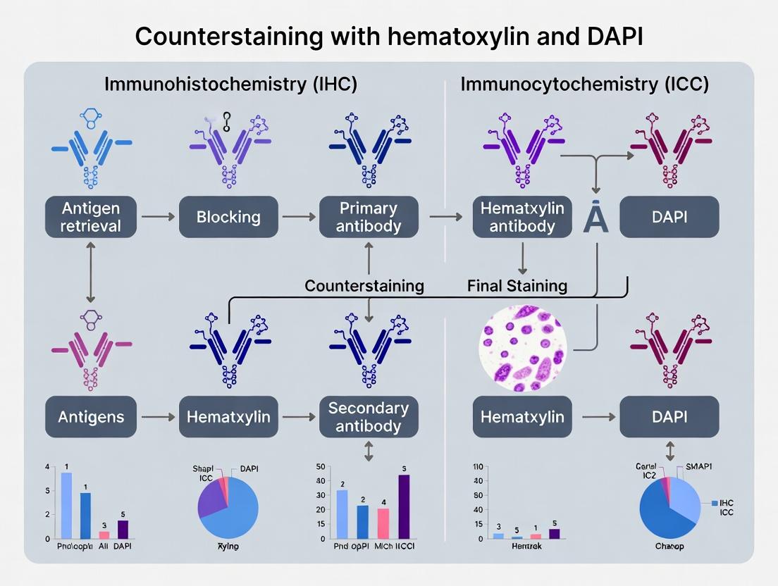
Step-by-Step Protocols: Integrating Hematoxylin or DAPI into Your IHC/ICC Workflow
Within the broader thesis examining nuclear counterstains (hematoxylin and DAPI) in IHC/ICC research, the sequential application of hematoxylin after chromogenic 3,3'-Diaminobenzidine (DAB) development is a cornerstone technique. Its primary function is to provide morphological context by staining non-target cell nuclei, allowing for precise cellular localization of the brown DAB precipitate. This protocol is essential for brightfield microscopy analysis, where contrast between the signal and cellular architecture is critical for accurate interpretation and quantitative analysis (e.g., determining labeling indices or assessing tissue pathology). Unlike fluorescent counterstains like DAPI, hematoxylin provides a permanent, stable stain compatible with routine histological mounting.
Key Research Reagent Solutions
| Reagent/Material | Function & Rationale |
|---|---|
| Hematoxylin Solution | A basic dye that complexes with anionic nuclear components (DNA/RNA), providing blue-purple nuclear contrast. Multiple formulations (e.g., Harris's, Mayer's, Gill's) exist with varying intensities. |
| DAB Chromogen Substrate | Enzymatic conversion by HRP produces an insoluble, brown precipitate at the antigen site. Provides the specific immunohistochemical signal. |
| Acid Alcohol (1% HCl in 70% EtOH) | Differentiating agent. Removes excess/unbound hematoxylin from cytoplasm and extracellular matrix, ensuring nuclear-specific staining. |
| Ammonia Water or Scott's Tap Water | Bluing agent. Converts the initial red hematoxylin complex to a stable blue-purple color by adjusting pH, enhancing contrast and permanence. |
| Phosphate-Buffered Saline (PBS) Buffer | Standard wash buffer for rinsing slides between steps, maintaining pH and removing residual reagents. |
| Aqueous Mounting Medium | Preserves the water-soluble hematoxylin stain under a coverslip for brightfield microscopy. |
Table 1: Impact of Hematoxylin Staining Duration on Stain Quality.
| Hematoxylin Type | Recommended Time | Over-staining Effect | Under-staining Effect |
|---|---|---|---|
| Mayer's | 1-3 minutes | High background, obscured DAB signal | Faint nuclei, poor morphology |
| Harris's | 30 seconds - 2 minutes | Excessive nuclear darkness, increased precipitate | Lack of contrast, difficult localization |
| Gill's | 1-4 minutes | Diffuse cytoplasmic staining | Incomplete nuclear detail |
Table 2: Comparison of Bluing Agents.
| Bluing Agent | Concentration | Time | Resultant Color Clarity |
|---|---|---|---|
| Scott's Tap Water | Ready-to-use | 1-2 minutes | Consistent, bright blue |
| Ammonium Hydroxide | 0.1% in water | 30-60 seconds | Rapid, strong blue |
| Lithium Carbonate | Saturated solution | 1 minute | Very crisp, high contrast |
Detailed Experimental Protocol
Title: Sequential Hematoxylin Counterstaining Post-DAB IHC.
Principle: Following the completion of DAB-based immunohistochemistry, slides are counterstained with hematoxylin to visualize nuclear morphology, differentiated to remove background, blued, dehydrated, cleared, and mounted.
Materials:
- IHC slides with developed DAB signal.
- Reagents listed in Section 2.
- Coplin jars or automated slide stainer.
- Dehydration series (70%, 95%, 100% Ethanol).
- Xylene or xylene-substitute clearing agent.
Methodology:
- Post-DAB Wash: After DAB reaction termination in water, rinse slides gently in running tap water for 1 minute.
- Hematoxylin Counterstaining: Immerse slides in filtered hematoxylin solution. Incubate for 30 seconds to 4 minutes (optimize based on hematoxylin type and fixation; see Table 1).
- Rinsing: Rinse slides thoroughly in running tap water for 1 minute to remove residual dye.
- Differentiation: Dip slides in 1% Acid Alcohol 3-5 times (approximately 5-10 seconds). Immediately rinse in tap water.
- Bluing: Immerse slides in bluing solution (e.g., Scott's Tap Water) for 1-2 minutes until nuclei turn blue-purple. Rinse in tap water.
- Dehydration & Clearing:
- Dip slides in 70% Ethanol (30 sec).
- Dip slides in 95% Ethanol (30 sec).
- Dip slides in 100% Ethanol twice (1 min each).
- Clear in xylene or substitute twice (2 min each).
- Mounting: Coverslip using a compatible, permanent mounting medium.
Visualization: Workflow Diagram
Diagram Title: Post-DAB Hematoxylin Staining & Mounting Workflow.
Diagram Title: Counterstain Selection Logic within IHC/ICC Thesis.
Within the framework of optimizing nuclear counterstaining for fluorescent multiplex imaging, this protocol addresses the critical need for DAPI application that ensures bright, specific nuclear labeling without compromising the detection of other fluorophores. Hematoxylin, while standard for brightfield IHC, is unsuitable for fluorescence multiplexing, making DAPI the indispensable nuclear counterstain. This protocol details a validated, optimized method for DAPI integration into ICC/IHC workflows, balancing signal intensity with minimal background and cross-talk.
DAPI Staining Optimization: Key Quantitative Findings
The following table summarizes experimental data comparing different DAPI staining conditions for multiplex fluorescence.
Table 1: Optimization Parameters for DAPI Staining in Multiplex ICC/IHC
| Parameter | Condition Tested | Optimal Value | Effect on Nuclear Signal (AU) | Effect on Background (AU) | Impact on Adjacent Fluorophore Signal (Cy3, % change) |
|---|---|---|---|---|---|
| Concentration | 50 ng/mL, 300 ng/mL, 1 µg/mL | 300 ng/mL | 15,250 ± 1,100 | 205 ± 45 | -2.5% |
| Incubation Time | 5 min, 10 min, 20 min | 5 min | 14,800 ± 950 | 190 ± 30 | -1.8% |
| Incubation Temp | 4°C, RT, 37°C | Room Temp (RT) | 15,100 ± 800 | 215 ± 40 | -3.1% |
| Wash Stringency | 1x PBS, 2x PBS, PBS-T | 2x PBS (5 min each) | 14,950 ± 900 | 165 ± 25 | -2.0% |
| Mounting Medium | Aqueous, Anti-fade w/ DAPI | Anti-fade without DAPI | 15,300 ± 1,050 | 180 ± 35 | N/A |
AU = Arbitrary Fluorescence Units. Data are mean ± SD from n=3 experiments.
Detailed Optimized Protocol
Part A: Sample Preparation and Primary/Secondary Staining
- Fixation & Permeabilization: Process cells or frozen sections as per standard multiplex ICC/IHC protocol (e.g., 4% PFA fixation, 0.1% Triton X-100 permeabilization).
- Blocking: Block with 5% normal serum/3% BSA in PBS for 1 hour at RT.
- Antibody Incubations: Perform primary antibody incubations (overnight, 4°C) and corresponding fluorescent secondary antibody incubations (1 hour, RT, in darkness) as required by your multiplex panel. Include all necessary wash steps.
Part B: Optimized DAPI Counterstaining and Mounting
- DAPI Solution Preparation: Dilute DAPI stock solution (e.g., 5 mg/mL) in PBS to a final concentration of 300 ng/mL. Filter through a 0.2 µm syringe filter.
- Staining Incubation: Apply the diluted DAPI solution to cover the sample entirely. Incubate for 5 minutes at room temperature in a covered, dark humidity chamber.
- Stringent Washing: Carefully aspirate the DAPI solution. Wash the sample twice with PBS for 5 minutes per wash with gentle agitation. Avoid using detergents (e.g., PBS-T) at this stage unless high background is persistent.
- Mounting: Briefly rinse sample with distilled water to remove salt crystals. Blot excess liquid and mount using a commercial, hardened, anti-fade mounting medium that does NOT contain DAPI (e.g., ProLong Diamond, Vectashield). Seal coverslips with clear nail polish.
- Curing & Imaging: Allow the mounting medium to cure as per manufacturer instructions (typically 24 hours at RT in the dark). Image using a DAPI/UV filter set (excitation ~358 nm, emission ~461 nm). Acquire DAPI channel first to minimize photobleaching of other fluorophores.
Visualization of the Protocol Workflow
Title: Workflow for Optimized DAPI Multiplex Staining
Visualization of DAPI's Role in Multiplex Analysis
Title: DAPI Enables Quantitative Spatial Analysis
The Scientist's Toolkit: Essential Research Reagents
Table 2: Key Reagents for Optimized Fluorescent Multiplex ICC/IHC
| Reagent | Function & Importance in Protocol | Example Product/Brand |
|---|---|---|
| High-Purity DAPI | DNA-specific fluorescent dye for definitive nuclear segmentation. Critical for multiplex analysis. | Thermo Fisher Scientific D1306, Sigma-Aldrich D9542 |
| Anti-fade Mounting Medium (DAPI-free) | Preserves fluorescence photostability. Using DAPI-free medium allows precise control over counterstain concentration. | ProLong Diamond (P36965), Vectashield H-1000 |
| Fluorophore-Conjugated Secondary Antibodies | Enable detection of multiple primary antibodies from different hosts. Must be spectrally distinct from DAPI. | Alexa Fluor 488, 555, 647 conjugates |
| Blocking Agent (BSA/Serum) | Reduces non-specific antibody binding, lowering background for all channels including DAPI. | Bovine Serum Albumin (BSA), Normal Donkey Serum |
| PBS (Phosphate Buffered Saline) | Washing and dilution buffer. Salt concentration and pH (7.4) critical for maintaining DAPI binding specificity. | Gibco DPBS |
| Microscope Filter Set for DAPI | Specific excitation/emission filters to isolate DAPI signal and prevent bleed-through into other channels. | Chroma 49000, Semrock DAPI-5060C |
Within the broader thesis on optimizing nuclear counterstaining for immunohistochemistry (IHC) and immunocytochemistry (ICC), the sequence and timing of the counterstain application are critical variables. Hematoxylin and DAPI are ubiquitous, yet their placement—before, after, or between detection steps—profoundly impacts signal clarity, background, and multiplexing capability. This application note synthesizes current methodologies and data to guide protocol design.
Comparative Analysis of Counterstain Sequencing
The optimal placement of the counterstain depends on the detection method (chromogenic vs. fluorescent), the target antigens, and the need for multiplexing. The table below summarizes the key advantages and disadvantages of each approach.
Table 1: Comparative Protocols for Counterstain Placement in IHC/ICC
| Sequence | Typical Workflow Order | Best For | Key Advantages | Key Risks & Considerations |
|---|---|---|---|---|
| Counterstain Last | 1. Primary Ab2. Detection3. Counterstain | Chromogenic IHC; Simple fluorescent ICC/IHC. | Prevents counterstain masking; Simplifies protocol; Standard for H&E-like visualization. | May mask weak target signals if too intense; DAPI can be quenched by mounting media if not sealed properly. |
| Counterstain First | 1. Counterstain2. Primary Ab3. Detection | Fluorescent multiplexing with nuclear antigens; Avoiding steric hindrance. | Unobstructed access to nuclear epitopes; Prevents antibody cross-reactivity with counterstain. | Risk of counterstain leaching or bleaching during subsequent steps; Not suitable for alcohol-soluble stains. |
| Counterstain Intermediate | 1. Primary Ab 12. Counterstain3. Primary Ab 2/Detection | Complex fluorescent multiplexing (>4 targets). | Provides fiduciary marker for subsequent imaging rounds; Can separate staining batches. | Requires rigorous wash steps to prevent carryover; Potential for signal crossover. |
Table 2: Quantitative Impact of DAPI Timing on Fluorescent Signal Integrity Data derived from recent studies on formalin-fixed paraffin-embedded (FFPE) tissues and cultured cells.
| DAPI Placement | Mean Nuclear Intensity (RFU) | Background (RFU) | Signal-to-Background Ratio | Observed Photostability (after 5 min illum.) |
|---|---|---|---|---|
| Before Primary Antibody | 15,200 ± 1,100 | 450 ± 80 | 33.8 | 78% retention |
| After Secondary Antibody | 14,800 ± 950 | 380 ± 60 | 38.9 | 92% retention |
| After Penultimate Tyramide Signal Amplification (TSA) | 16,500 ± 1,400 | 510 ± 90 | 32.4 | 85% retention |
Detailed Experimental Protocols
Protocol A: Standard IHC with Hematoxylin Counterstain (Sequenced Last)
Purpose: To visualize protein localization in tissue context with a classic H&E-like appearance. Key Reagent Solutions:
- Citrate Buffer (pH 6.0): For heat-induced epitope retrieval (HIER).
- Peroxidase Block: 3% H₂O₂ in methanol, to quench endogenous peroxidase activity.
- Protein Block: 5% normal serum from host of secondary antibody, to reduce nonspecific binding.
- Primary Antibody Diluent: TBS or PBS with 1% BSA and 0.1% sodium azide.
- HRP Polymer Detection System: Labeled polymer for signal amplification.
- DAB Chromogen: 3,3'-Diaminobenzidine, produces brown precipitate.
- Harris Hematoxylin: Modified for standardized nuclear staining.
- Scotch or Acid Alcohol: 1% HCl in 70% ethanol, for differentiation.
- Ammonia Water or Bluing Reagent: For pH shift to blue nuclear stain.
- Aqueous Mounting Medium: For preserving chromogenic signals.
Procedure:
- Perform deparaffinization and rehydration on FFPE sections.
- Perform HIER in citrate buffer using a pressure cooker or steamer (20 min, 95-100°C).
- Cool slides, wash in PBS, and apply peroxidase block for 10 min.
- Wash and apply protein block for 30 min at room temperature (RT).
- Apply primary antibody diluted in diluent overnight at 4°C.
- Wash and apply HRP-labeled polymer secondary for 30 min at RT.
- Wash and incubate with DAB chromogen for 5-10 min (monitor microscopically).
- Wash in distilled water and immerse in hematoxylin for 30-60 seconds.
- Rinse in tap water and differentiate in acid alcohol for 2-3 seconds.
- Rinse and immerse in bluing reagent for 1 min.
- Dehydrate through graded alcohols, clear in xylene, and mount with permanent medium.
Protocol B: Sequential Multiplex ICC with DAPI Counterstain First
Purpose: To enable high-dimensional imaging of co-localized nuclear and cytoplasmic targets. Procedure:
- Culture cells on chambered coverslips, fix with 4% PFA for 15 min, and permeabilize with 0.25% Triton X-100 for 10 min.
- Apply DAPI (300 nM in PBS) for 10 minutes at RT. Wash 3x with PBS.
- Apply primary antibody mix for Target 1 (nuclear) and Target 2 (cytoplasmic) overnight at 4°C.
- Wash and apply spectrally distinct fluorescent secondary antibodies for 1 hour at RT, protected from light.
- Wash, mount with anti-fade mounting medium, and image immediately.
- For subsequent multiplexing rounds, use gentle stripping buffer (e.g., glycine pH 2.0) to remove antibodies, preserving the initial DAPI signal as a registration marker.
Visualization: Counterstain Sequencing Decision Pathway
Counterstain Placement Decision Pathway
The Scientist's Toolkit: Essential Reagent Solutions
Table 3: Key Reagents for Optimized Counterstaining Protocols
| Reagent | Function & Role in Sequencing | Example Product/Specification |
|---|---|---|
| DAPI (4',6-Diamidino-2-Phenylindole) | AAT Bioquest DAPI Solution (300 nM in PBS), ready-to-use. | Blue-fluorescent DNA stain for fluorescent protocols. Timing affects photostability and background. |
| Harris Hematoxylin (Modified) | Sigma-Aldrich Harris Hematoxylin, Modified (alcoholic). | Standard nuclear counterstain for chromogenic IHC. Always applied last before dehydration. |
| Antibody Elution Buffer | Thermo Fisher Scientific Antibody Elution Buffer (pH 2.0). | Enables sequential multiplexing by stripping antibodies while preserving DAPI stained first. |
| Prolong Diamond Antifade Mountant | Invitrogen ProLong Diamond Antifade Mountant. | Critical for preserving fluorescence, especially when DAPI is applied early in protocol. |
| DAB Chromogen Substrate Kit | Agilent DAB+ Substrate Chromogen System. | HRP substrate for brown precipitate. Applied before hematoxylin counterstain. |
| Automated Stainer-Compatible Hematoxylin | Leica Biosystems Hematoxylin 560. | Formulated for consistency and timing in automated platforms, applied post-detection. |
Within the broader thesis on counterstaining in IHC/ICC research, the choice of mounting medium is a critical, yet often overlooked, determinant of experimental success and data integrity. Hematoxylin and DAPI serve distinct but complementary roles: hematoxylin provides morphological context for brightfield imaging, while DAPI (4',6-diamidino-2-phenylindole) offers high-sensitivity nuclear localization for fluorescence. Their optimal visualization is governed by opposing requirements for sample preservation, directly linked to the chemical nature of the mounting media—aqueous for DAPI and permanent resinous for hematoxylin. This application note details the rationale, protocols, and practical considerations for these two workflows, ensuring fidelity of both counterstains in multiplexed research and drug development studies.
Scientific Rationale and Data Comparison
Table 1: Core Properties and Requirements of DAPI vs. Hematoxylin Counterstains
| Parameter | DAPI (Fluorescence) | Hematoxylin (Brightfield) |
|---|---|---|
| Detection Mode | Fluorescence (Ex/Em ~358/461 nm) | Brightfield, colorimetric |
| Primary Function | Nuclear staining, often for co-localization with specific fluorescent markers. | Morphological nuclear staining for architectural context. |
| Mounting Imperative | Must be kept hydrated to maintain fluorophore conformation and signal intensity. | Must be dehydrated and sealed to preserve dye-metal complex and prevent fading/dissolution. |
| Compatible Medium | Aqueous-based (e.g., PBS/glycerol, polyvinyl alcohol with antifade). | Permanent resinous (e.g., synthetic toluene-based like DPX, or acrylic). |
| Key Vulnerability | Photobleaching under excitation light. | Aqueous fading ("washing out") and microbial growth. |
| Coverslip Sealing | Often sealed with nail polish to slow evaporation. | Inherently sealed as medium hardens; no additional sealant needed. |
| Long-Term Stability | Moderate to poor (weeks to months), even with antifade agents. | Excellent (years) when properly processed. |
Detailed Experimental Protocols
Protocol A: Aqueous Mounting for DAPI-Stained Fluorescence Samples (IHC/ICC)
This protocol assumes a fluorescently-labeled sample already stained with DAPI.
Materials:
- Fluorescently-stained, DAPI-counterstained slides, rinsed in buffer.
- Aqueous mounting medium (e.g., 90% glycerol in PBS, or commercial antifade media like ProLong Diamond Antifade Mountant, Vectashield).
- Microscope slides and #1.5 thickness coverslips.
- Nail polish or clear sealant.
- Absorbent tissue.
Procedure:
- Final Rinse: After the final wash post-DAPI staining, briefly drain the slide but do not allow it to dry completely.
- Mounting Application: Place a small drop (~25-50 µL) of aqueous mounting medium onto the tissue section or cell spot.
- Coverslipping: Gently lower a clean coverslip at a ~45-degree angle to avoid trapping air bubbles.
- Excess Removal: Carefully blot away any excess mounting medium that seeps out from the sides using absorbent tissue.
- Sealing (Optional but Recommended): Apply a thin bead of clear nail polish around the edges of the coverslip to seal it. This slows evaporation.
- Curing & Imaging: Allow the sealant to dry (5-10 minutes). For media like ProLong, follow manufacturer instructions for curing (often overnight in the dark). Image as soon as possible for optimal signal. Store slides flat, in the dark at 4°C.
Protocol B: Permanent Mounting for Hematoxylin-Stained Slides (IHC)
This protocol begins after hematoxylin staining and bluing steps.
Materials:
- Hematoxylin-stained slides.
- Dehydration series: 70% Ethanol, 90% Ethanol, 100% Ethanol (two changes), Xylene or Xylene substitute (two changes).
- Permanent mounting medium (e.g., DPX Mountant, Entellan).
- Microscope slides and coverslips.
- Fume hood.
Procedure:
- Dehydration: Pass the stained slide through an ascending ethanol series:
- 70% Ethanol – 1 minute.
- 90% Ethanol – 1 minute.
- 100% Ethanol I – 2 minutes.
- 100% Ethanol II – 2 minutes.
- Clearing: Transfer slide to a clearing agent to remove alcohol and render the tissue transparent:
- Xylene (or substitute) I – 3 minutes.
- Xylene (or substitute) II – 3 minutes. Perform all steps in a fume hood.
- Mounting: While the slide is still wet with xylene, place a drop of permanent mounting medium onto the tissue. Immediately lower a coverslip.
- Hardening: Gently press down on the coverslip to spread the medium and remove bubbles. Lay the slide flat in a fume hood and allow the medium to harden completely (several hours to overnight).
- Cleaning: Once hardened, use a razor blade and ethanol-dampened tissue to gently scrape off any excess dried medium from the slide surface. The slide is now permanently sealed and stable for years.
The Scientist's Toolkit: Research Reagent Solutions
Table 2: Essential Materials for Mounting Media Workflows
| Reagent/Material | Function/Description | Key Consideration |
|---|---|---|
| DAPI Stain | Fluorescent DNA intercalating dye for nuclear counterstain in fluorescence. | Light-sensitive; use antifade mounting media. |
| Hematoxylin (Harris's, Mayer's) | Basic dye forming complexes with nucleic acids, stained via metal (Al³⁺) mordant. | Requires "bluing" in slightly alkaline buffer (e.g., Scott's tap water) for optimal color. |
| Aqueous Antifade Mountant | Glycerol/PBS-based medium with additives (e.g., p-phenylenediamine, DABCO) to reduce photobleaching. | Critical for preserving DAPI and fluorophore signal over time. |
| DPX Mountant | Distyrene, Plasticizer, Xylene mixture. A classic synthetic resinous permanent medium. | Hardens to a clear, durable seal. Requires xylene clearing. Use in a hood. |
| Entellan | Rapidly hardening, toluene-based acrylic mounting medium. | A popular alternative to DPX with similar properties. |
| Xylene or Xylene Substitute | Clearing agent for dehydration prior to permanent mounting. | Xylene is toxic; substitutes are less hazardous but may require longer clearing times. |
| #1.5 Coverslips | High-precision glass coverslips (0.17mm thickness). | Essential for optimal high-resolution (63x/100x oil) microscopy to correct for spherical aberration. |
Visualized Workflows and Decision Pathways
Title: Mounting Media Selection Workflow for DAPI vs. Hematoxylin
Title: Aqueous Mounting Preserves DAPI Fluorescence
Title: Permanent Mounting Secures Hematoxylin Stain
Solving Common Counterstain Problems: Signal Masking, Background, and Fading
Within the broader thesis on counterstaining with hematoxylin and DAPI in IHC/ICC research, a critical technical challenge is the over-application of hematoxylin counterstain. Excessive nuclear staining can obscure weak but biologically significant 3,3'-Diaminobenzidine (DAB) signals, leading to false-negative interpretations. This application note details evidence-based protocols and dilution optimizations to resolve this issue, ensuring clear visualization of both nuclear detail and target antigen localization.
Table 1: Comparative Effects of Hematoxylin Protocols on DAB Signal Clarity (AU: Arbitrary Units)
| Protocol Variation | Hematoxylin Intensity (AU) | DAB Signal-to-Noise Ratio | Nuclear Detail | Optimal for Weak DAB |
|---|---|---|---|---|
| Standard 5-minute stain | 0.85 ± 0.10 | 1.5 ± 0.3 | Excellent | No |
| 1:2 Diluted, 2-minute stain | 0.45 ± 0.05 | 3.2 ± 0.4 | Very Good | Yes |
| 0.1% Acid Alcohol, 30-sec dip | 0.30 ± 0.08 | 3.8 ± 0.5 | Good (if controlled) | Yes (Risk of over-destaining) |
| Blueing in weak ammonia | 0.50 ± 0.06 | 3.0 ± 0.3 | Very Good | Yes |
Table 2: Recommended Dilution Optimization for Common Hematoxylins
| Hematoxylin Type | Standard Concentration | Starting Dilution for Weak DAB | Recommended Staining Time | Blueing Agent |
|---|---|---|---|---|
| Harris Hematoxylin | Undiluted | 1:2 in distilled H₂O | 1-2 minutes | Scott's Tap Water |
| Mayer's Hematoxylin | Undiluted | 1:3 in distilled H₂O | 2-3 minutes | 0.1% Ammonia Water |
| Gill's Hematoxylin | Formulation III | 1:1 in distilled H₂O | 1.5-2.5 minutes | Lithium Carbonate |
Detailed Experimental Protocols
Protocol 1: Optimized Sequential Staining for Weak DAB Signal Objective: To achieve a crisp, light nuclear counterstain that does not mask a weak DAB chromogen signal. Materials: IHC/ICC slides with developed DAB signal, diluted hematoxylin (see Table 2), acid alcohol (1% HCl in 70% ethanol), blueing solution, dehydration series, mounting medium. Procedure:
- Following DAB development and thorough rinsing in distilled water, place slides in diluted hematoxylin for the optimized time (e.g., 1.5 minutes for 1:2 Gill's).
- Rinse slides in several changes of tap water until clear.
- Differentiate briefly by dipping slides (1-3 quick dips) in 1% acid alcohol. Immediately rinse in tap water.
- Blue the nuclei by immersing slides in Scott's Tap Water or 0.1% ammonia water for 1 minute. Rinse in tap water.
- Dehydrate through a graded ethanol series (70%, 95%, 100%), clear in xylene, and mount with a non-aqueous, permanent mounting medium. Key Note: Differentiation (Step 3) is critical. Monitor under a microscope to remove excess hematoxylin from cytoplasm and ECM while retaining clear nuclear detail.
Protocol 2: Pilot Titration for Hematoxylin Dilution Objective: Empirically determine the ideal hematoxylin dilution for a specific experimental system. Materials: Test IHC slides with a range of DAB signal intensities, stock hematoxylin, phosphate-buffered saline (PBS), slide jars. Procedure:
- Prepare a series of hematoxylin dilutions in PBS (e.g., 1:1, 1:2, 1:4, 1:8).
- Apply each dilution to a replicate test slide for a fixed time (e.g., 90 seconds).
- Process all slides through identical brief differentiation and blueing steps.
- Evaluate slides by light microscopy. The optimal dilution provides definitive, crisp blue nuclei with no perceived diminishment of the weakest target DAB signal. Use this dilution for all subsequent experiments.
The Scientist's Toolkit: Essential Research Reagents
Table 3: Key Reagents for Optimized Hematoxylin Counterstaining
| Reagent/Material | Function & Importance |
|---|---|
| Gill's or Mayer's Hematoxylin | Progressive stains offering more controllable results than regressive stains like Harris. |
| 1% Acid Alcohol (Differentiator) | Selectively removes excess hematoxylin from non-nuclear components; concentration and time are key control variables. |
| Scott's Tap Water or Ammonia Water | Blueing agent; raises pH to convert hematein to its blue-colored form, completing the staining. |
| Weak DAB Chromogen Kit (Polymer-based) | High-sensitivity detection system to amplify weak antigen signals before counterstaining. |
| Aqueous Mounting Medium (for evaluation) | Allows temporary mounting for microscopic assessment of staining intensity before final dehydration and permanent mounting. |
Visualization: Workflow and Decision Pathway
Optimized Hematoxylin Staining & QC Workflow for IHC
Factors, Effects, and Solutions for DAB Masking
Achieving balance in IHC counterstaining is paramount for data accuracy. By systematically optimizing hematoxylin dilution, strictly controlling differentiation, and employing high-sensitivity DAB detection, researchers can prevent the masking of weak signals. These protocols, framed within the context of advanced counterstaining strategies, provide a reliable path to high-quality, publication-ready images that faithfully represent both nuclear and protein localization data.
Within the broader thesis on "Optimization of Nuclear Counterstaining in IHC/ICC for Quantitative Analysis," addressing high DAPI background is paramount. Hematoxylin provides robust, permanent nuclear contrast in brightfield IHC, while DAPI offers high sensitivity and specificity for fluorescence-based ICC and multiplex assays. Excessive DAPI background compromises the accuracy of nuclear segmentation, co-localization studies, and quantitative intensity measurements, leading to erroneous data interpretation in both basic research and drug development pipelines. This note details systematic troubleshooting of wash steps and DAPI concentration to restore assay fidelity.
Table 1: Impact of Wash Buffer Ionic Strength on DAPI Background Intensity (Relative Fluorescence Units, RFU)
| Wash Buffer Composition | pH | Nuclear Signal (RFU) | Background (RFU) | Signal-to-Background Ratio |
|---|---|---|---|---|
| 1X PBS | 7.4 | 15,000 | 2,500 | 6.0 |
| 1X PBS + 0.1% Triton X-100 | 7.4 | 14,800 | 3,100 | 4.8 |
| High-Salt PBS (0.5M NaCl) | 7.4 | 14,200 | 950 | 14.9 |
| TBST (0.05% Tween-20) | 7.6 | 13,500 | 1,800 | 7.5 |
Table 2: Optimizing DAPI Concentration and Exposure Time
| DAPI Concentration | Incubation Time | Mounting Media | Optimal Camera Exposure (ms) | Background Assessment |
|---|---|---|---|---|
| 1 µg/mL | 10 min | Aqueous | 100 | High, diffuse |
| 300 ng/mL | 5 min | Aqueous | 150 | Moderate |
| 100 ng/mL | 3 min | Aqueous | 200 | Low, crisp nuclei |
| 100 ng/mL | 3 min | Anti-fade | 250 | Lowest, stable |
Detailed Experimental Protocols
Protocol 1: High-Stringency Washes to Reduce Non-Specific DAPI Binding
- Objective: Remove excess and non-specifically bound DAPI through ionic competition.
- Materials: High-salt wash buffer (1X PBS with 0.5M NaCl, pH 7.4), standard wash buffer (1X PBS or TBST), humidified chamber.
- Method:
- Following primary/secondary antibody incubations and final washes in IHC/ICC, prepare the high-salt wash buffer.
- Wash the sample slide or coverslip with high-salt buffer three times, for 5 minutes each, with gentle agitation.
- Proceed to DAPI staining at a reduced concentration (see Protocol 2).
- Post-DAPI staining, perform one final 2-minute wash in high-salt buffer before mounting.
- Rationale: High ionic strength competes with DAPI for low-affinity binding to anionic sites on cytoskeletal elements and phospholipids, reducing background.
Protocol 2: Titration of DAPI for Optimal Nuclear Staining
- Objective: Establish the minimal DAPI concentration yielding saturated nuclear signal with minimal background.
- Materials: DAPI stock solution (e.g., 5 mg/mL in water), wash buffer, mounting medium.
- Method:
- Prepare a dilution series of DAPI: 1 µg/mL, 300 ng/mL, 100 ng/mL, and 30 ng/mL in your standard assay buffer (e.g., PBS or TBST).
- Apply each dilution to replicate samples (e.g., fixed HeLa or tissue sections). Incubate for 5 minutes at room temperature, protected from light.
- Wash all samples identically with 2 x 5 minute washes in buffer.
- Mount and image using identical microscope and camera settings (especially exposure time and gain).
- Quantify mean nuclear intensity and extra-nuclear background fluorescence using image analysis software (e.g., ImageJ). Select the concentration yielding the highest signal-to-background ratio.
Diagrams
Title: DAPI Background Troubleshooting Decision Flowchart
Title: Mechanisms of DAPI Specific vs. Non-Specific Binding
The Scientist's Toolkit: Research Reagent Solutions
Table 3: Essential Reagents for Mitigating DAPI Background
| Reagent/Solution | Function & Rationale | Key Consideration |
|---|---|---|
| DAPI (4',6-diamidino-2-phenylindole) | Fluorescent DNA intercalator; binds AT-rich regions. Use at 100-300 ng/mL. | Aliquot stock to avoid freeze-thaw cycles; protect from light. |
| High-Salt Wash Buffer (PBS + 0.2-0.5M NaCl) | Competitively elutes DAPI bound to anionic cellular components via ionic interference. | Check antibody stability in high salt. Adjust pH to 7.4 post-addition. |
| Triton X-100 or Tween-20 (0.1-0.5%) | Non-ionic detergent for permeabilization and reducing hydrophobic interactions. | Can increase background if used after DAPI staining; use pre-staining. |
| Commercial Anti-fade Mounting Media (e.g., with p-phenylenediamine or propyl gallate) | Reduces photobleaching and may contain agents that limit diffusion of unbound DAPI. | Some are not compatible with certain tissues or plastic substrates. |
| Bovine Serum Albumin (BSA) | Used in wash/incubation buffers to block non-specific binding sites. | Use at 1-5% in wash buffers to reduce overall non-specific staining. |
| DNase I | Control enzyme. Pre-treatment eliminates specific nuclear DAPI signal, confirming staining specificity. | Use on a control slide to distinguish specific from non-specific signal. |
Within the broader thesis on counterstaining with hematoxylin and DAPI in IHC/ICC research, a critical technical challenge is the optimization of visual contrast. Effective counterstaining provides essential histological or cellular context without overshadowing the primary signal of interest. These application notes provide a detailed framework and protocols for achieving this balance, ensuring accurate, quantitative, and reproducible data interpretation in research and drug development.
Table 1: Comparative Properties of Common Counterstains
| Counterstain | Target (IHC/ICC) | Optimal Excitation/Emission (nm) | Typical Incubation Time | Compatible Primary Signal Colors | Key Consideration |
|---|---|---|---|---|---|
| Hematoxylin | DNA (nuclei) | N/A (broad spectrum light) | 30 seconds - 10 minutes | Red (AEC), Brown (DAB), Pink/Red (Fluorophores) | Requires differentiation; intensity varies with bluing. |
| DAPI | DNA (nuclei) | 358/461 | 5 - 30 minutes | Green (FITC, Alexa 488), Red (Texas Red, Cy3), Far-Red (Cy5) | Can bleed into other channels; requires UV/DAPI filter. |
| Hoechst 33342 | DNA (live/fixed) | 350/461 | 5 - 30 minutes | Green, Red, Far-Red | More permeable than DAPI; used for live-cell imaging. |
| DRAQ5 | DNA (nuclei) | 646/681 | 5 - 15 minutes | Green, Orange | Far-red emission; suitable for multiplexing with common fluorophores. |
Table 2: Impact of Counterstain Concentration on Signal-to-Noise Ratio (SNR) Data derived from model ICC experiment with a Cy3-labeled primary target (λex/em: 550/570nm).
| Counterstain | Concentration | Nuclear Signal Intensity (Mean Gray Value) | Cytoplasmic Background (Mean Gray Value) | Calculated SNR (Target/Background) | Recommended for Quantitative Work? |
|---|---|---|---|---|---|
| DAPI | 300 nM | 1850 | 120 | 15.4 | Yes (Optimal) |
| DAPI | 1 µM | 4200 | 450 | 9.3 | No (Too intense, high background) |
| DAPI | 100 nM | 650 | 85 | 7.6 | Yes (Weaker but sufficient) |
| Hoechst 33342 | 2 µM | 2100 | 200 | 10.5 | Yes |
| Hematoxylin (1:10 dil.) | - | Subjectively "Optimal" | Low | N/A (Brightfield) | Yes (Visual assessment critical) |
Detailed Application Protocols
Protocol 1: Optimizing Hematoxylin Counterstaining for DAB-Based IHC
Objective: To achieve crisp, light blue-gray nuclei that provide context without competing with a brown DAB chromogen signal.
Materials:
- Deparaffinized and antigen-retrieved tissue sections
- Blocked and primary/secondary antibody-labeled slides (DAB developed)
- Mayer’s or Harris Hematoxylin
- Acid Alcohol (1% HCl in 70% ethanol) or differentiation solution
- Bluing solution (0.1% ammonium hydroxide or Scott’s tap water substitute)
- Ethanol series and xylenes
- Mounting medium (non-aqueous, permanent)
Methodology:
- Following DAB development and water rinse, immerse slides in hematoxylin for 30 seconds to 2 minutes. Start with 1 minute as a standard.
- Rinse thoroughly in running tap water for 1 minute.
- Differentiate: Dip slides in acid alcohol 3-5 times (1-5 seconds total). This step removes excess hematoxylin from the cytoplasm. Rinse immediately in tap water.
- Bluing: Immerse slides in bluing solution for 1 minute to convert the hematoxylin color to a stable blue. Rinse in tap water.
- Dehydrate through an ethanol series (70%, 95%, 100%), clear in xylenes, and mount with a permanent medium.
- Optimization Notes: If nuclei are too dark, shorten hematoxylin time or increase differentiation. If too light, lengthen hematoxylin time or reduce differentiation. The ideal result yields clearly visible nuclear detail without a dark background that obscures DAB signal.
Protocol 2: Titrating DAPI for Multiplex Fluorescence ICC
Objective: To identify the minimum concentration of DAPI that provides robust nuclear segmentation without spectral bleed-through into adjacent fluorescence channels.
Materials:
- Fixed and permeabilized cells on coverslips
- Blocking buffer (e.g., 3% BSA/PBS)
- Primary and fluorescent secondary antibodies
- DAPI stock solution (5 mg/mL or 14.3 mM in water)
- Antifade mounting medium (e.g., ProLong Diamond)
- PBS (pH 7.4)
Methodology:
- Complete immunostaining with your primary and secondary antibodies. Perform final washes in PBS.
- Prepare a dilution series of DAPI in PBS (e.g., 300 nM, 500 nM, 1 µM, 2 µM) from the stock.
- Incubate stained coverslips with each DAPI concentration for 10 minutes at room temperature in the dark.
- Rinse gently but thoroughly with PBS (2 x 5 minutes).
- Mount coverslips on slides using antifade mounting medium. Seal edges with nail polish if necessary.
- Image using consistent acquisition settings (especially laser power and gain for the DAPI channel) across all samples.
- Analysis: Use image analysis software to measure the mean intensity of nuclear DAPI signal and the cytoplasmic background in a non-fluorescent channel. Calculate SNR. Visually inspect for bleed-through into the green (FITC) and red (TRITC) channels. The concentration yielding a high SNR with no visible bleed-through is optimal.
Visualizations
Title: Hematoxylin Staining & Differentiation Workflow
Title: Signal-Counterstain Balance Logic
The Scientist's Toolkit
Table 3: Essential Research Reagent Solutions
| Item | Function & Role in Optimization | Example Product/Brand |
|---|---|---|
| Hematoxylin (Mayer's) | A well-defined, progressive nuclear stain. Provides consistent, moderate-intensity staining ideal for quantitative IHC. | Sigma-Aldrich Mayer's Hematoxylin |
| DAPI (4',6-diamidino-2-phenylindole) | A fluorescent DNA intercalator. Standard nuclear counterstain for fluorescence microscopy; concentration must be titrated. | Thermo Fisher Scientific DAPI |
| Hoechst 33342 | Cell-permeable DNA stain. Used for live-cell nuclear labeling and fixed-cell work; often brighter than DAPI. | Invitrogen Hoechst 33342 |
| Antifade Mounting Medium | Preserves fluorescence signal and reduces photobleaching. Critical for maintaining optimized contrast over time. | Vector Laboratories Vectashield |
| DAB Chromogen Kit | Produces an insoluble brown precipitate at the antigen site. The intensity must be balanced against the hematoxylin. | Agilent DAB+ Substrate Kit |
| Acid Alcohol (1% HCl/70% EtOH) | Differentiator for hematoxylin. Removes excess stain from cytoplasm; timing controls counterstain intensity. | Lab-prepared or commercial |
| Bluing Solution | Alkalizes the slide, converting hematoxylin to its final blue color. Stabilizes the nuclear stain. | Leica Biosystems Bluing Reagent |
| TrueBlack Lipofuscin Autofluorescence Quencher | Reduces tissue autofluorescence, improving the SNR of specific signals against counterstains. | Biotium TrueBlack |
Within the framework of a thesis on counterstaining in IHC/ICC research, maintaining signal integrity is paramount. Hematoxylin provides nuclear contrast and tissue architecture, while DAPI is the ubiquitous nuclear counterstain for fluorescence. Both are susceptible to fading, compromising data reproducibility and quantitative analysis. This application note details protocols to mitigate photobleaching of DAPI and ensure permanence of hematoxylin through optimized coverslipping.
Antifade Reagents for DAPI: Quantitative Comparison
DAPI, when excited by UV light, undergoes rapid photobleaching. Antifade mounting media significantly retard this process. The following table summarizes key performance data for common commercial antifade reagents.
Table 1: Comparison of Antifade Mounting Media for DAPI Preservation
| Reagent Name | Primary Active Component(s) | Recommended For | DAPI Signal Half-Life (Relative to PBS-Glycerol) | Compatibility with Other Fluorophores (e.g., Alexa Fluors) | Hardening/Sealing Requirement |
|---|---|---|---|---|---|
| Prolong Diamond | Patented tris-triazole-based formulation | Long-term preservation, multimodal imaging | >20x | Excellent (broad spectrum) | Slow-curing polymer; does not require nail polish |
| Vectashield Vibrance | Proprietary antifade with DAPI-specific enhancers | Fluorescence microscopy, especially DAPI/blue dyes | >15x | Very Good | Non-hardening; requires sealed edges |
| Fluoromount-G | p-Phenylenediamine (PPD) & Glycerol | General use, cost-effective | 8-10x | Good (may quench some green dyes) | Non-hardening; requires sealed edges |
| SlowFade Gold | Trolox-based (vitamin E analog) | Live-cell imaging & photostability | >12x | Excellent | Slow-curing; seal recommended for long-term |
| Glycerol-PBS (Control) | None | Temporary mounting only | 1x | Baseline | Evaporates; requires immediate imaging/sealing |
Protocols
Protocol 3.1: Immunofluorescence Staining with Optimized DAPI Mounting
Aim: To preserve DAPI and fluorophore signals in ICC/IF samples for long-term archival. Workflow:
- Sample Preparation: Culture cells on coverslips or prepare tissue cryosections. Fix (e.g., 4% PFA, 10 min), permeabilize, and block.
- Primary/Secondary Incubation: Apply primary antibody (diluted in blocking buffer), wash (3x PBS), apply fluorophore-conjugated secondary antibody, wash.
- DAPI Counterstaining: Incubate samples in DAPI (e.g., 1 µg/mL in PBS) for 5 minutes at room temperature. Wash in PBS (3 x 5 min).
- Mounting with Antifade:
- For Prolong Diamond/SlowFade Gold: Apply a small drop (~10-20 µL) to a clean glass slide. Invert the coverslip (sample-side down) onto the drop. Avoid bubbles. Let it cure in the dark at room temperature for 24 hours before imaging for optimal hardening.
- For Non-Hardening Media (Vectashield, Fluoromount-G): Apply mounting medium. Lower coverslip. Seal the edges with clear nail polish or a commercial sealant to prevent evaporation and compression artifacts.
- Storage: Store slides flat, in the dark, at 4°C (or -20°C for very long-term storage).
Diagram Title: Optimized Immunofluorescence Workflow with DAPI & Antifade
Protocol 3.2: Coverslipping for Permanent Hematoxylin Staining
Aim: To ensure dehydration, clearing, and permanent mounting of H&E or IHC-DAB samples with hematoxylin counterstain, preventing fade and aqueous damage. Workflow:
- Post-Staining Rinse: After hematoxylin staining, differentiation, and bluing, rinse slides in distilled water.
- Dehydration:
- Dip slides sequentially in a graded ethanol series: 70% ethanol (1 min), 95% ethanol (1 min), 100% ethanol (2 x 2 min).
- Clearing:
- Transfer slides to a xylene substitute (e.g., Histo-Clear) or pure xylene (2 x 3 min) until completely clear. This step is critical for optical clarity and mounting medium compatibility.
- Coverslipping:
- Using a Non-Aqueous, Hardening Mountant (e.g., Cytoseal, Permount):
- Place a drop of mounting medium on the coverslip or slide.
- Gently lower the slide onto the coverslip (or vice versa) at an angle to avoid bubbles.
- Let slides cure flat in a fume hood for 24-48 hours. Wipe away excess medium with a xylene-dampened cloth.
- Using a Non-Aqueous, Hardening Mountant (e.g., Cytoseal, Permount):
Diagram Title: Permanent Coverslipping Protocol for Hematoxylin
The Scientist's Toolkit: Essential Reagent Solutions
Table 2: Key Reagents for Fade Prevention in Counterstaining
| Item | Function & Rationale |
|---|---|
| Prolong Diamond Antifade Mountant | A hardening polymer-based mountant providing exceptional photostability across the visible and far-red spectrum. Ideal for multi-label fluorescence and long-term storage. |
| Vectashield Vibrance | Non-hardening mountant specifically formulated to enhance and preserve DAPI/blue fluorescent signals while maintaining compatibility with other dyes. |
| Histo-Clear or Xylene | Clearing agent. Removes alcohol, makes tissue transparent, and is miscible with resinous mounting media. Essential for permanent brightfield slides. |
| Cytoseal 60 or DPX Mountant | A synthetic resin-based, xylene-soluble mounting medium. Dries to a hard finish, permanently sealing the coverslip for hematoxylin-based slides. |
| #1.5 Precision Coverslips (0.17 mm thickness) | High-quality glass coverslips with the optimal thickness for high-resolution oil-immersion microscopy objectives. |
| Clear Nail Polish or VALAP Sealant | Used to create a physical seal around the edges of non-hardening aqueous mounting media, preventing evaporation and sample degradation. |
Head-to-Head Comparison: Choosing the Right Counterstain for Your Experimental Goals
Within the context of advanced immunohistochemistry (IHC) and immunocytochemistry (ICC) research, counterstaining serves a dual purpose: providing architectural context and facilitating multiplexed analysis. This application note provides a comparative analysis of two principal nuclear counterstains—hematoxylin and 4',6-diamidino-2-phenylindole (DAPI)—focusing on critical parameters of signal permanence, multiplexing capability, and associated equipment needs. The insights are framed to support a broader thesis on optimizing detection strategies in drug development and basic research.
Comparative Quantitative Analysis
Table 1: Core Characteristics of Hematoxylin vs. DAPI
| Parameter | Hematoxylin | DAPI |
|---|---|---|
| Chemical Nature | Metal-complexed dye (oxidized hematoxylin + Al³⁺/Fe³⁺) | Fluorescent intercalating agent |
| Excitation/Emission Max | Broad spectrum, visible light (~550-650 nm) | ~358 nm / ~461 nm |
| Signal Permanence | High (permanent, alcohol & xylene resistant) | Moderate to Low (prone to photobleaching) |
| Multiplexing Capability | Low (broad absorption, interferes with chromogen detection) | High (distinct UV/blue channel, minimal spectral overlap) |
| Compatibility | Chromogenic IHC/ICC (brightfield) | Immunofluorescence (IF), Fluorescence in situ hybridization (FISH) |
| Primary Equipment Need | Standard brightfield microscope | Epifluorescence/confocal microscope with UV/DAPI filter set |
| Typical Cost (per slide) | Very Low (~$0.10) | Low (~$0.50) |
| Quantitative Potential | Low (subjective intensity, non-linear) | High (linear fluorescence intensity, suitable for image cytometry) |
Table 2: Suitability Assessment for Research Applications
| Application Context | Recommended Counterstain | Rationale |
|---|---|---|
| Diagnostic Pathology, Archival Slides | Hematoxylin | Permanent record, standard for H&E-like context. |
| Multiplex Immunofluorescence (≥4 markers) | DAPI | Clean channel segregation, enables spectral unmixing. |
| Live-Cell Imaging or Kinetic Studies | DAPI (or live-cell variants like Hoechst) | Compatibility with live cells (permeant), though toxic over time. |
| Combined IHC-IF Workflows | Sequential: Hematoxylin first, then IF with DAPI | Preserves permanent chromogenic signal, adds fluorescent nuclear detail. |
| High-Throughput Screening (HTS) | DAPI | Amenable to automated image acquisition and nuclear segmentation. |
| Co-localization Studies with Blue-Emitting Probes | Hematoxylin | Avoids spectral conflict with DAPI channel. |
Experimental Protocols
Protocol 2.1: Combined Hematoxylin Counterstaining for Chromogenic IHC
Objective: To provide a permanent, high-contrast nuclear counterstain following chromogen development (e.g., DAB).
- Post-Chromogen Wash: After chromogen development and stopping, rinse slides in deionized water for 2 minutes.
- Hematoxylin Application: Immerse slides in filtered Mayer’s or Harris’s hematoxylin for 30-90 seconds. Optimization Tip: Duration depends on hematoxylin age and desired intensity.
- Rinsing: Rinse in running tap water for 1 minute.
- Differentiation (Optional): For over-stained slides, briefly dip (1-3 seconds) in 1% acid alcohol (1% HCl in 70% ethanol), then immediately rinse in tap water.
- Bluing: Immerse slides in a bluing solution (e.g., 0.1% ammonia water or Scott’s tap water substitute) for 1 minute to convert hematein to blue-purple.
- Dehydration & Mounting: Dehydrate through graded alcohols (70%, 95%, 100%), clear in xylene or xylene substitute, and mount with permanent mounting medium (e.g., resinous).
Protocol 2.2: DAPI Counterstaining for Immunofluorescence (IF)
Objective: To provide a high-contrast nuclear stain for multiplex fluorescence imaging.
- Post-Antibody Washes: After secondary antibody incubation, wash slides 3 x 5 minutes in phosphate-buffered saline (PBS) or Tris-buffered saline (TBS).
- DAPI Solution Preparation: Prepare a working solution of DAPI (typically 1-5 µg/mL) in PBS or an antifade mounting medium. Note: Lower concentrations reduce background.
- Application and Incubation: Apply sufficient DAPI solution to cover the specimen. Incubate for 5-10 minutes at room temperature in the dark.
- Final Wash: Wash slides 2 x 5 minutes in PBS in the dark to remove excess, unbound DAPI.
- Mounting: Briefly drain excess PBS and mount with a commercial antifade mounting medium (e.g., ProLong Gold, Vectashield). Seal coverslip with clear nail polish.
- Storage: Store slides at 4°C in the dark. Image promptly to minimize photobleaching.
Protocol 2.3: Sequential Hematoxylin and DAPI Staining for Correlative Analysis
Objective: To enable correlative brightfield and fluorescence analysis on the same specimen.
- Perform Chromogenic IHC: Complete the IHC protocol through Protocol 2.1, Step 5 (Bluing). Do not dehydrate or mount.
- Rinse: Rinse slides in PBS for 5 minutes to remove salts.
- Perform Immunofluorescence: Proceed with standard IF protocol (primary/secondary antibodies) on the hematoxylin-stained slide.
- DAPI Counterstain: Apply DAPI as per Protocol 2.2.
- Aqueous Mounting: Mount with an aqueous-based antifade medium. Critical: Avoid organic solvents or resinous mounts which will dissolve hematoxylin.
- Imaging: First capture brightfield images for IHC/chromogen + hematoxylin. Then capture fluorescence images for IF signals + DAPI.
The Scientist's Toolkit: Research Reagent Solutions
| Item | Function & Relevance |
|---|---|
| Mayer's Hematoxylin | A ready-to-use, aluminum-based hematoxylin offering precise nuclear staining with low background. Essential for brightfield context. |
| DAPI (Dihydrochloride or Dilactate) | Cell-permeant fluorescent nuclear stain. The cornerstone for nuclear segmentation in multiplex IF. |
| Antifade Mounting Medium (e.g., ProLong Gold) | Contains reagents that retard photobleaching of fluorophores (including DAPI), preserving signal for quantitative analysis. |
| Fast Red/Vector Blue AP Substrates | Chromogens compatible with subsequent hematoxylin counterstaining, providing strong contrast. |
| DAB Chromogen Kit | The most common peroxidase substrate, yielding a permanent brown precipitate. Requires careful hematoxylin timing to avoid masking. |
| Multi-Bandpass Fluorescence Filter Set | Enables simultaneous visualization of DAPI and common fluorophores (e.g., FITC, TRITC), crucial for multiplex capability assessment. |
| Automated Slide Stainer | Standardizes hematoxylin application time and washing, critical for reproducible signal permanence in high-throughput studies. |
| Confocal Microscope with Spectral Unmixing | Advanced equipment needed to fully exploit DAPI's multiplexing capability by separating overlapping emission spectra. |
Visualizations
Title: Decision Workflow for Counterstain Selection
Title: Signal Properties & Research Trade-offs
Application Notes
Counterstaining is a fundamental step in both immunohistochemistry (IHC) and immunocytochemistry (ICC), providing cellular context and morphological detail. Hematoxylin (H&E) and 4',6-diamidino-2-phenylindole (DAPI) are two dominant counterstains, each with distinct applications rooted in their inherent properties.
Hematoxylin (Diagnostic Pathology): Primarily used in formalin-fixed, paraffin-embedded (FFPE) tissue sections. Hematoxylin, a basic dye, binds to nucleic acids (DNA/RNA) and acidic components in the cytoplasm and extracellular matrix (e.g., ribosomes, rough ER), staining them blue-purple. Its polychromatic nature provides exceptional detail of tissue architecture, nuclear morphology, and cytoplasmic features. This makes it indispensable for diagnostic histopathology, where nuanced grading of disease states (e.g., cancer grading, inflammatory scoring) is required. It is paired with eosin, a cytoplasmic counterstain. Quantitative analysis is often morphometric (nuclear size, shape, texture) rather than purely fluorescent intensity.
DAPI (High-Content Screening - HCS): A fluorescent, AT-selective DNA minor groove binder. DAPI is the counterstain of choice for fluorescent-based IHC/ICC, particularly in high-content screening and analysis (HCA). It provides a high-contrast, specific nuclear signal upon UV excitation (~358 nm excitation, ~461 nm emission). Its function is primarily to identify and segment individual nuclei in automated image analysis pipelines, enabling metrics like cell count, nuclear intensity of target markers, and spatial relationships. It is ideal for rapid, multiplexed assays in cell culture models or frozen sections where architectural detail is secondary to quantitative, single-cell data extraction.
Quantitative Comparison Table
| Parameter | Hematoxylin (H&E) | DAPI |
|---|---|---|
| Primary Use Case | Diagnostic histopathology, tissue morphology, clinical grading | High-content screening, automated image analysis, multiplex fluorescence |
| Staining Target | Nucleic acids (DNA/RNA) & acidic cellular components (e.g., ribosomes) | AT-rich regions of dsDNA |
| Signal Type | Broad-spectrum, brightfield, polychromatic | Fluorescent, monochromatic (blue) |
| Excitation/Emission (nm) | N/A (broad white light absorption) | ~358 / ~461 |
| Compatible Mounting | Aqueous or organic mounting media (non-fluorescent) | Antifade mounting media (e.g., ProLong Gold, VECTASHIELD) |
| Sample Compatibility | Best for FFPE tissues; requires differentiation/bluing steps | Excellent for cells, frozen sections, and FFPE (post-fluorescence compatibility check) |
| Quantitative Output | Morphometric analysis (area, shape, texture) | Pixel intensity, nuclear segmentation masks, object counts |
| Typical Imaging | Brightfield microscopy, slide scanners | Fluorescence/confocal microscopy, automated HCS platforms |
| Multiplexing Capacity | Low (typically with one IHC chromogen) | High (with red, green, far-red fluorophores) |
| Protocol Duration | ~5-20 minutes (after IHC) | ~2-10 minutes (often concurrent with final wash) |
Experimental Protocols
Protocol 1: Hematoxylin Counterstaining for IHC (FFPE Tissue Sections)
Application: Diagnostic pathology and morphological assessment post-IHC chromogen development (e.g., DAB).
Materials:
- Mayer’s or Harris Hematoxylin
- 1% Acid Alcohol (1% HCl in 70% ethanol) or differentiation solution.
- 0.1% Ammonia Water or Scott’s Tap Water Substitute (bluing agent).
- Ethanol (70%, 95%, 100%), Xylene or clearing substitute.
- Aqueous mounting medium (e.g., Glycergel).
Method:
- Following IHC chromogen development and final rinse in distilled water, immerse slides in hematoxylin for 30 seconds to 2 minutes. Duration depends on hematoxylin strength and desired intensity.
- Rinse slides thoroughly in running tap water for 1 minute to remove excess stain.
- Differentiate: Dip slides 1-3 times in 1% acid alcohol (or commercial differentiator) to remove non-specific cytoplasmic staining. Rinse immediately in tap water.
- Bluing: Immerse slides in 0.1% ammonia water or Scott’s Tap Water for 1 minute to convert hematein to a blue-purple color. Rinse in tap water.
- Dehydrate & Clear: Sequentially pass slides through 70% ethanol (1 min), 95% ethanol (1 min), 100% ethanol (1 min, twice), and clearing agent (xylene or substitute, 2 min, twice).
- Mount: Coverslip using an aqueous, non-fluorescent mounting medium.
Protocol 2: DAPI Counterstaining for ICC/HCS
Application: Nuclear segmentation for high-content analysis in cell-based assays or fluorescent IHC.
Materials:
- DAPI stock solution (e.g., 5 mg/mL in water or DMSO). Store at -20°C protected from light.
- Phosphate-Buffered Saline (PBS) or washing buffer.
- Antifade mounting medium (e.g., ProLong Diamond, VECTASHIELD).
- Nail polish or sealant.
Method:
- Following the final wash after secondary antibody incubation, prepare a DAPI working solution (typically 1 µg/mL, e.g., 1:5000 dilution from 5 mg/mL stock) in PBS or your wash buffer.
- Apply DAPI: Remove slides/coverslips from wash buffer. Apply enough DAPI working solution to cover the sample. Incubate at room temperature for 5-10 minutes protected from light.
- Rinse: Briefly rinse the sample 2-3 times with PBS to remove excess DAPI. Do not over-rinse.
- Mount: For cells on coverslips, invert onto a drop of antifade mounting medium on a glass slide. For tissue sections on slides, apply mounting medium and gently lower a coverslip. Avoid bubbles.
- Seal: Seal the edges of the coverslip with clear nail polish if required for long-term storage.
- Image: Store slides in the dark at 4°C. Image using a UV/DAPI filter set. For HCS, optimize exposure time to avoid saturation.
Visualizations
Title: Decision Flowchart: Hematoxylin vs. DAPI Selection
Title: Comparative Experimental Workflows for H&E and DAPI
The Scientist's Toolkit: Key Research Reagent Solutions
| Item | Function & Application | Example Product/Brand |
|---|---|---|
| Mayer's Hematoxylin | A ready-to-use, progressive aluminum-based hematoxylin for consistent nuclear staining in histology. Does not require differentiation in most cases. | Sigma-Aldrich Mayer's Hematoxylin, Dako |
| DAPI Stain Solution (Ready-to-Use) | A pre-diluted, stable solution for fluorescent nuclear counterstaining, optimized for convenience in HCS workflows. | Thermo Fisher Scientific DAPI (1 mg/mL), Invitrogen |
| Antifade Mounting Medium | Preserves fluorescence by reducing photobleaching. Critical for long-term storage and imaging of DAPI-stained samples. | Vector Laboratories VECTASHIELD, ProLong Diamond |
| Automated Stainers | Platforms for consistent, high-throughput hematoxylin staining of FFPE tissue sections, essential for clinical pathology. | Leica BOND, Ventana Symphony |
| High-Content Screening Imagers | Automated microscopes with environmental control for kinetic live-cell imaging and high-throughput fixed-cell analysis. | PerkinElmer Opera Phenix, Molecular Devices ImageXpress |
| Nuclear Segmentation Software | AI/ML-based image analysis tools for identifying DAPI-stained nuclei and extracting quantitative single-cell data. | CellProfiler, HALO, IN Carta |
| Acid Alcohol Differentiator | Removes excess hematoxylin from cytoplasm and connective tissue to ensure nuclear specificity in H&E staining. | 1% HCl in 70% Ethanol (standard lab prep) |
| Bluing Solution | Alkaline solution (e.g., ammonia water) that converts the reddish hematein complex to a stable blue color. | Scott's Tap Water Substitute, 0.1% Ammonium Hydroxide |
Within the broader context of a thesis on counterstaining with hematoxylin and DAPI in IHC/ICC research, a critical yet often overlooked validation step is confirming that the nuclear counterstain does not interfere with the specific detection of target antigen epitopes. Non-specific interactions or steric hindrance can lead to false-negative results or inaccurate quantification, compromising data integrity in drug development and biomedical research.
Quantitative Data on Counterstain Interference
Recent studies have investigated the potential for common counterstains to affect antibody-antigen binding, particularly with weakly expressed or small epitopes.
Table 1: Impact of Counterstaining Sequence on Signal Intensity (Quantitative Fluorescence)
| Target Antigen (Cell Line) | Primary Antibody Clone | Counterstain & Sequence | Mean Fluorescence Intensity (A.U.) | % Signal Change vs. No Counterstain | P-value |
|---|---|---|---|---|---|
| Phospho-Histone H3 (A549) | D2C8 | DAPI (Post-primary) | 15,240 ± 1,200 | -12% | 0.03 |
| Phospho-Histone H3 (A549) | D2C8 | Hematoxylin (Pre-primary) | 8,540 ± 950 | -51% | <0.01 |
| Ki-67 (MCF-7) | MIB-1 | DAPI (Post-secondary) | 42,500 ± 3,100 | +3% (NS) | 0.45 |
| Ki-67 (MCF-7) | MIB-1 | Hematoxylin (Post-secondary) | 40,800 ± 2,800 | -1% (NS) | 0.62 |
| FOXP3 (Jurkat) | 236A/E7 | DAPI (Post-primary) | 9,850 ± 1,100 | -28% | 0.01 |
Table 2: Recommended Counterstain Protocols by Antigen Class
| Antigen Class | Recommended Counterstain | Optimal Sequencing | Rationale | Risk of Interference |
|---|---|---|---|---|
| Nuclear Phospho-Proteins | DAPI | After primary antibody incubation | Avoids hematoxylin's metal ions disrupting phospho-epitopes | High with hematoxylin |
| Nuclear Transcription Factors | DAPI | After secondary antibody, before mounting | Minimizes any ionic interaction with DNA-binding domains | Moderate |
| Proliferation Markers (e.g., Ki-67) | Hematoxylin or DAPI | After all IHC/ICC steps | Robust epitopes; minimal risk | Low |
| Cytoplasmic & Membrane | Hematoxylin | Standard post-staining | No direct contact with target | Very Low |
Experimental Protocols
Protocol 1: Validating Counterstain Specificity via Sequential Fluorescence Intensity Assay Objective: To determine if counterstain application order affects target antigen signal.
- Cell Culture & Fixation: Seed appropriate cells (e.g., A549, MCF-7) on chamber slides. At 70% confluency, fix with 4% PFA for 15 min and permeabilize with 0.1% Triton X-100 for 10 min.
- Experimental Grouping: Divide slides into 5 treatment groups:
- Group A: Primary Antibody (α-Target) → Secondary Antibody → DAPI.
- Group B: DAPI → Primary Antibody (α-Target) → Secondary Antibody.
- Group C: Hematoxylin (30 sec) → Rinse → Primary Antibody → Secondary Antibody.
- Group D: Primary Antibody → Secondary Antibody → Hematoxylin.
- Group E (Control): Primary Antibody → Secondary Antibody (No Counterstain).
- Immunostaining: Perform standard IHC/ICC. Use consistent antibody concentrations and incubation times (e.g., primary Ab: 1:200, 1h RT; secondary: 1:500, 45 min RT).
- Imaging & Quantification: Image using a calibrated fluorescence/light microscope under identical exposure settings per channel. For each group (n≥20 cells/group), measure the mean fluorescence intensity (MFI) of the target signal within a defined region.
- Analysis: Perform one-way ANOVA comparing Groups A-D to Control (Group E). A significant decrease (>15%) indicates interference.
Protocol 2: Competitive Binding Assay (Fluorophore-Conjugated Counterstain) Objective: To test for direct competition between counterstain and antibody for epitope access.
- Conjugate Labeling: Chemically conjugate DAPI with a non-interfering fluorophore (e.g., DAPI-Atto550) using a commercial labeling kit. Verify conjugation via spectrometry.
- Competitive Staining: Incubate fixed/permeabilized cells with a mixture of:
- Unlabeled primary antibody at its standard working concentration.
- Increasing concentrations (0x, 0.5x, 1x, 2x) of the conjugated counterstain (DAPI-Atto550).
- Detection: Apply appropriate secondary antibody.
- Analysis: Image and quantify the target signal (secondary Ab channel) and the conjugated counterstain signal (Atto550 channel). Plot target MFI vs. counterstain concentration. A downward slope indicates competitive binding.
Visualizations
Title: Counterstain Interference Validation Workflow
Title: Mechanisms of Counterstain Interference
The Scientist's Toolkit: Research Reagent Solutions
Table 3: Essential Materials for Specificity Validation
| Item | Function & Relevance |
|---|---|
| Validated Primary Antibodies | Clones known for specific nuclear localization (e.g., for phospho-histones, transcription factors). Critical as positive controls. |
| DAPI (Fluorophore-Conjugated) | For competitive binding assays. Allows direct visualization of counterstain localization relative to target signal. |
| Mayer's or Gill's Hematoxylin | Standard alum-based formulations. Must be used consistently to control for variable metal ion content. |
| Antigen Retrieval Buffer (pH 6.0 & 9.0) | To evaluate if interference is pH-dependent or can be overcome with optimized retrieval. |
| Fluorescence Mounting Medium with Anti-fade | Preserves signal for quantitative comparison across validation groups. Essential for intensity assays. |
| Automated Cell Counter or Imaging Cytometer | Enables high-throughput, quantitative analysis of mean fluorescence intensity across large cell populations. |
| Microscope Slide Chamber System | Allows identical treatment across all experimental groups on the same slide, minimizing variability. |
| Blocking Serum (from secondary host species) | Reduces non-specific background, ensuring any signal loss is due to epitope interference, not background noise. |
In the context of a broader thesis on counterstaining in immunohistochemistry (IHC) and immunocytochemistry (ICC), the choice of nuclear stain is a critical pre-analytical variable. Hematoxylin, the ubiquitous histological stain, and 4',6-diamidino-2-phenylindole (DAPI), the fluorescent DNA intercalator, serve the same fundamental purpose: to delineate nuclei for segmentation and quantitative analysis. However, their distinct physicochemical properties necessitate different imaging modalities (brightfield vs. fluorescence) and present unique challenges and opportunities for automated image analysis pipelines in drug development and biomarker discovery.
Quantitative Comparison of Key Properties
Table 1: Core Characteristics of DAPI and Hematoxylin for Nuclear Segmentation
| Characteristic | DAPI (Fluorescence) | Hematoxylin (Brightfield) |
|---|---|---|
| Binding Mechanism | Intercalates into A-T rich regions of dsDNA. | Complexes with anionic substrates (e.g., DNA, RNA) via oxidized hematein and metal mordant. |
| Excitation/Emission Max (nm) | ~358 / ~461 | Broad spectrum absorption; peak ~560-580nm (absorbance). |
| Imaging Modality | Epifluorescence, Confocal, High-Content Screening. | Brightfield, Brightfield slide scanning. |
| Signal Specificity | Very high for dsDNA. | Moderate; can stain cytoplasmic RNA and extracellular matrix components. |
| Signal-to-Background (Typical) | High (dark background). | Variable; dependent on tissue type, staining condition, and eosin counterstain intensity. |
| Photostability | Low to moderate; prone to photobleaching. | High; permanent stain. |
| Compatibility with IHC/ICC | Excellent for multiplex immunofluorescence; cannot be used with chromogenic IHC on same slide. | Standard for chromogenic IHC; requires deconvolution from chromogen signal for analysis. |
| Segmentation Complexity | Generally lower; thresholding-based methods often sufficient. | Generally higher; requires color deconvolution, advanced algorithms to separate nuclei from debris and chromogen. |
| Quantitative Potential | High for DNA content and nuclear texture; intensity can be affected by staining variability. | High for morphological features; stain intensity is non-linear and influenced by many factors. |
Table 2: Performance Metrics in Automated Segmentation (Representative Data)
| Metric | DAPI-based Segmentation | Hematoxylin-based Segmentation |
|---|---|---|
| Segmentation Accuracy (F1-Score) | Typically >0.95 on cultured cells. | Typically 0.85-0.92 on FFPE tissue; varies with tissue architecture. |
| Processing Speed | Faster (direct intensity thresholding). | Slower (requires color deconvolution and often machine learning). |
| Key Challenge | Over-segmentation of clustered nuclei; photobleaching. | Under-segmentation of touching nuclei; stain heterogeneity. |
| Optimal Use Case | High-content screening, multiplex fluorescence, fresh/frozen samples. | Histopathology, clinical diagnostics, chromogenic IHC companion. |
Detailed Experimental Protocols
Protocol 1: Nuclear Segmentation with DAPI in Immunofluorescence (ICC)
Title: High-Content Analysis of Cultured Cells Stained with DAPI. Objective: To segment nuclei from fluorescence images for quantification of cell count, nuclear area, and integrated DAPI intensity. Materials: See "Scientist's Toolkit" below. Procedure:
- Cell Seeding & Fixation: Seed cells in a 96-well imaging plate. At assay endpoint, fix cells with 4% paraformaldehyde (PFA) for 15 min at RT. Permeabilize with 0.1% Triton X-100 for 10 min.
- Staining: Prepare DAPI working solution (e.g., 1 µg/mL in PBS or antibody diluent). Incubate cells with DAPI for 10-15 min at RT, protected from light.
- Imaging: Acquire images using a high-content scanner or automated microscope with a DAPI filter set (e.g., EX 360/40, EM 460/50). Use a 10x or 20x objective. Ensure exposure time is below saturation.
- Image Analysis (Workflow Diagram A):
- Load 16-bit grayscale DAPI image.
- Apply a mild Gaussian blur (σ=1-2) to reduce noise.
- Perform background subtraction (rolling ball algorithm).
- Apply an adaptive or global threshold (e.g., Otsu's method) to create a binary mask.
- Execute a watershed algorithm to separate touching nuclei (seeds from distance transform).
- Measure parameters (count, area, intensity) on the segmented objects.
Protocol 2: Nuclear Segmentation with Hematoxylin in Digital Pathology (IHC)
Title: Nuclei Segmentation from Hematoxylin and Eosin (H&E) or IHC Whole-Slide Images. Objective: To isolate hematoxylin-stained nuclei from brightfield images for quantitative tissue analysis. Materials: See "Scientist's Toolkit" below. Procedure:
- Slide Preparation & Scanning: Perform standard H&E or chromogenic IHC staining on FFPE tissue sections. Scan slides using a whole-slide scanner at 20x or 40x magnification, saving in a pyramidal file format (e.g., .svs, .ndpi).
- Tissue Detection & Tiling: Use a tissue detection algorithm to identify the region of interest (ROI) on the slide. Extract non-overlapping image tiles (e.g., 512x512 px) from the ROI for processing.
- Color Deconvolution (Workflow Diagram B):
- Load an RGB image tile.
- Apply color normalization (optional, to correct for stain variability).
- Perform color deconvolution using the Ruifrok & Johnston method with predefined vectors for Hematoxylin and Eosin (or DAB for IHC). This separates the H-channel image representing hematoxylin optical density.
- Nuclear Segmentation:
- On the H-channel image, apply adaptive thresholding or a pre-trained machine learning model (e.g., U-Net, Cellpose) to generate a probability map of nuclei.
- Threshold the probability map and apply morphological operations (hole filling, erosion) to refine the binary mask.
- Separate touching nuclei using a marker-controlled watershed algorithm.
- Extract features (morphology, texture) for downstream analysis.
Visualization: Experimental Workflows
Diagram A: DAPI Nuclear Segmentation Workflow
Diagram B: Hematoxylin Nuclear Segmentation Workflow
The Scientist's Toolkit: Research Reagent Solutions
Table 3: Essential Materials for Nuclear Segmentation Studies
| Item / Reagent | Function / Explanation |
|---|---|
| DAPI (Solid or Solution) | Fluorescent nuclear counterstain for fluorescence microscopy. Binds dsDNA. |
| Hematoxylin Solution (Harris, Mayer) | Standard histological nuclear stain for brightfield microscopy. Requires differentiation for optimal specificity. |
| Eosin Y Solution | Cytoplasmic counterstain used with hematoxylin to provide tissue structure contrast in H&E. |
| Mounting Media (Permanent, Aqueous) | For slides. Antifade mounting media (e.g., with PPD) is critical for DAPI to retard photobleaching. |
| Cell Culture-Treated Imaging Plates | Optically clear, black-walled plates ideal for high-content screening with minimal background fluorescence. |
| Formalin/PAF (4%, Buffered) | Standard fixative for preserving tissue architecture and cellular antigens. |
| Triton X-100 or Saponin | Permeabilization agents to allow antibody and DAPI penetration into cells for ICC. |
| Whole-Slide Scanner | Automated microscope for digitizing entire histology slides at high resolution, enabling digital pathology analysis. |
| Image Analysis Software (e.g., QuPath, HALO, CellProfiler, Fiji) | Platforms containing algorithms for color deconvolution, thresholding, watershed, and machine learning-based segmentation. |
| Color Deconvolution Plugin (Ruifrok) | Essential algorithmic tool for separating the hematoxylin signal from other stains in brightfield RGB images. |
Conclusion
The strategic choice between hematoxylin and DAPI is fundamental to successful IHC/ICC, extending beyond simple nuclear visualization to impact experimental validation, multiplexing capability, and data analysis. Hematoxylin remains the gold standard for permanent archival samples and diagnostic pathology, providing excellent morphological context with brightfield microscopy. DAPI is indispensable for fluorescent multiplexing, high-content analysis, and confocal imaging, where precise nuclear segmentation is required. The optimal selection depends on the detection method, desired permanence, equipment, and final application. Future directions include the development of novel counterstains compatible with tissue clearing and 3D imaging, as well as AI-assisted tools for automated nuclear quantification in both chromogenic and fluorescent paradigms. Mastering these counterstaining techniques ensures robust, reproducible, and publication-ready data that strengthens the interpretation of spatial biology in drug development and basic research.